Abstract
Background
Myasthenia gravis (MG) is an acquired autoimmune disease of the neuromuscular junction. As immunosuppressive agents used to treat MG have a significant impact on the growth and development of children, treatment is extremely challenging. Jianpi Yiqi Bugan Yishen Decoction (JYBYD) has been developed to treat MG and has achieved satisfactory results in clinical practice. This study aimed to explore its action mechanism and evaluate its active ingredients and potential therapeutic targets.
Methods
Single-cell transcriptome sequencing of peripheral blood immune cells of children with MG was performed to reveal the changes in immune cell profiles before and after JYBYD treatment. Lewis rats were included in the model, with classic MG induced by subcutaneous injection of the immunogen acetylcholine receptor (AChR). Twenty rats were divided into two groups and administered normal saline and JYBYD by gavage daily.
Results
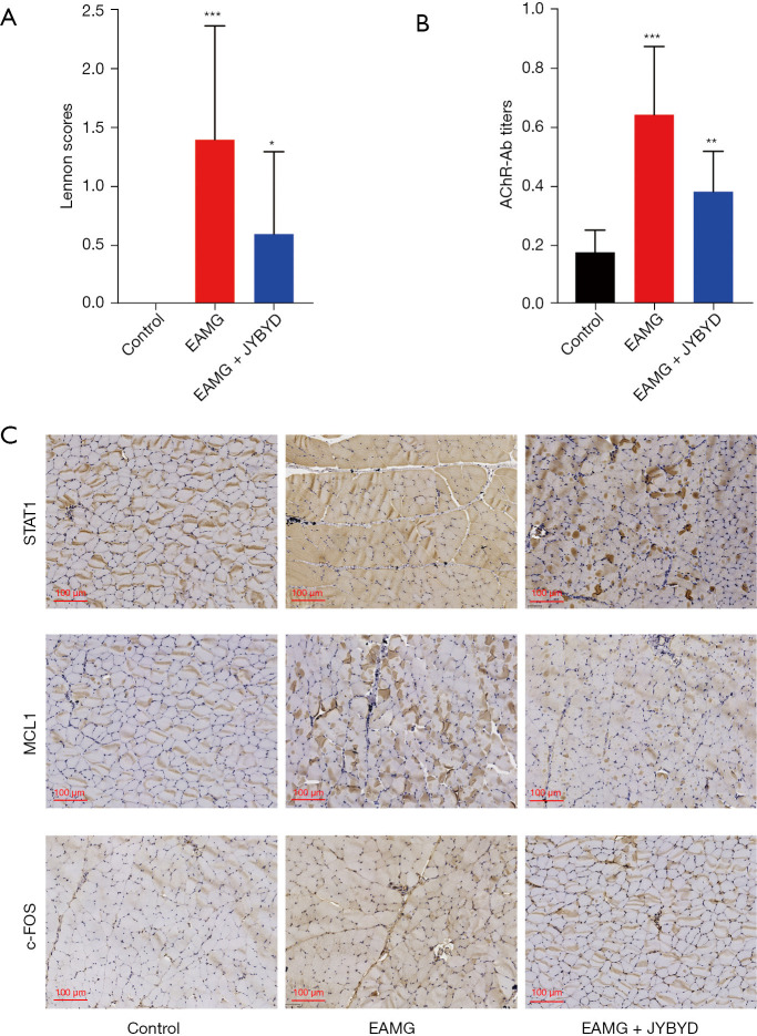
An increase in cell populations characterized by cortactin expression was observed, which has a potential effect on the recovery of lesions at the neuromuscular junction in patients with MG. Based on the differential expression of genes in various immune cells and the predicted targets of traditional Chinese medicine (TCM) compounds, the possible therapeutic targets of JYBYD in different cell subsets were identified, among which STAT1, MCL1, and FOS were the most frequent. Comprehensive network pharmacological analysis suggested quercetin, luteolin, and resveratrol as important active ingredients of JYBYD for the treatment of children with MG. JYBYD could relieve myasthenia symptoms and reduce the AChR-Ab titer in the rat model. Immunohistochemistry results of the muscle showed that JYBYD treatment decreased the expression of STAT1, MCL1, and c-FOS proteins in the muscles of MG rat models.
Conclusions
The results of this study are of significance for the clinical application of JYBYD and drug development against MG in children.
Keywords: Traditional Chinese medicine (TCM), myasthenia gravis (MG), cortactin, Jianpi Yiqi Bugan Yishen Decoction (JYBYD)
Highlight box.
Key findings
• STAT1, MCL1, and FOS may be key targets of JYBYD in the treatment of children with myasthenia gravis.
What is known and what is new?
• Chinese herbal medicines have been widely used in clinical practice as a treatment for myasthenia gravis.
• Our study suggests possible targets and active ingredients of JYBYD for treating myasthenia gravis in children.
What is the implication, and what should change now?
• The results of this study are of significance for the clinical application of JYBYD and drug development against myasthenia gravis in children.
Introduction
Myasthenia gravis (MG) is an acquired autoimmune disease of the neuromuscular junction (1). MG can be subdivided into ocular and generalized forms, according to whether symptoms are confined to the ocular muscles. Anti-acetylcholine receptor antibody (AChR-Ab) can be detected in approximately 80% of patients with MG, whereas anti-muscle-specific tyrosine kinase antibody or anti-low-density lipoprotein receptor-related protein 4 antibody can be detected in a small proportion of patients (2). Epidemiological studies have shown the global prevalence of MG is approximately 100–350/million, while the annual incidence is approximately 10–29/million (3). In contrast, the incidence of MG in China is approximately 0.68/100,000 people, with a slightly higher incidence in females (4). China has a higher proportion of patients with MG than Europe and North America, with a median age of patients below 15 (5). Approximately 80% of children with MG in China show eye muscle involvement (6). Children and adults with MG present with a wide spectrum of characteristics, including variations in symptoms, clinical severity, and antibody titers (7).
Currently, symptomatic treatment with cholinesterase inhibitors and non-specific immunosuppressants (mainly glucocorticoids) are the preferred treatment methods for patients in China (8), although these have serious side effects that may affect the growth and development of children, and patients are prone to relapse after drug withdrawal. Therefore, it is important to identify new therapeutic targets for MG.
Traditional Chinese medicine (TCM) dates back thousands of years, and Chinese herbal medicines have been widely used in clinical practice as a treatment for MG (9). While holding definite curative effects and few adverse effects, there has been little in-depth research on the mechanism of action of Chinese herbal medicine in MG, which has become one of the main barriers to its routine application and popularization. Jianpi Yiqi Bugan Yishen Decoction (JYBYD) mainly consists of Astragalus membranaceus (Fisch.) Bunge (Huangqi), Codonopsis pilosula (Franch.) Nannf. (Dangshen), Atractylis lancea var. chinensis (Bunge) Kitam. (Baizhu), Bupleurum chinense DC (Chaihu), Cimicifuga foetida L. (Shengma), Angelica sinensis (Oliv.) Diels (Danggui), Polygonum multiflorum Thunb. (Heshouwu), Cornus officinalis Siebold & Zucc. (Shanzhuyu), Lycium chinense Mill. (Gouqizi), Morinda officinalis F.C.How (Bajitian), Spatholobus suberectus Dunn (Jixueteng), Glycyrrhiza uralensis Fisch. (Gancao), Cuscuta chinensis Lam. (Tusizi), and Citrus reticulata Blanco (Chenpi), and is clinically effective in the treatment of children with MG (10). Our previous clinical study has shown the clinical remission rate of children with MG treated with JYBYD can reach 96.2% within one year, in both AChR-Ab-positive and -negative children (10). In AChR-Ab-positive children with ocular muscle MG, the AChR-Ab value can drop to the normal range after clinical remission, although its target and immunological mechanism remain unclear.
Network pharmacology is an emerging discipline based on network theory and system biology principles and is considered an effective tool to systematically uncover complex network relationships among drugs, ingredients, targets, and diseases. While it provides a new perspective for analyzing and predicting the pharmacological mechanism of drugs (11,12), TCM and its prescriptions are multi-component and multi-target and display multi-way synergy. Therefore, the research concept of network pharmacology highly matches the intended in-depth analysis of the overall treatment concept of TCM, which provides a new opportunity for in-depth study of the action mechanism of various TCM compounds (13).
Heterogeneity exists among different subsets of immune cells, resulting in indistinguishable responses to therapeutic drugs when assessed using traditional research methods. With the advent of single-cell RNA sequencing (scRNA-seq), it has become possible to accurately identify different subsets of immune cells and further evaluate the heterogeneity of gene expression changes in these subsets before and after drug treatment. In this study, network pharmacology combined with scRNA-seq technology was used to explore the mechanisms of action of TCM in the treatment of children with MG. scRNA-seq was used to detect the peripheral blood cells before and after treatment with TCM, and the transcriptome of peripheral blood immune cells was established to identify the differential gene expression in different immune cells. Furthermore, the targets of TCM compounds were predicted by combining systematic network pharmacology, and the possible therapeutic targets of TCM compounds were identified. Our findings may accelerate the discovery of the mechanisms of drug action and development of new therapeutic targets for MG. We present the following article in accordance with the ARRIVE reporting checklist (available at https://tp.amegroups.com/article/view/10.21037/tp-22-593/rc).
Methods
Screening of active ingredients and possible targets of JYBYD
The active ingredients of JYBYD were identified using the TCM systematic pharmacology database and analysis platform (http://lsp.nwu.edu.cn/tcmsp.php) and the BATMAN-TCM database (http://bionet.ncpsb.org/batman-tcm/). The parameter standards for the identification of the active ingredients were set as oral bioavailability ≥30% and drug-likeness ≥0.18. Using the target prediction function in the database, relationship pairs of active ingredients and target genes in JYBYD were obtained for further analysis of drug targets.
Patient inclusion criteria and sample preparation
This study was conducted at the Diagnosis and Treatment Center for Myasthenia Gravis, People’s Hospital of Shijiazhuang affiliated to Hebei Medical University. The study was conducted in accordance with the Declaration of Helsinki (as revised in 2013). This study complied with Chinese regulations and was approved by the Ethics Committee of the People’s Hospital of Shijiazhuang affiliated to Hebei Medical University (No. [2019]085). Informed consent was taken from all the patients’ legal guardians. scRNA-seq analysis was performed on the peripheral white blood cells of both a child with MG before and after Chinese medicine treatment and a healthy
control. We determined the drug dosing according to the results of our previous clinical study (10), which did not observe significant side effects in the clinic. According to the clinical classification of the American Myasthenia Gravis Foundation, the child was clinically graded as grade I (14) (ocular muscle weakness, possible weakness in closing the eyes, and normal strength of all other muscles). The included children met the following criteria: (I) 3–6 years old, (II) not receiving any medical treatment, (III) no family history or illness after birth, and (IV) no complications of other diseases.
Peripheral blood (3 mL) was collected from each patient in an ethylenediaminetetraacetic acid anticoagulant tube, and 6 mL of red blood cell lysate (Sigma, Cat: R7757) was added to each test tube, mixed, and centrifuged at 500 ×g for 5 min at 4 ℃ to remove the supernatant. Subsequently, the cells were resuspended in PBS, assayed for activity by trypan blue staining, and counted using an automated Cell Counter (CountessTM 3 FL, Thermo Fisher Scientific, Waltham, USA).
Library preparation and sequencing
scRNA-seq libraries were prepared using the Chromium Single Cell 3' Library & Single Cell 3' v3 Gel Beads kit (10× Genomics) according to the manufacturer’s instructions. The single-cell suspension was loaded onto a chromium single-cell controller (10× Genomics) to generate droplet structures of single-cell gel in emulsion, with approximately 12,000 cells loaded into each channel. A reverse transcription reaction was used to generate the full-length complementary DNA (cDNA) of the barcode, and the emulsion was destroyed with a restorative agent. cDNA was cleaned using DynaBead Myone Silane Beads (Thermo Fisher Scientific), then amplified, fragmented, end-repaired, A-tailed, and ligated to the index adapter. The final individual sample libraries were evaluated on an Agilent Bioanalyzer using a highly sensitive DNA kit (Agilent Technologies). Every library was sequenced on a HiSeq X Ten platform (Illumina), and a pair-wise end reading of 150 bp was generated.
scRNA-seq data processing
Data statistics, data quality evaluation, and reference genome comparison were performed on the original data using Cell Ranger, an official software of 10x genomics. The software distinguished each cell by identifying the barcode sequence and each molecule by identifying the unique molecular identifier to generate data on the number of detected cells, the median value of genes, and the sequencing saturation. Based on the preliminary quality control results of the cell ranger, the Seurat software package was used to further control and process the data to obtain a high-quality cell population for subsequent dimensionality reduction and clustering analysis.
Differential gene and enrichment analyses
Differential gene expression screening was performed using the Seurat software package. Significant differential gene expression was defined by a P value less than 0.05 and fold difference greater than 1.5. Gene Ontology and Kyoto Encyclopedia of Genes and Genomes enrichment analyses of the differentially expressed genes were performed using a hypergeometric distribution test. Bar charts were drawn using the GraphPad software, and bubble charts were drawn using the ggplot software package.
Protein-protein interaction (PPI) network construction
A list of genes related to ocular muscle MG was downloaded from the DisGeNET database (https://www.disgenet.org/). The STRING database (Version 11.5; https://cn.string-db.org/) was used to analyze the interaction between MG-related genes and target genes of the active ingredients of TCM, and Cytoscape software (Version 3.7.2) was used to draw the network diagram.
Construction of medicinal material-compound-target network
The relationship pairs of Chinese medicinal material compounds and compound target genes were constructed and imported into the Cytoscape Version 3.7.2 software to construct the network of “Chinese medicinal materials-compounds-targets”.
Component-target molecular docking
The crystal structures of the target protein were retrieved from the UniProt database, the 3D structures were obtained from the PubChem compound database, and the energy was minimized using AVOGADR 1.2.0 under the MMFF94 force field.
The AutoDock Vina 1.1.2 software was used for molecular docking, and all receptor proteins were hydrotreated with PyMol prior to docking. Furthermore, ADFRsuite 1.0 was used to convert all the processed small molecules and receptor proteins into the PDBQT format necessary for AutoDock Vina 1.1.2 docking. Before docking, appropriate X, Y, and Z side lengths were adjusted with the centroid of the protein as the center of the box to construct it and fully wrap the entire protein. For docking, the grid box and PDBQT files of processed proteins and small molecules were used as input files, and Vina was used for docking. The exhaustive degree of the global search for docking was set to 32, and other parameters were kept at default settings. Finally, the output docking conformation with the highest score was considered as the binding conformation, and the docking results were visually analyzed using PyMol.
Construction of MG rat model and drug experiments
All animal care and experimental procedures in this study were in accordance with the National Institute of Health Guidelines for the Care and Use of Laboratory Animals and were approved by the Animal Ethics Committee of the People’s Hospital of Shijiazhuang affiliated to Hebei Medical University (No. 2022-002). Thirty specific pathogen-free (SPF) female Lewis rats (the sample size was determined by statistical analysis of pre-experimental data) aged 4–6 weeks (Vital River, Beijing) were selected and raised in an SPF environment (the rats were randomly selected for the subsequent experiments). Ten rats were used as normal controls, and the other 20 were included in the model with classic MG induced by subcutaneous injection of the immunogen acetylcholine receptor (AChR) into the footpad, abdomen, and back. The 20 rats were divided into two groups and administered normal saline and JYBYD (after conversion to pediatric patient dosing, 5 g/mL, 2 mL) by gavage daily (30 days in total). Modeling was performed in the following five steps: (I) 1 mg/mL AchR2α protein was mixed with the same volume of Freund’s complete adjuvant to form a water-in-oil emulsion; (II) the rats were anesthetized with isoflurane by injecting the emulsion subcutaneously into the footpad, abdomen, and back; (III) 1 mg/mL AchR2α protein was mixed with the same volume of Freund’s incomplete adjuvant to form a water-in-oil emulsion, and the above emulsion was injected again on the 20th day to immunize the rats; and (IV) 1 mg/mL AchR2α protein was mixed with the same volume of Freund’s complete adjuvant to form a water-in-oil emulsion, and the above emulsion was injected again on the 30th day to enhance immunization. All surgeries were performed under isoflurane anesthesia.
On the 35th day, Lennon’s scoring method was used to score muscle strength (15), and the peripheral blood and muscle tissue of rats were collected for follow-up experiments. Serum AChR-Ab titers (AchR2α polyAb Rat, njbiogene, Lot. 17001367) were detected using ELISA (three repetitions)., and STAT1 (CST, Cat. 14994, Lot. 1), MCL1 (HUABIO, Cat. ET1606-14), and c-FOS (HUABIO, Cat. ET1701-95) immunohistochemical staining was performed on the muscle tissue of the rats. The rats used in our study were euthanized painlessly by decapitation after the experiments. The protocols used in the study were prepared before the study without registration.
Statistical analysis
A t-test was used to evaluate the difference between the two groups statistically, and data were expressed as mean ± standard deviation (SD). The statistical significance criteria used were *P<0.05, **P<0.01, ***P<0.001 (GraphPad Prism 9.0).
Results
Target prediction analysis of JYBYD
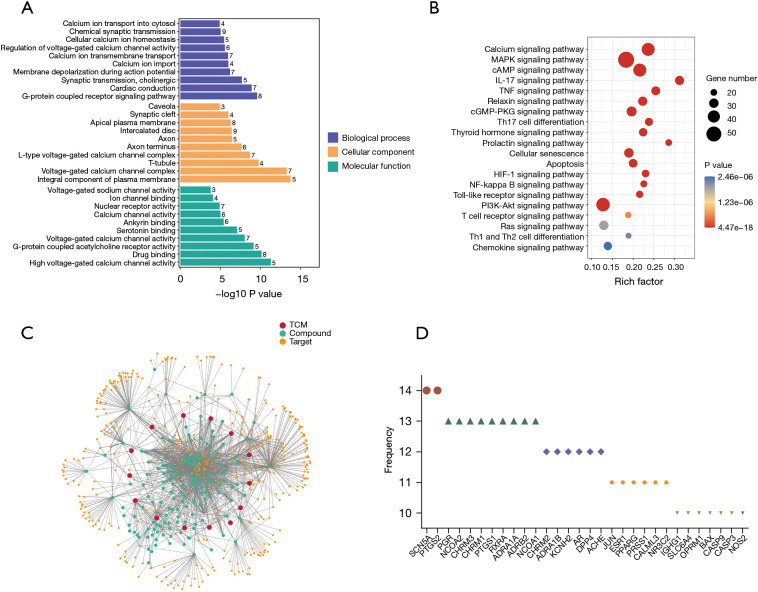
In the TCM systematic pharmacology database and analysis platform and Batman-TCM databases, the active components of JYBYD and their corresponding targets were retrieved with the screening conditions of oral bioavailability ≥30% and drug-likeness ≥0.18. A total of 161 compounds were obtained, targeting 426 genes (Table 1). The results of functional enrichment analysis of drug target genes revealed JYBYD may influence the functions of the G protein-coupled receptor signaling pathway, voltage-gated calcium channel activation, calcium channel activation, calcium signaling pathway, mitogen-activated protein kinase signaling pathway, and tumor necrosis factor (TNF) signaling pathway (Figure 1A,1B). For an in-depth analysis of the relationship between TCMs, compounds, and gene targets, a network diagram of the three was constructed (Figure 1C), and the statistical results indicated many target genes existed in at least 10 types of TCM action networks (Figure 1D).
Table 1. Compounds and target genes corresponding to the 14 components of JYBYD.
| TCM | Compound | Target |
|---|---|---|
| Jixueteng | 23 | 122 |
| Gouqizi | 35 | 180 |
| Chenpi | 5 | 61 |
| Shengma | 8 | 51 |
| Shanzhuyu | 13 | 55 |
| Huangqi | 15 | 186 |
| Chaihu | 12 | 171 |
| Dangshen | 17 | 100 |
| Tusizi | 10 | 191 |
| Gancao | 23 | 51 |
| Bajitian | 16 | 47 |
| Baizhu | 4 | 16 |
| Danggui | 2 | 43 |
| Heshouwu | 16 | 203 |
JYBYD, Jianpi Yiqi Bugan Yishen Decoction; TCM, traditional Chinese medicine.
Figure 1.
JYBYD target analysis. (A) Gene Ontology enrichment analysis results presented as bar chart for JYBYD target genes. (B) Kyoto Encyclopedia of Genes and Genomes enrichment analysis results presented as bubble chart for JYBYD target genes. (C) The traditional Chinese medicine-compound-target gene network diagram for JYBYD. (D) Frequency statistics of top target genes. TCM, traditional Chinese medicine; JYBYD, Jianpi Yiqi Bugan Yishen Decoction.
Cellular composition changes after JYBYD treatment
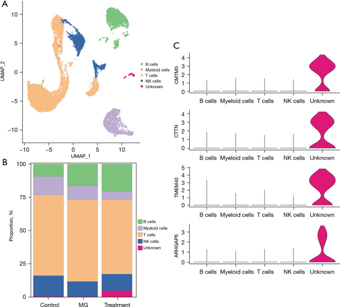
To investigate the changes in cellular composition in children with MG after JYBYD treatment, 10× genomic single-cell transcriptome sequencing was performed on peripheral blood leukocytes from a healthy control (control group) and a child with MG before and after JYBYD treatment (MG and treatment groups, respectively). We identified T, B, myeloid, and natural killer (NK) cells using classical cell markers (Figure 2A), including CD2, CD3D, CD3E, and CD3G for T cells; CD79A, BANK1, and MS4A1 for B cells; CD14, CD68, and CD300E for myeloid cells; and NKG7, GZMA, GZMB, and NCR1 for NK cells. Further, while the proportions of these four cell types changed slightly after JYBYD treatment (Figure 2B), the number of a small group of new cells significantly increased in the treatment samples. Further analysis of this group revealed the following characteristics: (I) a total of 154 cells; (II) a relatively significant increase in the proportion after JYBYD treatment (only 0.2%–0.3% in the control group and MG group, and 4.6% in the treatment group); and (III) the cells did not express any specific markers for T, B, myeloid, or NK cells but expressed other specific markers, such as CMTM5, CTTN, TMEM40, and ARHGAP6 (Figure 2C).
Figure 2.
Single-cell transcriptome sequencing revealed changes in cell profile after JYBYD treatment. (A) UMAP dimensionality reduction clustering diagram. (B) Histogram of the proportion of each type of cells in the sample. (C) Violin map of marker genes in unknown cells. UMAP, uniform manifold approximation and projection; NK, natural killer; MG, myasthenia gravis; JYBYD, Jianpi Yiqi Bugan Yishen Decoction.
Potential gene targets in myeloid cells after JYBYD treatment
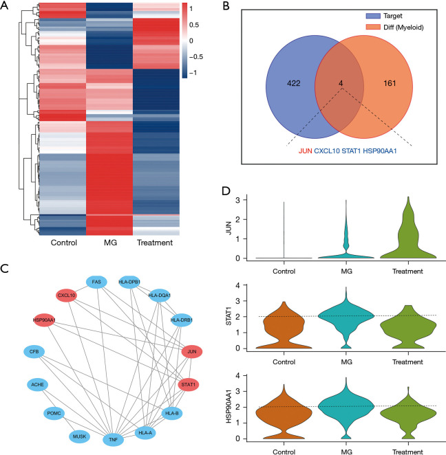
The gene expression in myeloid cells of children with MG before and after treatment was assessed, and a significant difference was found in 165 genes, of which 54 were upregulated and 111 downregulated. Interestingly, expression of all 54 upregulated genes in the control, MG, and treatment groups, characteristically decreased after illness and increased after treatment, whereas most of the 111 downregulated genes showed the opposite trend (Figure 3A). These results show JYBYD may play a role in the treatment of MG in children by regulating gene expression in myeloid cells. To clarify the potential targets of JYBYD in myeloid cells, Venn analysis was performed on its targets and the differential genes before and after myeloid cell treatment, revealing four intersecting genes: JUN, CXCL10, STAT1, and HSP90AA1. Among these, the expression of JUN was upregulated while that of CXCL10, STAT1, and HSP90AA1 was downregulated after treatment (Figure 3B). Additionally, PPI networks of the four potential target genes and MG-related genes in the network database were constructed, and the results showed the four aforementioned genes had simultaneous interactions with MG-related genes, including a relationship between CXCL10 and FAS, JUN and FAS, STAT1 and TNF, HLA-A and HLA-B, and HSP90AA1 and TNF (Figure 3C). Furthermore, a violin plot was used to visualize the expression of JUN, STAT1, and HSP90AA1 in myeloid cells (the relative expression of CXCL10 was low and is not shown). The results showed that after JYBYD treatment, the expression of JUN increased significantly, and that of STAT1 and HSP90AA1 decreased (Figure 3D). These findings suggest JUN, CXCL10, STAT1, and HSP90AA1 are possible targets of JYBYD in myeloid cells.
Figure 3.
Differential expression of myeloid cells and JYBYD target analysis. (A) Differential gene of medullary cells. Color bar, expression level of genes, z-score. (B) Venn analysis of JYBYD target genes and differentially expressed genes in medullary cells. (C) PPI network map of MG-related and drug target genes in the eye muscle. (D) Violin map of drug target gene expression in medullary cells. MG, myasthenia gravis; JYBYD, Jianpi Yiqi Bugan Yishen Decoction; PPI, protein-protein interaction.
Potential gene targets in NK cells after JYBYD treatment
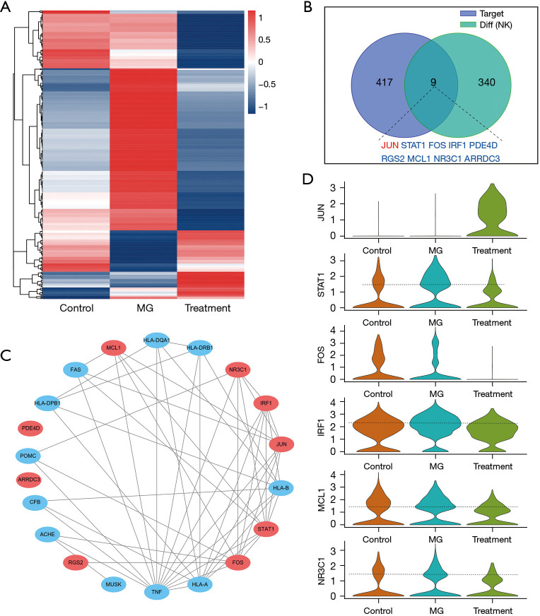
The investigation of NK cells, an important immune cell population in the human body, is crucial to determine the targets of JYBYD. The gene expression in NK cells of children with MG before and after treatment was first compared, and a significant difference was found in 349 genes, of which 79 were upregulated and 270 downregulated. Similarly, many abnormally expressed genes restored their normal expression after JYBYD treatment, including those in the MG group, where genes whose expression was significantly increased were downregulated, and genes whose expression was significantly decreased were upregulated) (Figure 4A). Venn analysis showed nine possible target genes of JYBYD in NK cells during MG treatment: JUN, STAT1, FOS, IRF1, PDE4D, RGS2, MCL1, NR3C1, and ARRDC3 (Figure 4B), and except for JUN, the other eight genes were significantly downregulated after treatment. The results of the PPI network analysis with MG-related genes showed that, except for PDE4D and ARRDC3, the other seven genes had direct or indirect interactions with MG-related genes, including the relationship between MCL1 and TNF, NR3C1 and POMC, IRF1 and TNF, and HLA-A and HLA-B (Figure 4C). Comparison of the expression of several potential target genes in MG before and after treatment revealed that, similar to the expression pattern in myeloid cells, the expression of JUN after JYBYD treatment was significantly increased, while that of FOS was significantly decreased in NK cells. STAT1, IRF1, MCL1, and NR3C1 also showed lower expression in NK cells (RGS2 is not shown as it had overall low expression) (Figure 4D). These results suggest JUN, STAT1, FOS, IRF1, PDE4D, RGS2, MCL1, NR3C1, and ARRDC3 are possible targets of JYBYD in NK cells.
Figure 4.
Differential expression of NK cells and JYBYD target analysis. (A) NK cell differential gene thermomap. Color bar, expression level of genes, z-score. (B) Venn analysis of JYBYD target genes and differentially expressed genes in NK cells. (C) PPI network map of MG-related genes and drug target genes in the eye muscle. (D) Violin map of drug target gene expression in NK cells. MG, myasthenia gravis; NK, natural killer; JYBYD, Jianpi Yiqi Bugan Yishen Decoction; PPI, protein-protein interaction.
Potential gene targets in B cells after JYBYD treatment
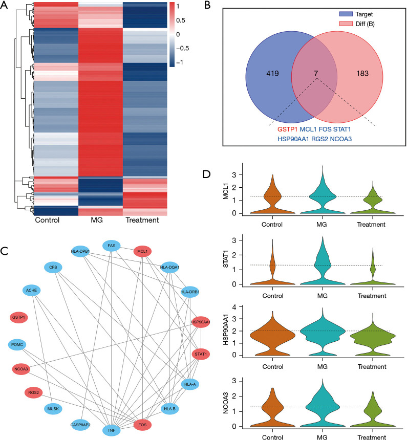
B cells play a direct role in the development of MG. The gene expression in B cells of children with MG before and after treatment was first compared, and a significant difference was found in 190 genes, including 27 upregulated and 163 downregulated genes. Although many genes in B cells showed significant upregulation, after JYBYD treatment, most restored a relatively low expression (Figure 5A). Among the genes showing significant differential expression after treatment, seven were identified as potential target genes of JYBYD: GSTP1, MCL1, FOS, STAT1, HSP90AA1, RGS2, and NCOA3 (Figure 5B). All genes other than GSTP1 could directly or indirectly form a PPI network with MG-related genes (Figure 5C), providing further evidence of them being target genes of JYBYD in B cells. Furthermore, a violin plot was used to visualize changes in the expression of MCL1, FOS, STAT1, HSP90AA1, RGS2, and NCOA3 in the B cells of children with MG after JYBYD treatment. The results showed MCL1, STAT1, HSP90AA1, and NCOA3 expression were slightly upregulated in the MG group but downregulated after JYBYD treatment (FOS and RGS2 are not shown because their overall expression was low) (Figure 5D). These results suggest GSTP1, MCL1, FOS, STAT1, HSP90AA1, RGS2, and NCOA3 are drug targets of JYBYD in B cells.
Figure 5.
Differential expression of B cells and JYBYD target analysis. (A) B cell differential gene thermomap. Color bar, expression level of genes, z-score. (B) Venn analysis of JYBYD target genes and differentially expressed genes in B cells. (C) PPI network map of MG-related and drug target genes in the eye muscle. (D) Violin map of drug target gene expression in B cells. MG, myasthenia gravis; JYBYD, Jianpi Yiqi Bugan Yishen Decoction; PPI, protein-protein interaction.
Potential drug targets in T cells
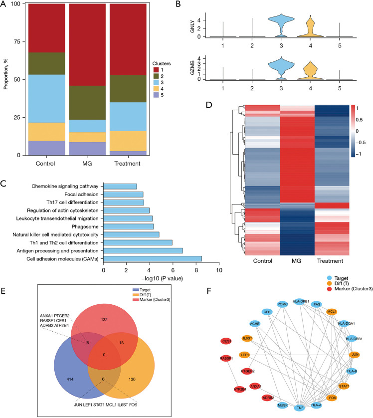
MG is a T-cell-dependent immune disease, and the possible role of JYBYD on T cells in its treatment was investigated. T cells were clustered using uniform manifold approximation and projection dimensionality reduction and divided into five subgroups Changes in the five T-cell subset proportions before and after JYBYD treatment were then analyzed. The results showed slight changes in the proportions of subgroups 1 and 2, a decrease in the proportion of subgroup 5, and a partial recovery of the decreased proportions of subgroups 3 and 4 in the MG group, among which the proportion of cells in subgroup 3 changed significantly. Therefore, changes in subgroup 3 were the focus of subsequent parts of the study (Figure 6A). Subgroup 3 T cells showed high expression of GNLY and GZMB, suggesting they may serve as effector T cells (Figure 6B). Functional enrichment analysis of the top marker genes of subgroup 3 showed cell adhesion molecules, Th1 and Th2 cell differentiation, Th17 cell differentiation, the chemokine signaling pathway, and other functions were enriched (Figure 6C). As the proportion of subgroup 3 T cells showed the largest difference, this subgroup was considered the key T-cell subgroup responsible for the effects of JYBYD, and the top marker genes of T cells in this subgroup were included in subsequent analysis of its potential targets. Gene expression changes in T cells after JYBYD treatment were also studied (Figure 6D). After JYBYD treatment, the expression of 154 genes changed significantly in T cells, including 54 upregulated and 100 downregulated genes. Venn analysis was then conducted on the JYBYD target gene, top marker genes of subgroup 3, and differentially expressed genes of T cells. Among the top marker genes of T cells in subgroup 3, six candidate genes were identified: ANXA1, PTGER2, RASSF1, CES1, ADRB2, and ATP2B4, and among the differentially expressed genes, six additional candidate genes were identified: JUN, LEF1, STAT1, MCL1, IL6ST, and FOS (Figure 6E). Twelve candidate genes and MG-related genes were constructed to obtain a PPI network (Figure 6F), and nine (except CES1, RASSF1, and ATP2B4) showed obvious interactions with MG-related genes. These results suggest six marker genes and six differentially expressed genes in subgroup 3 T cells are potential targets of JYBYD in T cells.
Figure 6.
Target analysis of JYBYD in T cells. (A) Histogram showing T-cell subset proportions. (B) Marker gene violin map. (C) Bar chart showing results of functional enrichment analysis of top marker genes in subgroup 3 T-cell subset. (D) T cell differential gene thermomap. Color bar, expression level of genes, z-score. (E) Venn analysis of JYBYD target genes, differentially expressed genes, and subgroup 3 marker genes in T cells. (F) PPI network map of MG-related genes and drug target genes in the eye muscle. MG, myasthenia gravis; JYBYD, Jianpi Yiqi Bugan Yishen Decoction; PPI, protein-protein interaction.
Comprehensive analysis of targets in various cells of children with MG treated with JYBYD
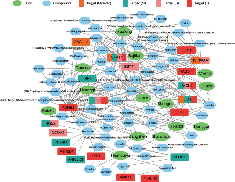
Based on the above analysis of the potential targets of JYBYD in myeloid cells, NK cells, B cells, and T cells, an in-depth comprehensive summary analysis was conducted on the 14 TCMs contained in JYBYD, their corresponding compounds, and the target genes discussed above (Figure 7).
Figure 7.
Summary and analysis of JYBYD-compound-target genes. TCM, traditional Chinese medicine; NK, natural killer; JYBYD, Jianpi Yiqi Bugan Yishen Decoction.
Results obtained for myeloid cells showed 12 TCMs could act on JUN, CXCL10, STAT1, and HSP90AA1, while 13 medicines were found to act on NK cells, and ten could directly act on B cells. In contrast, all 14 TCMs contained in JYBYD could exert curative effects on T cells via different target genes.
Of the compounds contained in the 14 kinds of TCMs, 60 compounds acted on 21 target genes in four types of cells to varying degrees. Among these genes, JUN, HSP90AA1, and ADRB2 could be simultaneously activated by more than 20 compounds.
STAT1 is a common target in myeloid, NK, B, and T cells, while MCL1 and FOS are common targets in NK, B, and T cells, and JUN is a common target in myeloid, NK, and T cells.
Quercetin (from Gouqizi, Huangqi, Chaihu, and Tusizi), luteolin (from Dangshen and Jixueteng), and resveratrol (from Heshouwu) can target more than five genes at the same time, which indicates the important role of these compounds and their corresponding TCMs in treating children with MG.
Overall, we preliminarily identified the key compounds of JYBYD and the possible target genes in each cell type in the process of treating children with MG by combining a pharmacological analysis of the TCM network and scRNA-seq sequencing.
Active ingredients of JYBYD target proteins associated with childhood MG
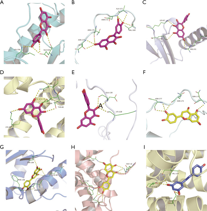
As indicated by the above results, quercetin, luteolin, and resveratrol may be important active ingredients of JYBYD for the treatment of children with MG, and to verify the relationship between these ingredients and the target proteins related to the above study, molecular docking was performed (Figure 8). The results suggest the binding energies of quercetin, luteolin, and resveratrol to related proteins are all lower than −5 kcal/mol, indicating these compounds and proteins have ideal potential activity effects (Table 2).
Figure 8.
Compound docking with the target protein molecule. The yellow dotted line shows hydrogen bonding, and the green line shows amino acids that form hydrogen bonds with the compound. (A) STAT1—Quercetin. (B) JUN—Quercetin. (C) CXCL10—Quercetin. (D) ADRB2—Quercetin. (E) FOS—Quercetin. (F) JUN—Luteolin. (G) HSP90AA1—Luteolin. (H) MCL1—Luteolin. (I) ADRB2—Resveratrol.
Table 2. Components docked with key targets.
| Components | Gene | Binding energy (kcal/moL) |
|---|---|---|
| Quercetin | STAT1 | −7.2 |
| Quercetin | JUN | −5.6 |
| Quercetin | CXCL10 | −6.6 |
| Quercetin | ADRB2 | −7.1 |
| Quercetin | FOS | −5.5 |
| Luteolin | JUN | −5.7 |
| Luteolin | HSP90AA1 | −9.3 |
| Luteolin | MCL1 | −7.3 |
| Resveratrol | ADRB2 | −9.1 |
JYBYD improved MG symptoms in experimental autoimmune MG
To verify the therapeutic effect of JYBYD on MG and its effect on the above targets, a rat model of MG was constructed, and the results suggested JYBYD could relieve myasthenia symptoms and reduce the AChR-Ab titer (Figure 9A,9B). Immunohistochemistry results of the muscle showed JYBYD treatment decreased the expression of STAT1, MCL1, and c-FOS proteins in the muscles of MG rat models (Figure 9C).
Figure 9.
Preventive effect of JYBYD on EAMG. (A) Lennon scores of controls (n=10), EAMG (n=10), and EAMG + JYBYD (n=10). (B) AChR-Ab titers of control (n=10), EAMG (n=10), and EAMG + JYBYD (n=10). (C) Target protein expression in muscle tissues (IHC) of control, EAMG, and EAMG + JYBYD. *, P<0.05, **, P<0.01, ***, P<0.001. EAMG, experimental autoimmune myasthenia gravis; JYBYD, Jianpi Yiqi Bugan Yishen Decoction; EAMG + JYBYD, EAMG with JYBYD treatment; AChR-Ab, anti-acetylcholine receptor antibody; IHC, immunohistochemistry.
Discussion
MG is an autoimmune disease, and most patients with MG have specific autoantibodies (2). Antibody production depends on CD4+ T cells, which are necessary to induce subsequent B-cell responses. Innate CD4+ T cells differentiate into different subsets of T helper (Th) cells, including Th1, Th2, Th17, T follicular helper (Tfh) cells, and regulatory T (Treg) cells. Abnormalities in these cells are involved in the pathogenic mechanisms of MG (16). Because of the high connectivity, heterogeneity, and plasticity of the immune system, traditional marker-based and bulk technologies left key questions unanswered. Single-cell genomics changes the way we characterize complex immune cell assemblies and study their spatial organization, dynamics, clonal distribution, pathways, function, and crosstalks (17). For reasons of cost, standardization and calculation tools, its full potential to revolutionize immunology and immunotherapy has yet to be fully realized.
Identification of disease targets in network pharmacology research is of paramount importance. Given the complexity of biomolecular interaction networks, in-depth research is required to uncover the mechanism of action of drugs on different cell subsets.In contrast to the study of adult MG (18), we conducted the first s scRNA-seq study of peripheral blood cell in pediatric MG patients to the best of our knowledge. Based on previous clinical research on JYBYD in treating children with MG, scRNA-seq was used to detect the peripheral blood cells before and after treatment with TCM compounds and to analyze changes in different cell groups. Combining these results with the network pharmacology method of target prediction and network analysis, the possible mechanism of action of JYBYD in the treatment of children with MG was evaluated, and its potential targets and material basis were analyzed.
After JYBYD treatment, a small group of new cells was added to the treatment samples, and as a specific marker of this group, CTTN deserves attention. Cortactin, a protein expressed by CTTN, is a postsynaptic neuromuscular junction intracellular protein (19) which acts downstream of agrin/LRP4/MuSK and promotes AChR aggregation (20). Cortactin is considered a potential antigen in MG pathogenesis because of its important role in neuromuscular transmission (21). Previous study has detected anti-cortactin autoantibodies in patients with MG, especially in double-seronegative patients (negative for AChR-Ab and anti-muscle-specific tyrosine kinase antibody) (22). In the present study, after JYBYD treatment, the number of cells with specific expression of cortactin increased, suggesting JYBYD may promote the aggregation of AChR through cortactin and ameliorate myasthenia symptoms.
Our results showed the target genes STAT1, MCL1, and FOS were upregulated in all cell subsets, and their abundance changed significantly from the normal control and pre-treatment group compared to their levels after treatment, indicating their importance in the remission of MG. STATs are also downstream targets of Janus tyrosine kinases, and the Janus tyrosine kinase/STAT signal transduction pathway mediates a variety of immune-related cytokine signals (23,24). Among these, the type-I interferon (IFN-I) signaling pathway depends on the induction of STAT1 (25), and IFN-I has been found to increase the risk of MG (26). Patients with MG exhibit high levels of IFN-I and expression of various IFN-I-induced genes in the thymus (27), and targeted STAT1 therapy may hold promise for its treatment. Thymic abnormalities are considered the key to the pathogenesis of MG (28), and several previous studies have demonstrated an abnormal expression of apoptosis-related genes, such as FAS and BCL-2, in the thymus of patients with the disease (29). MCL1 is a member of the BCL-2 family which can inhibit cell apoptosis by blocking the activation of the pro-apoptotic proteins Bax and Bak, and the overexpression of MCL1 may aggravate autoimmune nephropathy in lpr mice (30). Belonging to the activator protein 1 family, FOS is a key transcription factor that regulates cell proliferation, differentiation, and apoptosis (31). Recent study have correlated the dysfunction of activator protein 1 activity with the occurrence of several autoimmune diseases (32). A previous study indicated the expression of FRA1, a FOS member, is higher in the thymus of patients with MG than in the normal thymus, and that the overexpression of FRA1 interferes with the secretion of inflammatory cytokines in mTEC in these patients (33).
The results of the present study suggest three compounds; quercetin, luteolin, and resveratrol, may play a significant role in the treatment of children with MG. Quercetin is a flavonoid widely found in plants and has many pharmacological activities, including antitumor, antioxidant, anti-inflammatory, antiviral, and immunomodulatory activities (34). A study has shown that quercetin has a significant inhibitory effect on nitric oxide, TNF-α, interleukin 6 (IL-6), and IL-1β release by RAW264.7 cells induced by lipopolysaccharides (35). In a randomized, double-blind, placebo-controlled clinical study, morning stiffness and pain were significantly relieved by quercetin in patients with rheumatoid arthritis (RA) (36). In in vivo experiments, quercetin significantly reduced the arthritis score and improved the symptoms of RA in mice by inhibiting neutrophil infiltration and reducing the levels of pro-inflammatory cytokines, such as TNF-α, IL-6, and IL-17 (37,38). Luteolin has been shown to have anti-inflammatory activity both in vitro and in vivo (39,40) and can also inhibit the phosphorylation of STAT1 (41). In addition, luteolin, as an anti-inflammatory drug, has been shown to be effective in other autoimmune diseases, such as multiple sclerosis (42,43) and experimental autoimmune encephalomyelitis (44). Resveratrol can inhibit T-cell differentiation, particularly by inhibiting key cytokines, such as TNF-α, IL-17, IL-6, and IL-1β (45), and its therapeutic effects on autoimmune diseases have been demonstrated in previous study (46). These active compounds are mainly derived from Fructus lycii, Astragalus membranaceus, Bupleurum chinense, Semen cuscutae, Codonopsis pilosula, Spatholobus suberectus, and Polygonum multiflorum Thunb, which also indicates the essential role of these TCMs in the treatment of children with MG.
Our results provide theoretical support for further target function validation experiments and small molecule therapeutic experiments for therapeutic targets while providing a basis for optimizing clinical applications and developing novel therapeutic agents. Nevertheless, this study has several limitations. First, the sample size we tested was small, and we did not consider variations in peripheral blood lymphocytes across normal childhood growth and development. Second, further experiments are needed to verify the function of key regulatory genes and the causality of the TCM–compound–target network. Third, as this study selected children with ocular MG, the therapeutic targets of children with systemic MG may differ, although these patients are relatively rare.
Conclusions
In conclusion, this study demonstrated the heterogeneity of gene transcription in different immune cell subsets in children with MG before and after JYBYD treatment and identified its possible targets through a joint network pharmacological analysis and a vital material basis. The results of this study lay the foundation for the optimization of clinical prescriptions and the development of novel therapeutic drugs for MG.
Supplementary
The article’s supplementary files as
Acknowledgments
Funding: This work was supported by the Hebei Provincial Natural Science Foundation (No. H2019106063); the S&T Program of Hebei (No. 19277701D); and the TCM Research Program of Hebei (No. 2018251). The funding sources had no role in the study design, data collection, data analysis, data interpretation, and writing of the report.
Ethical Statement: The authors are accountable for all aspects of the work in ensuring that questions related to the accuracy or integrity of any part of the work are appropriately investigated and resolved. The study was conducted in accordance with the Declaration of Helsinki (as revised in 2013). This study complied with Chinese regulations and was approved by the Ethics Committee of the People’s Hospital of Shijiazhuang affiliated to Hebei Medical University (No. [2019]085). Informed consent was taken from all the patients’ legal guardians. All animal care and experimental procedures in this study were in accordance with the National Institute of Health Guidelines for the Care and Use of Laboratory Animals and were approved by the Animal Ethics Committee of the People’s Hospital of Shijiazhuang affiliated to Hebei Medical University (No. 2022-002).
Reporting Checklist: The authors have completed the ARRIVE reporting checklist. Available at https://tp.amegroups.com/article/view/10.21037/tp-22-593/rc
Data Sharing Statement: Available at https://tp.amegroups.com/article/view/10.21037/tp-22-593/dss
Conflicts of Interest: All authors have completed the ICMJE uniform disclosure form (available at https://tp.amegroups.com/article/view/10.21037/tp-22-593/coif). The authors have no conflicts of interest to declare.
(English Language Editor: B. Draper)
References
- 1.Gilhus NE, Tzartos S, Evoli A, et al. Myasthenia gravis. Nat Rev Dis Primers 2019;5:30. 10.1038/s41572-019-0079-y [DOI] [PubMed] [Google Scholar]
- 2.Gilhus NE, Verschuuren JJ. Myasthenia gravis: subgroup classification and therapeutic strategies. Lancet Neurol 2015;14:1023-36. 10.1016/S1474-4422(15)00145-3 [DOI] [PubMed] [Google Scholar]
- 3.Punga AR, Maddison P, Heckmann JM, et al. Epidemiology, diagnostics, and biomarkers of autoimmune neuromuscular junction disorders. Lancet Neurol 2022;21:176-88. 10.1016/S1474-4422(21)00297-0 [DOI] [PubMed] [Google Scholar]
- 4.Chen J, Tian DC, Zhang C, et al. Incidence, mortality, and economic burden of myasthenia gravis in China: A nationwide population-based study. Lancet Reg Health West Pac 2020;5:100063. 10.1016/j.lanwpc.2020.100063 [DOI] [PMC free article] [PubMed] [Google Scholar]
- 5.Zhang X, Yang M, Xu J, et al. Clinical and serological study of myasthenia gravis in HuBei Province, China. J Neurol Neurosurg Psychiatry 2007;78:386-90. 10.1136/jnnp.2006.100545 [DOI] [PMC free article] [PubMed] [Google Scholar]
- 6.Huang X, Li Y, Feng H, et al. Clinical Characteristics of Juvenile Myasthenia Gravis in Southern China. Front Neurol 2018;9:77. 10.3389/fneur.2018.00077 [DOI] [PMC free article] [PubMed] [Google Scholar]
- 7.Heckmann JM, Europa TA, Soni AJ, et al. The Epidemiology and Phenotypes of Ocular Manifestations in Childhood and Juvenile Myasthenia Gravis: A Review. Front Neurol 2022;13:834212. 10.3389/fneur.2022.834212 [DOI] [PMC free article] [PubMed] [Google Scholar]
- 8.Narayanaswami P, Sanders DB, Wolfe G, et al. International Consensus Guidance for Management of Myasthenia Gravis: 2020 Update. Neurology 2021;96:114-22. 10.1212/WNL.0000000000011124 [DOI] [PMC free article] [PubMed] [Google Scholar]
- 9.Chen S, Xu MB, Zhou XL, et al. Chinese Herbal Medicine for Myasthenia Gravis: A Systematic Review and Meta-Analysis. Front Pharmacol 2018;9:969. 10.3389/fphar.2018.00969 [DOI] [PMC free article] [PubMed] [Google Scholar]
- 10.Qi G, Liu P, Gu S, et al. Effect of Jianpiyiqi granule on ocular myasthenia gravis in children prepared with a formula from Traditional Chinese Medicine. J Tradit Chin Med 2017;37:546-53. [PubMed] [Google Scholar]
- 11.Yu G, Luo Z, Zhou Y, et al. Uncovering the pharmacological mechanism of Carthamus tinctorius L. on cardiovascular disease by a systems pharmacology approach. Biomed Pharmacother 2019;117:109094. 10.1016/j.biopha.2019.109094 [DOI] [PubMed] [Google Scholar]
- 12.Ge Q, Chen L, Yuan Y, et al. Network Pharmacology-Based Dissection of the Anti-diabetic Mechanism of Lobelia chinensis. Front Pharmacol 2020;11:347. 10.3389/fphar.2020.00347 [DOI] [PMC free article] [PubMed] [Google Scholar]
- 13.Zhu B, Zhang W, Lu Y, et al. Network pharmacology-based identification of protective mechanism of Panax Notoginseng Saponins on aspirin induced gastrointestinal injury. Biomed Pharmacother 2018;105:159-66. 10.1016/j.biopha.2018.04.054 [DOI] [PubMed] [Google Scholar]
- 14.Jaretzki A, 3rd, Barohn RJ, Ernstoff RM, et al. Myasthenia gravis: recommendations for clinical research standards. Task Force of the Medical Scientific Advisory Board of the Myasthenia Gravis Foundation of America. Neurology 2000;55:16-23. 10.1212/wnl.55.1.16 [DOI] [PubMed] [Google Scholar]
- 15.Lennon VA, Lindstrom JM, Seybold ME. Experimental autoimmune myasthenia: A model of myasthenia gravis in rats and guinea pigs. J Exp Med 1975;141:1365-75. 10.1084/jem.141.6.1365 [DOI] [PMC free article] [PubMed] [Google Scholar]
- 16.Behin A, Le Panse R. New Pathways and Therapeutic Targets in Autoimmune Myasthenia Gravis. J Neuromuscul Dis. 2018;5:265-277. 10.3233/JND-170294 [DOI] [PMC free article] [PubMed] [Google Scholar]
- 17.Ginhoux F, Yalin A, Dutertre CA, Amit I. Single-cell immunology: Past, present, and future. Immunity. 2022;55:393-404. 10.1016/j.immuni.2022.02.006 [DOI] [PubMed] [Google Scholar]
- 18.Jin W, Yang Q, Peng Y, et al. Single-cell RNA-Seq reveals transcriptional heterogeneity and immune subtypes associated with disease activity in human myasthenia gravis. Cell Discov 2021;7:85. 10.1038/s41421-021-00314-w [DOI] [PMC free article] [PubMed] [Google Scholar]
- 19.Peng HB, Xie H, Dai Z. Association of cortactin with developing neuromuscular specializations. J Neurocytol 1997;26:637-50. 10.1023/a:1018545827369 [DOI] [PubMed] [Google Scholar]
- 20.Madhavan R, Gong ZL, Ma JJ, et al. The function of cortactin in the clustering of acetylcholine receptors at the vertebrate neuromuscular junction. PLoS One 2009;4:e8478. 10.1371/journal.pone.0008478 [DOI] [PMC free article] [PubMed] [Google Scholar]
- 21.Gallardo E, Martínez-Hernández E, Titulaer MJ, et al. Cortactin autoantibodies in myasthenia gravis. Autoimmun Rev 2014;13:1003-7. 10.1016/j.autrev.2014.08.039 [DOI] [PubMed] [Google Scholar]
- 22.Cortés-Vicente E, Gallardo E, Martínez MÁ, et al. Clinical Characteristics of Patients With Double-Seronegative Myasthenia Gravis and Antibodies to Cortactin. JAMA Neurol 2016;73:1099-104. 10.1001/jamaneurol.2016.2032 [DOI] [PubMed] [Google Scholar]
- 23.Goropevšek A, Holcar M, Avčin T. The Role of STAT Signaling Pathways in the Pathogenesis of Systemic Lupus Erythematosus. Clin Rev Allergy Immunol 2017;52:164-81. 10.1007/s12016-016-8550-y [DOI] [PubMed] [Google Scholar]
- 24.Kang YH, Biswas A, Field M, et al. STAT1 signaling shields T cells from NK cell-mediated cytotoxicity. Nat Commun 2019;10:912. 10.1038/s41467-019-08743-8 [DOI] [PMC free article] [PubMed] [Google Scholar]
- 25.Fanunza E, Grandi N, Quartu M, et al. INMI1 Zika Virus NS4B Antagonizes the Interferon Signaling by Suppressing STAT1 Phosphorylation. Viruses 2021;13:2448. 10.3390/v13122448 [DOI] [PMC free article] [PubMed] [Google Scholar]
- 26.Payet CA, You A, Fayet OM, et al. Myasthenia Gravis: An Acquired Interferonopathy? Cells 2022;11:1218. 10.3390/cells11071218 [DOI] [PMC free article] [PubMed] [Google Scholar]
- 27.Cizeron-Clairac G, Le Panse R, Frenkian-Cuvelier M, et al. Thymus and Myasthenia Gravis: what can we learn from DNA microarrays? J Neuroimmunol 2008;201-202:57-63. 10.1016/j.jneuroim.2008.06.028 [DOI] [PubMed] [Google Scholar]
- 28.Pinal-Fernandez I, Pak K, Gil-Vila A, et al. Anti-Cortactin Autoantibodies Are Associated With Key Clinical Features in Adult Myositis But Are Rarely Present in Juvenile Myositis. Arthritis Rheumatol 2022;74:358-64. 10.1002/art.41931 [DOI] [PMC free article] [PubMed] [Google Scholar]
- 29.Onodera J, Nakamura S, Nagano I, et al. Upregulation of Bcl-2 protein in the myasthenic thymus. Ann Neurol 1996;39:521-8. 10.1002/ana.410390414 [DOI] [PubMed] [Google Scholar]
- 30.Anstee NS, Vandenberg CJ, Campbell KJ, et al. Overexpression of Mcl-1 exacerbates lymphocyte accumulation and autoimmune kidney disease in lpr mice. Cell Death Differ 2017;24:397-408. 10.1038/cdd.2016.125 [DOI] [PMC free article] [PubMed] [Google Scholar]
- 31.He YY, Zhou HF, Chen L, et al. The Fra-1: Novel role in regulating extensive immune cell states and affecting inflammatory diseases. Front Immunol 2022;13:954744. 10.3389/fimmu.2022.954744 [DOI] [PMC free article] [PubMed] [Google Scholar]
- 32.Trop-Steinberg S, Azar Y. AP-1 Expression and its Clinical Relevance in Immune Disorders and Cancer. Am J Med Sci 2017;353:474-83. 10.1016/j.amjms.2017.01.019 [DOI] [PubMed] [Google Scholar]
- 33.Li QR, Ni WP, Lei NJ, et al. The overexpression of Fra1 disorders the inflammatory cytokine secretion by mTEC of myasthenia gravis thymus. Scand J Immunol 2018;88:e12676. 10.1111/sji.12676 [DOI] [PubMed] [Google Scholar]
- 34.Shen P, Lin W, Deng X, et al. Potential Implications of Quercetin in Autoimmune Diseases. Front Immunol 2021;12:689044. 10.3389/fimmu.2021.689044 [DOI] [PMC free article] [PubMed] [Google Scholar]
- 35.Cao H, Jia Q, Yan L, et al. Quercetin Suppresses the Progression of Atherosclerosis by Regulating MST1-Mediated Autophagy in ox-LDL-Induced RAW264.7 Macrophage Foam Cells. Int J Mol Sci 2019;20:6093. 10.3390/ijms20236093 [DOI] [PMC free article] [PubMed] [Google Scholar]
- 36.Javadi F, Ahmadzadeh A, Eghtesadi S, et al. The Effect of Quercetin on Inflammatory Factors and Clinical Symptoms in Women with Rheumatoid Arthritis: A Double-Blind, Randomized Controlled Trial. J Am Coll Nutr 2017;36:9-15. 10.1080/07315724.2016.1140093 [DOI] [PubMed] [Google Scholar]
- 37.Haleagrahara N, Miranda-Hernandez S, Alim MA, et al. Therapeutic effect of quercetin in collagen-induced arthritis. Biomed Pharmacother 2017;90:38-46. 10.1016/j.biopha.2017.03.026 [DOI] [PubMed] [Google Scholar]
- 38.Kawaguchi K, Kaneko M, Miyake R, et al. Potent Inhibitory Effects of Quercetin on Inflammatory Responses of Collagen-Induced Arthritis in Mice. Endocr Metab Immune Disord Drug Targets 2019;19:308-15. 10.2174/1871530319666190206225034 [DOI] [PubMed] [Google Scholar]
- 39.Caporali S, De Stefano A, Calabrese C, et al. Anti-Inflammatory and Active Biological Properties of the Plant-Derived Bioactive Compounds Luteolin and Luteolin 7-Glucoside. Nutrients 2022;14:1155. 10.3390/nu14061155 [DOI] [PMC free article] [PubMed] [Google Scholar]
- 40.Kritas SK, Saggini A, Varvara G, et al. Luteolin inhibits mast cell-mediated allergic inflammation. J Biol Regul Homeost Agents 2013;27:955-9. [PubMed] [Google Scholar]
- 41.Rezai-Zadeh K, Ehrhart J, Bai Y, et al. Apigenin and luteolin modulate microglial activation via inhibition of STAT1-induced CD40 expression. J Neuroinflammation 2008;5:41. 10.1186/1742-2094-5-41 [DOI] [PMC free article] [PubMed] [Google Scholar]
- 42.Theoharides TC. Luteolin as a therapeutic option for multiple sclerosis. J Neuroinflammation 2009;6:29. 10.1186/1742-2094-6-29 [DOI] [PMC free article] [PubMed] [Google Scholar]
- 43.Skaper SD, Barbierato M, Facci L, et al. Co-Ultramicronized Palmitoylethanolamide/Luteolin Facilitates the Development of Differentiating and Undifferentiated Rat Oligodendrocyte Progenitor Cells. Mol Neurobiol 2018;55:103-14. 10.1007/s12035-017-0722-0 [DOI] [PubMed] [Google Scholar]
- 44.Contarini G, Franceschini D, Facci L, et al. A co-ultramicronized palmitoylethanolamide/luteolin composite mitigates clinical score and disease-relevant molecular markers in a mouse model of experimental autoimmune encephalomyelitis. J Neuroinflammation 2019;16:126. 10.1186/s12974-019-1514-4 [DOI] [PMC free article] [PubMed] [Google Scholar]
- 45.Meng T, Xiao D, Muhammed A, et al. Anti-Inflammatory Action and Mechanisms of Resveratrol. Molecules 2021;26:229. 10.3390/molecules26010229 [DOI] [PMC free article] [PubMed] [Google Scholar]
- 46.Oliveira ALB, Monteiro VVS, Navegantes-Lima KC, et al. Resveratrol Role in Autoimmune Disease-A Mini-Review. Nutrients 2017;9:1306. 10.3390/nu9121306 [DOI] [PMC free article] [PubMed] [Google Scholar]
Associated Data
This section collects any data citations, data availability statements, or supplementary materials included in this article.
Supplementary Materials
The article’s supplementary files as


