Abstract
Background
Human nasal epithelial (HNE) cells can be sampled noninvasively and cultured to provide a model of the airway epithelium that reflects cystic fibrosis (CF) pathophysiology. We hypothesised that in vitro measures of HNE cell physiology would correlate directly with in vivo measures of lung physiology and therapeutic response, providing a framework for using HNE cells for therapeutic development and precision medicine.
Methods
We sampled nasal cells from participants with CF (CF group, n=26), healthy controls (HC group, n=14) and single CF transmembrane conductance regulator (CFTR) mutation carrier parents of the CF group (CR group, n=16). Participants underwent lung physiology and sweat chloride testing, and nuclear imaging-based measurement of mucociliary clearance (MCC) and small-molecule absorption (ABS). CF participants completed a second imaging day that included hypertonic saline (HS) inhalation to assess therapeutic response in terms of MCC. HNE measurements included Ussing chamber electrophysiology, small-molecule and liquid absorption rates, and particle diffusion rates through the HNE airway surface liquid (ASL) measured using fluorescence recovery after photobleaching (FRAP).
Results
Long FRAP diffusion times were associated with increased MCC response to HS in CF. This implies a strong relationship between inherent factors affecting ASL mucin concentration and therapeutic response to a hydrating therapy. MCC decreased with age in the CR group, which had a larger range of ages than the other two groups. Likely this indicates a general age-related effect that may be accentuated in this group. Measures of lung ABS correlated with sweat chloride in both the HC and CF groups, indicating that CFTR function drives this measure of paracellular small-molecule probe absorption.
Conclusions
Our results demonstrate the utility of HNE cultures for assessing therapeutic response for hydrating therapies. In vitro measurements of FRAP were particularly useful for predicting response and for characterising important properties of ASL mucus that were ultimately reflected in lung physiology.
Short abstract
Measurements in nasal cell cultures can be used to predict therapeutic response in the lungs of cystic fibrosis patients and pulmonary function in healthy controls https://bit.ly/3ClgpbB
Introduction
Human bronchial and nasal epithelial (HNE) cell cultures have been used to study cystic fibrosis (CF) pathophysiology and to develop therapies for CF. HNE cells can be sampled from the nose through minimally invasive procedures and demonstrate the characteristic pathophysiology of CF airways disease [1, 2]. They also provide a means of comparing cell- and organ-level physiology and therapeutic response within the same subjects. HNE cells have received previous use for personalisation of therapies [3].
We hypothesised that in vitro measures of HNE cell physiology would correlate directly with in vivo measures of therapeutic response in people with CF, providing a framework for using HNE cells for therapeutic development and precision medicine. We collected and cultured HNE cells from participants with CF who then performed assessments of lung physiology at baseline and after the inhalation of 7% hypertonic saline (HS). HS is a common therapy applied in CF to hydrate secretions and improve mucus clearance. We also sought to determine whether there were direct correlations between measures of in vivo lung physiology and in vitro measures of HNE cell physiology. To obtain a wide range for physiological assessments we also performed HNE cell sampling and lung physiology measurements with healthy controls (HC group) and some of the parents of the enrolled CF subjects who were carriers of a single disease-causing CF transmembrane conductance regulator (CFTR) mutation (CR group).
HNE physiology measures included Ussing chamber assessments of sodium (Na+) and chloride (Cl−) currents, and transepithelial resistance (TER), particle diffusion rates through the HNE airway surface liquid (ASL) made using fluorescence recovery after photobleaching (FRAP) [4, 5], optical measures of ASL absorption rate [6] and measurements of small-molecule absorption made using radiolabelled probes (cell ABS) [7].
Lung and systemic physiology measurements included sweat chloride, spirometry, multiple-breath washout (MBW) testing, and nuclear imaging-based measurements of mucociliary clearance (MCC) [8] and small-molecule absorption (ABS) [9, 10]. MCC measures the clearance rate of an inhaled radiolabelled nonabsorbable probe (technetium-99m sulfur colloid) from the lungs as a surrogate for mucus clearance. Therapeutic response was assessed as the increase in MCC after the inhalation of HS versus baseline. ABS measures the absorption rate of a small-molecule radiolabelled probe (indium-111-DTPA) from the lung. This measure was initially developed as an in vivo surrogate for detecting changes in ASL absorption. It provides a measure of paracellular transport likely affected by both paracellular liquid absorption and permeability. Previous studies have demonstrated that ABS is increased in the CF lung and increases proportionally with sweat chloride [9].
Methods
General study procedures
We enrolled participants with CF from our regional centre in Pittsburgh (PA, USA) who were ≥12 years old with forced expiratory volume in 1 s (FEV1) % pred ≥30% (CF group), their single CFTR mutation carrier biological parents (CR group) and healthy controls who were ≥18 years old with FEV1 % pred ≥70% (HC group). Subjects were excluded if they were pregnant or nursing, smokers, or using e-cigarettes. Subjects in the HC group provided blood samples and were excluded if they carried one of 144 known disease-causing CFTR mutations. Recruitment occurred from early 2017 through late 2019.
The HC and CR groups performed a single study visit that included nasal cell sampling, pulmonary function testing and MBW, sweat chloride measurement, and a two-probe nuclear scan to assess MCC/ABS. Nebulised isotonic saline (IS) was delivered for 10 min after the first 10 min of the MCC/ABS scan as a stimulus for liquid absorption in the airways. Participants in the CF group performed one study day where they inhaled nebulised IS and a second study day where they inhaled nebulised HS during the MCC/ABS measurement. The order of the HS and IS days was randomised and the studies were separated by 7–14 days. The study was approved by the University of Pittsburgh Institutional Review Board and registered at ClinicalTrials.gov with identifier number NCT02947126.
Nasal cell sampling, culturing and testing
During nasal cell sampling, a standard otoscope was used to visualise the inferior turbinate and a cytology brush was used to collect cells from along the lower aspect of the turbinate in both nostrils. Culturing methods are described in the supplementary material. Ussing chamber studies were performed on the HNE cultures, including measurements of Na+ and Cl− currents and transepithelial resistance (TER). Na+ current was determined using amiloride to block the epithelial sodium channel. Cl− current was measured after forskolin activation of CFTR. We also measured the absorption rate of the γ-emitting small molecule technetium-99m-DTPA from the apical surface of the HNE cells (cell ABS). This in vitro measurement parallels the in vivo measurement of indium-111-DTPA absorption in the MCC/ABS scans. HNE cultures were not treated with any CFTR modulators and thus the in vitro measurements do not reflect modulator use. More detailed methods are included in the supplementary material.
FRAP was used to measure the diffusion rate of 70-kDa FITC-labelled dextran through the ASL layer. Procedures were performed as previously described [4, 11]. Detailed FRAP methods are included in the supplementary material. The physiological relevance of this measurement has been previously described [5]. FRAP is presented as a ratio of the ASL diffusion time relative to that of saline.
Measurements of lung physiology and sweat chloride
Participants performed nuclear scans to measure MCC and ABS. Subjects inhaled a combination of technetium-99 m sulfur colloid and indium-111-DTPA in a nebulised liquid aerosol and sequential γ-camera images were collected for 80 min. All participants inhaled nebulised IS for 10 min starting 10 min into the imaging period while imaging continued. On a separate study day (order randomised) the CF group inhaled 7% HS (Pulmosal) during this period instead of IS.
Previous studies have demonstrated that differences in the initial distribution of the deposited radioisotope aerosol in the lung can have a confounding effect on measurements of MCC [10]. To facilitate comparisons of MCC measurements on different days with potentially different aerosol distributions, we calculated a measure of MCC that was adjusted based on the initial deposition of the radioisotope aerosol (MCCadjusted). More details on the imaging methods and MCC adjustment are included in the supplementary material. Therapeutic response was defined as the increase in MCCadjusted on the HS day versus the baseline IS day.
Details of sweat chloride measurement are included in the supplementary material. MBW methods are included in the supplementary material and Lung Clearance Index (LCI) data are presented.
Statistical analysis
In vitro and in vivo continuous variables were compared between the CF, CR and HC groups using Kruskal–Wallis (nonparametric) testing including all three groups followed by individual group comparisons by Dunn's test with Holm adjustment (nonparametric, multiple comparisons). A similar analysis was done to compare the effects of the use of CFTR modulators within the CF group. Sex and culture success rates were compared with the Chi-squared test. HS versus IS comparisons of imaging outcomes in the CF group were performed using the Wilcoxon matched-pairs signed-ranks test (nonparametric, paired). Multivariable linear regression was used to determine the effects of aerosol distribution and testing group on MCC. Univariant regression was used to assess relationships between in vitro and in vivo variables and therapeutic response. Univariant regression was used to assess relationships between in vitro and in vivo variables and baseline MCC. Multivariable regression was used to model FEV1 % pred in the CF group. For all regressions, a robust variance estimator was used and the normality of the residuals was verified with the Shapiro–Wilk W-test. Analysis was performed using Stata/IC version 14.0 (StataCorp, College Station, TX, USA).
Results
Participant demographics, pulmonary function, MBW and sweat chloride measurements are presented in table 1. As expected, CF was associated with increased sweat chloride, decreased pulmonary function and increased LCI. Sweat chloride in the single-mutation carrier group (CR group) was similar to previous reports [12]. No subjects in the CR group had sweat chloride measurements >60 mmol·L−1. The CR group was significantly older than the other groups. 11 participants in the CF group had chronic Pseudomonas aeruginosa infection, defined here as two or more positive throat or sputum cultures in the previous year.
TABLE 1.
Demographics of study participants along with pulmonary function, Lung Clearance Index (LCI) and sweat chloride data
| CF (n=26) | CR (n=16) | HC (n=14) | p-value |
p-value
(CF vs HC) |
p-value
(CF vs CR) |
p-value
(CR vs HC) |
|
| Age (years) | 26.5 (19–39) | 48.0 (44–63.5) | 22.5 (20–23) | 0.0001 | 0.09 | <0.0001 | <0.0001 |
| Female/male (n) | 14/12 | 10/6 | 6/8 | 0.56 | 0.51 | 0.29 | 0.43 |
| FEV1 % pred | 70 (50–93) | 97 (93–109) | 102 (95.5–113) (n=12) | 0.0003 | 0.0008 | 0.0014 | 0.30 |
| FVC % pred | 94.5 (76–104) | 104.0 (96–109) | 107.5 (100–116) (n=12) | 0.02 | 0.01 | 0.05 | 0.23 |
| FEF25–75% % pred | 40.5 (22–66) | 100.5 (74–119.5) | 83.0 (68–99.5) (n=12) | 0.0001 | 0.004 | 0.0002 | 0.29 |
| LCI | 9.0 (7.6–13) (n=23) | 7.8 (7.1–8.3) | 7.2 (6.7–7.5) (n=11) | 0.0007 | 0.0005 | 0.01 | 0.10 |
| Sweat chloride (mmol·L−1) | 101 (91–110) | 36 (21–53) (n=14) | 22 (10–29) (n=12) | 0.0001 | <0.0001 | 0.0002 | 0.11 |
Data are presented as median (interquartile range), unless otherwise stated. CF: cystic fibrosis; CR: single CF transmembrane conductance regulator mutation carrier; HC: healthy control; FEV1: forced expiratory volume in 1 s; FVC: forced vital capacity; FEF25–75%: forced expiratory flow at 25–75% of FVC; LCI: Lung Clearance Index (from multiple-breath washout testing). p-values comparing all groups by Kruskal–Wallis (nonparametric) except sex which is Chi-squared. Group comparisons by Dunn's test with Holm adjustment (nonparametric, multiple comparisons).
Table 2 compares cell physiology and electrophysiology in the CF, CR and HC nasal cell cultures. Culturing success in the CR group was limited, with just over 50% of cell samples producing successful cultures. CF subjects demonstrated the expected low Cl− currents along with increased liquid absorption rates, cell ABS and FRAP versus HC. Na+ currents were significantly lower in the CR group compared with the HC group. There were no significant differences in Cl− currents, liquid absorption rate, cell ABS or FRAP when comparing the CR group with the HC group. TER was similar in all three groups.
TABLE 2.
In vitro measures of human nasal epithelial (HNE) cell physiology across the groups
| CF (n=26) | CR (n=16) | HC (n=14) | p-value |
p-value
(CF vs HC) |
p-value
(CF vs CR) |
p-value
(CR vs HC) |
|
| Culture success/failed (n) | 23/3 | 9/7 | 14/0 | 0.004 | 0.18 | 0.004 | 0.001 |
| Cl− current (µA·cm−2) | 0.21 (−0.2–0.5) (n=16) | 4.0 (1.5–5.2) (n=7) | 5.4 (4.9–10.2) (n=13) | 0.0001 | <0.0001 | 0.003 | 0.13 |
| Na+ current (µA·cm−2) | 22.1 (7.8–41.3) (n=16) | 9.3 (2.5–18.4) (n=7) | 37.2 (26.0–46.9) (n=13) | 0.012 | 0.10 | 0.04 | 0.004 |
|
i-ratio (Na+ current/
Cl− current) |
29.4 (−3.9–112.3) (n=16) | 1.2 (0.7–3.9) (n=7) | 6.0 (4.2–8.0) (n=13) | 0.03 | 0.06 | 0.02 | 0.18 |
| TER (ohm·cm2) | 651 (453–807) (n=16) | 717 (205–1066) (n=7) | 556 (499–673) (n=13) | 0.63 | 0.50 | 0.37 | 0.66 |
| Cell ABS (% cleared per 24 h) | 49.7 (38.2–54.1) (n=23) | 42.4 (35.6–54.3) (n=9) | 36.8 (27.1–45.2) (n=14) | 0.03 | 0.01 | 0.19 | 0.22 |
| Normalised liquid absorption rate (% per 24 h) | 68.4 (61.2–80.2) (n=17) | 53.7 (39.5–72.5) (n=7) | 59.4 (37.2–66.5) (n=13) | 0.03 | 0.02 | 0.11 | 0.31 |
| HNE ASL FRAP diffusion time (τ/τsaline) | 3.5 (2.8–4.0) (n=10) | 2.7 (1.2–3.0) (n=5) | 2.1 (1.3–3.1) (n=13) | 0.03 | 0.02 | 0.08 | 0.40 |
Data are presented as median (interquartile range), unless otherwise stated. CF: cystic fibrosis; CR: single CF transmembrane conductance regulator mutation carrier; HC: healthy control; TER: transepithelial resistance; ABS: technetium-99m-DTPA absorption rate; ASL: airway surface liquid; FRAP: fluorescence recovery after photobleaching. Cl− and Na+ currents and TER were measured using Ussing chamber assessments. Cell ABS is the absorption rate of technetium-99m-DTPA from the apical surface of the cultures after addition in a 10 µL volume. Liquid absorption is measured via an optical technique [6] based on changes in ASL volume after 10 µL volume addition. Not all sampled cultures were viable and available for all measurements. Data presented graphically in supplementary figure S3. p-values comparing all groups by Kruskal–Wallis (nonparametric) except for culture success/failure which is Chi-squared. Group comparisons by Dunn's test with Holm adjustment (nonparametric, multiple comparisons). The number of individual cell donors is indicated. A minimum of three cultures is included in each measurement.
Table 3 compares in vivo measurements of MCC and ABS in the CF, HC and CR groups made after IS saline inhalation. MCC measurements in table 3 are not adjusted for aerosol distribution. MCC was similar in all three groups. ABS was higher in the CF group versus the HC group, matching previous results [13, 14]. The CF subjects also performed a second study day where they inhaled 7% HS during the MCC/ABS measurement. As anticipated, HS inhalation increased MCC. Whole lung ABS did not decrease with HS inhalation as it had in previous studies [14], but peripheral lung ABS did decrease with HS use.
TABLE 3.
Imaging-based measurements across the groups
| CF IS (n=26) | CR (n=16) | HC (n=12) | CF HS (n=26) | p-value |
p-value
(CF vs HC) |
p-value
(CF vs CR) |
p-value
(CR vs HC) |
p-value
(CF HS vs CF IS) |
|
| MCC | |||||||||
| Whole lung | 38 (26–49) | 36 (30–43) | 36 (26–47) | 55 (35–70) | 0.97 | 0.49 | 1.00 | 0.83 | 0.0001 |
| Peripheral lung | 36 (16–43) | 35 (31–39) | 35 (28–43) | 54 (35–62) | 0.81 | 0.84 | 0.63 | 0.44 | <0.0001 |
| ABS | |||||||||
| Whole lung | 21 (8–26) | 13 (7–24) | 6 (0–13) | 18 (6–25) | 0.03 | 0.01 | 0.14 | 0.14 | 0.21 |
| Peripheral lung | 20 (11–32) | 17 (5–22) | 7 (0–18) | 14 (8–25) | 0.05 | 0.03 | 0.15 | 0.16 | 0.03 |
| Cen% | 51 (47–57) | 52 (48–57) | 49 (46–51) | 52 (48–56) | 0.22 | 0.14 | 0.43 | 0.16 | 0.83 |
Data are presented as median (interquartile range), unless otherwise stated. CF: cystic fibrosis; IS: isotonic saline; CR: single CF transmembrane conductance regulator mutation carrier; HC: healthy control; HS: hypertonic saline; MCC: mucociliary clearance rate; ABS: technetium-99m-DTPA absorption rate; Cen%: percentage of radioactive counts deposited in the central lung zone (see supplementary material). All groups inhaled IS during the MCC/ABS scan. CF subjects performed an additional study day where they inhaled 7% HS during the scan. Data are presented graphically in supplementary figures S4 and S5 (CF HS versus CF IS). p-values comparing all groups by Kruskal–Wallis (nonparametric). Group comparisons by Dunn's test with Holm adjustment (nonparametric, multiple comparisons). HS versus IS comparison for CF group by Wilcoxon matched-pairs signed-ranks test (nonparametric, paired).
A comparison of the in vitro and in vivo variables based on CFTR modulator use is included in supplementary table S1. Four subjects were using ivacaftor, three were using lumacaftor/ivacaftor, four were using tezacaftor/ivacaftor, 14 did not use modulators and one had unknown status. The study pre-dated the approval of elexacaftor. We compared subjects in three groups: 1) ivacaftor (n=4), 2), lumacaftor or tezacaftor (n=7) and 3) no CFTR modulator (n=14). Sweat chloride was significantly lower in the ivacaftor group compared with those not using modulators.
In previous studies radioisotope aerosol distribution has been shown to affect the MCC measurement with high central lung deposition resulting in higher MCC measurements. In supplementary table S2 we show the results of multivariable regression models demonstrating this effect in whole lung and peripheral lung MCC measurements. Central lung deposition percentage was used as a measure of aerosol distribution. Both whole lung and peripheral lung MCC increased with central deposition percentage (p<0.001 and p=0.02, respectively). MCC did not significantly vary by group (CR or CF versus HC).
Therapeutic response was defined here as the increase in MCC after inhalation of HS versus a baseline measurement made after IS inhalation. To facilitate comparisons of therapeutic response we calculated an “adjusted” whole lung MCC value that accounts for the effect of aerosol distribution patterns, thus allowing for easier comparisons between different days with potentially different initial aerosol distributions. Measures of central aerosol deposition percentage are used to adjust MCC. Details of this calculation are included in the supplementary material and values of MCCadjusted are shown in supplementary table S3. Median therapeutic response (MCCadjusted HS day minus MCCadjusted IS day) was 17% (interquartile range 8–28%; range −24–50%).
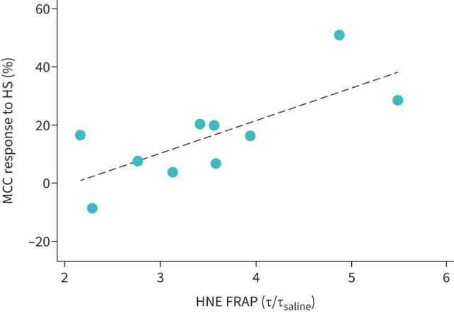
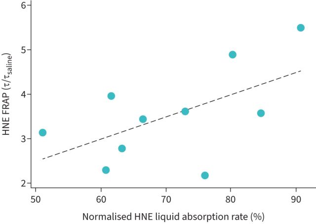
We considered whether any in vivo measurements associated with therapeutic response. Not surprisingly, subjects with lower baseline MCC values had more therapeutic response to HS inhalation. There was no relationship between response and age, FEV1, forced vital capacity (FVC), forced expiratory flow at 25–75% of FVC (FEF25–75%), LCI, sweat chloride or ABS. We also considered whether any in vitro measurements were associated with therapeutic response. Cl− current, Na+ current, i-ratio, TER, cell ABS and HNE normalised liquid absorption rate were not. Therapeutic response increased with FRAP as shown in figure 1. FRAP is a measure of particle diffusion through the ASL. Longer FRAP diffusion times have been associated with higher ASL solids concentrations [5]. Our result implies a strong relationship between inherent factors affecting ASL mucin concentration, as assessed in vitro, and in vivo therapeutic response to a hydrating therapy. Normalised liquid absorption rate was weakly associated with FRAP in the CF HNE cell cultures (R2=0.34, p=0.07) as shown in figure 2. Factors in addition to dehydration may also contribute to increasing FRAP. Model coefficients and associated R2 and p-values are included in table 4.
FIGURE 1.
In vivo mucociliary clearance (MMC) response to hypertonic saline (HS) inhalation correlates with in vitro measures of fluorescence recovery after photobleaching (FRAP) diffusion time measured in human nasal epithelial (HNE) cell cultures from the cystic fibrosis (CF) participants (R2=0.55, p=0.04). Only a portion of the CF group had FRAP measurements available (n=10).
FIGURE 2.
Relationship between fluorescence recovery after photobleaching (FRAP) diffusion time and airway surface liquid absorption rate in cystic fibrosis (CF) human nasal epithelial (HNE) cell cultures (R2=0.34, p=0.07). Only a portion of the CF group had FRAP measurements available (n=10).
TABLE 4.
Variables that correlated with therapeutic response to hypertonic saline (HS) in the cystic fibrosis group
| β0 | β1 | R2 | p-value | |
| Baseline MCCadjusted | 31.62 | −0.44 | 0.20 | 0.01 |
| HNE ASL FRAP diffusion time | −23.52 | 11.23 | 0.55 | 0.04 |
MCCadjusted: mucociliary clearance adjusted based on aerosol distribution; HNE: human nasal epithelial; ASL: airway surface liquid; FRAP: fluorescence recovery after photobleaching. Therapeutic response (=β1 (listed variable)+β0) is the improvement in MCCadjusted after inhaling HS compared with a baseline measurement made after isotonic saline inhalation.
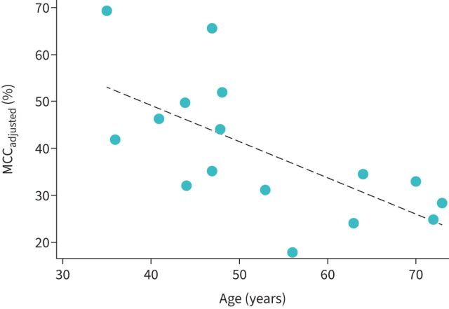
Comparing in vitro and in vivo physiology, we considered whether any in vivo measures were associated with baseline MCCadjusted in any of the three groups. There was no relationship with FEV1, FVC, FEF25-75, LCI or sweat chloride in any of the groups. There was no difference in MCCadjusted based on sex in any of the groups. Contrary to previous outcomes [8, 10, 15], there was no difference in MCCadjusted based on chronic P. aeruginosa in the CF group. MCCadjusted decreased significantly with age in the CR group, which had a wider range of ages than the CR or HC groups (range 35–73 years) (figure 3). Decreases in MCC with age have been reported previously in HC [16]. There was no association between MCCadjusted and Cl− current, Na+ current, i-ratio, FRAP, TER or HNE normalised liquid absorption rate in any of the groups.
FIGURE 3.
Mucociliary clearance (MCC) rate decreases with age in single cystic fibrosis transmembrane conductance regulator mutation carriers (CR group) (R2=0.44, p=0.002; n=16).
We considered multivariable models of FEV1 % pred in the CF group in table 5. We included baseline factors of age, chronic P. aeruginosa infection, gender and sweat chloride, which reflects both the baseline severity of CFTR dysfunction and correction of this defect with CFTR modulators. The baseline model accounted for 66% of the variation in FEV1 % pred in a highly significant model (model 1: R2=0.66). The only in vitro variable that provided substantial improvement of this correlation was FRAP. Since not all subjects had FRAP measurements, this effectively decreased the size of the dataset to n=10 but did increase R2 to 0.91 (model 2). The baseline model run with the same 10 subjects yielded an R2=0.76 (model 3).
TABLE 5.
Multivariable regression models of forced expiratory volume in 1 s (FEV1) % pred in the cystic fibrosis (CF) group
| Model | Group | Model of | Age (years) | Chronic Pseudomonas aeruginosa# | Sex (female) | Sweat chloride (mmol·L−1) | HNE ASL FRAP | Model | |||||||
| β1 | p-value | β2 | p-value | β3 | p-value | β4 | p-value | β5 | p-value | R2 | p-value | β0 | |||
| 1 | CF | FEV1 % pred (n=25) | −1.1 | 0.001 | −22.7 | 0.007 | −10.9 | 0.12 | −0.26 | 0.05 | 0.66 | <0.0001 | 145 | ||
| 2 | CF | FEV1 % pred (n=10)¶ | −0.85 | 0.11 | −30.0 | 0.09 | −27.8 | 0.03 | −0.21 | 0.45 | −13.8 | 0.02 | 0.91 | 0.01 | 187 |
| 3 | CF | FEV1 % pred (n=10)¶ | −1.1 | 0.09 | −7.95 | 0.70 | −24.8 | 0.07 | −0.25 | 0.57 | 0.76 | 0.03 | 142 | ||
MCC: mucociliary clearance; P. aeruginosa: Pseudomonas aeruginosa; HNE: human nasal epithelial; ASL: airway surface liquid; FRAP: fluorescence recovery after photobleaching. FEV1 % pred=β1(age)+β2(P. aeruginosa)+β3(sex)+β4(sweat chloride)+β5(FRAP)+β0. #: chronic P. aeruginosa is defined as two or more positive throat or sputum cultures in the previous year; ¶: only a portion of the CF group had FRAP measurements available (n=10).
There were no relationships between lung ABS and measured in vitro variables (cell ABS, Cl− current, normalised liquid absorption rate or TER) in any of the groups. Cell ABS increased with ASL absorption rate in vitro (R2=0.14, p=0.002; n=37) in agreement with previous reports [7]. There was a correlation between lung ABS and sweat chloride in both the CF (R2=0.19, p=0.002) and HC groups (R2=0.60, p=0.01). Our group had previously reported similar correlations in CF [9]. There was no correlation in the CR group. Here much of the relationship in the CF group was driven by subjects with low ABS and sweat chloride values associated with the use of CFTR modulators. ABS is a measure of the paracellular absorption of a small-molecule radiolabelled probe in the lung. This result indicates that this process is highly influenced by CFTR function in both healthy and CF lungs, likely moderated by paracellular liquid absorption.
Discussion
Here we hypothesised that in vitro measures of HNE cell physiology would correlate with in vivo measures of therapeutic response and lung physiology, providing a framework for using HNE cells for therapeutic development and personalisation.
In vivo therapeutic response to HS inhalation increased with FRAP diffusion time in the CF group. FRAP was measured using 70-kDa fluorescent dextran particles that are significantly smaller than the mesh size of the mucin network in the ASL. Previous studies have associated the diffusion time of small-particle probes with the viscosity of the solvent component of the mucus gel [5]. Here FRAP is likely driven by ASL solids concentrations, which could be affected by mucus secretion or hydration. FRAP was only weakly correlated with ASL absorption rate, suggesting that differences in mucus secretion may also be involved. Differences in mucin binding could also play a role. Previous studies have demonstrated a pH effect on FRAP associated with changes in electrostatic bonds between mucins. These studies also demonstrated Ca+-dependent effects on FRAP [17]. Our results indicate that the inherent airway mucus composition and hydration characteristics of a patient can be characterised through measurements of HNE FRAP and used to predict the utility of hydrating therapies like HS. This relationship exists independent of CFTR modulator use, which is not reflected in HNE cultures.
We sought to determine whether any in vitro measures correlated directly with in vivo measures or organ-level physiology in the lungs in any of the three participant groups. No in vitro measures consistently correlated with baseline MCC. In considering the relationship between other patient variables and MCC we noted clear relationship between age and MCC in the CR group that included the CF carrier parents of the CF group. It is unknown if this effect is unique to this group or a more general effect of ageing illustrated here based on the wide range of ages within the CR group. Previous studies have noted decreases in MCC with age in humans and mice [16, 18–20]. Studies have indicated that single CFTR mutation carriers may be at higher risk of conditions such as pancreatitis, bronchiectasis, diabetes, constipation and cholelithiasis [21], as well as bronchitis and lung cancer [22]. The relationship between age and decreasing MCC merits further examination since this important host defence prevents obstruction and limits exposure to inhaled pathogens and toxins (including cigarette smoke). A decrease in MCC with age would indicate increased vulnerability to these effects.
We considered whether any in vitro measures correlated with measures of FEV1 % pred in the CF group using multivariable models including factors known to affect pulmonary function in this group: age, chronic P. aeruginosa infection and gender [23]. We also included sweat chloride as a measure of both the severity of the baseline CFTR defect and to capture the function correction associated with CFTR modulators. This baseline model accounted for 66% of the variation in FEV1 % pred. Addition of FRAP to this model significantly improved correlation, although only a very limited dataset was available for use (n=10).
Our imaging measurements included a measurement of small-molecule absorption (ABS). This technique was developed to detect changes in ASL absorption in the airways and has been previously described. These studies confirmed previously described increases in ABS in CF and correlation between ABS and sweat chloride in CF [9, 10, 13]. We also noted correlation between ABS and sweat chloride in the HC group. These results indicate an influence of CFTR-driven liquid absorption on this imaging biomarker in both CF patients and the HC group.
Limitations of our study include the small numbers of assessments in some groups. Culturing cells from the CR group was particularly challenging and limited our ability to assess this group. Our study also did not include a validation dataset, which limits the utility of the models presented.
Overall, our results demonstrate the utility of HNE cultures for assessing therapeutic response for hydrating therapies. In vitro measurements of FRAP were particularly useful for predicting response and for characterising important properties of ASL mucus that were ultimately reflected in lung physiology. Further studies are required to determine the applicability of FRAP for assessing other therapies and to better understand the pathophysiological mechanisms reflected in the measurement.
Supplementary material
Please note: supplementary material is not edited by the Editorial Office, and is uploaded as it has been supplied by the author.
Supplementary material 00382-2022.SUPPLEMENT (1MB, pdf)
Footnotes
Provenance: Submitted article, peer reviewed.
This study is registered at ClinicalTrials.gov with identifier number NCT02947126. We will share data describing participant characteristics and outcome measures. These data will be provided as an Excel spreadsheet.
Conflict of interest: The authors have nothing to disclose.
Support statement: This study was supported by National Heart, Lung, and Blood Institute grant 1 UO1 HL131046-01. Funding information for this article has been deposited with the Crossref Funder Registry.
References
- 1.de Courcey F, Zholos AV, Atherton-Watson H, et al. Development of primary human nasal epithelial cell cultures for the study of cystic fibrosis pathophysiology. Am J Physiol Cell Physiol 2012; 303: C1173–C1179. doi: 10.1152/ajpcell.00384.2011 [DOI] [PubMed] [Google Scholar]
- 2.van Meegen MA, Terheggen-Lagro SW, Koymans KJ, et al. Apical CFTR expression in human nasal epithelium correlates with lung disease in cystic fibrosis. PLoS One 2013; 8: e57617. doi: 10.1371/journal.pone.0057617 [DOI] [PMC free article] [PubMed] [Google Scholar]
- 3.McCarthy C, Brewington JJ, Harkness B, et al. Personalised CFTR pharmacotherapeutic response testing and therapy of cystic fibrosis. Eur Respir J 2018; 51: 1702457. doi: 10.1183/13993003.02457-2017 [DOI] [PubMed] [Google Scholar]
- 4.Lennox AT, Coburn SL, Leech JA, et al. ATP12A promotes mucus dysfunction during type 2 airway inflammation. Sci Rep 2018; 8: 2109. doi: 10.1038/s41598-018-20444-8 [DOI] [PMC free article] [PubMed] [Google Scholar]
- 5.Hill DB, Long RF, Kissner WJ, et al. Pathological mucus and impaired mucus clearance in cystic fibrosis patients result from increased concentration, not altered pH. Eur Respir J 2018; 52: 1801297. doi: 10.1183/13993003.01297-2018 [DOI] [PMC free article] [PubMed] [Google Scholar]
- 6.Harvey PR, Tarran R, Garoff S, et al. Measurement of the airway surface liquid volume with simple light refraction microscopy. Am J Respir Cell Mol Biol 2011; 45: 592–599. doi: 10.1165/rcmb.2010-0484OC [DOI] [PMC free article] [PubMed] [Google Scholar]
- 7.Corcoran TE, Thomas KM, Brown S, et al. Liquid hyper-absorption as a cause of increased DTPA clearance in the cystic fibrosis airway. EJNMMI Res 2013; 3: 14. doi: 10.1186/2191-219X-3-14 [DOI] [PMC free article] [PubMed] [Google Scholar]
- 8.Donaldson SH, Laube BL, Corcoran TE, et al. Effect of ivacaftor on mucociliary clearance and clinical outcomes in cystic fibrosis patients with G551D-CFTR. JCI Insight 2018; 3: e122695. doi: 10.1172/jci.insight.122695 [DOI] [PMC free article] [PubMed] [Google Scholar]
- 9.Corcoran TE, Huber AS, Myerburg MM, et al. Multiprobe nuclear imaging of the cystic fibrosis lung as a biomarker of therapeutic effect. J Aerosol Med Pulm Drug Deliv 2019; 32: 242–249. doi: 10.1089/jamp.2018.1491 [DOI] [PMC free article] [PubMed] [Google Scholar]
- 10.Locke LW, Myerburg MM, Weiner DJ, et al. Pseudomonas infection and mucociliary and absorptive clearance in the cystic fibrosis lung. Eur Respir J 2016; 47: 1392–1401. doi: 10.1183/13993003.01880-2015 [DOI] [PMC free article] [PubMed] [Google Scholar]
- 11.Derichs N, Jin BJ, Song Y, et al. Hyperviscous airway periciliary and mucous liquid layers in cystic fibrosis measured by confocal fluorescence photobleaching. FASEB J 2011; 25: 2325–2332. doi: 10.1096/fj.10-179549 [DOI] [PMC free article] [PubMed] [Google Scholar]
- 12.Colin AA, Sawyer SM, Mickle JE, et al. Pulmonary function and clinical observations in men with congenital bilateral absence of the vas deferens. Chest 1996; 110: 440–445. doi: 10.1378/chest.110.2.440 [DOI] [PubMed] [Google Scholar]
- 13.Corcoran TE, Thomas KM, Myerburg MM, et al. Absorptive clearance of DTPA as an aerosol-based biomarker in the cystic fibrosis airway. Eur Respir J 2010; 35: 781–786. doi: 10.1183/09031936.00059009 [DOI] [PMC free article] [PubMed] [Google Scholar]
- 14.Locke LW, Myerburg MM, Markovetz MR, et al. Quantitative imaging of airway liquid absorption in cystic fibrosis. Eur Respir J 2014; 44: 675–684. doi: 10.1183/09031936.00220513 [DOI] [PMC free article] [PubMed] [Google Scholar]
- 15.Laube BL, Sharpless G, Benson J, et al. Mucus removal is impaired in children with cystic fibrosis who have been infected by Pseudomonas aeruginosa. J Pediatr 2014; 164: 839–845. doi: 10.1016/j.jpeds.2013.11.031 [DOI] [PubMed] [Google Scholar]
- 16.Svartengren M, Falk R, Philipson K. Long-term clearance from small airways decreases with age. Eur Respir J 2005; 26: 609–615. doi: 10.1183/09031936.05.00002105 [DOI] [PubMed] [Google Scholar]
- 17.Tang XX, Ostedgaard LS, Hoegger MJ, et al. Acidic pH increases airway surface liquid viscosity in cystic fibrosis. J Clin Invest 2016; 126: 879–891. doi: 10.1172/JCI83922 [DOI] [PMC free article] [PubMed] [Google Scholar]
- 18.Grubb BR, Livraghi-Butrico A, Rogers TD, et al. Reduced mucociliary clearance in old mice is associated with a decrease in Muc5b mucin. Am J Physiol Lung Cell Mol Physiol 2016; 310: L860–L867. doi: 10.1152/ajplung.00015.2016 [DOI] [PMC free article] [PubMed] [Google Scholar]
- 19.Proenca de Oliveira-Maul J, Barbosa de Carvalho H, Goto DM, et al. Aging, diabetes, and hypertension are associated with decreased nasal mucociliary clearance. Chest 2013; 143: 1091–1097. doi: 10.1378/chest.12-1183 [DOI] [PubMed] [Google Scholar]
- 20.Bailey KL, Bonasera SJ, Wilderdyke M, et al. Aging causes a slowing in ciliary beat frequency, mediated by PKCepsilon. Am J Physiol Lung Cell Mol Physiol 2014; 306: L584–L589. doi: 10.1152/ajplung.00175.2013 [DOI] [PMC free article] [PubMed] [Google Scholar]
- 21.Miller AC, Comellas AP, Hornick DB, et al. Cystic fibrosis carriers are at increased risk for a wide range of cystic fibrosis-related conditions. Proc Natl Acad Sci USA 2020; 117: 1621–1627. doi: 10.1073/pnas.1914912117 [DOI] [PMC free article] [PubMed] [Google Scholar]
- 22.Colak Y, Nordestgaard BG, Afzal S. Morbidity and mortality in carriers of the cystic fibrosis mutation CFTR Phe508del in the general population. Eur Respir J 2020; 56: 2000558. doi: 10.1183/13993003.00558-2020 [DOI] [PubMed] [Google Scholar]
- 23.Gecili E, Brokamp C, Palipana A, et al. Seasonal variation of lung function in cystic fibrosis: longitudinal modeling to compare a Midwest US cohort to international populations. Sci Total Environ 2021; 776: 145905. doi: 10.1016/j.scitotenv.2021.145905 [DOI] [PMC free article] [PubMed] [Google Scholar]
Associated Data
This section collects any data citations, data availability statements, or supplementary materials included in this article.
Supplementary Materials
Please note: supplementary material is not edited by the Editorial Office, and is uploaded as it has been supplied by the author.
Supplementary material 00382-2022.SUPPLEMENT (1MB, pdf)


