

Abstract
Background
The corticalis group is one of most diverse species-group in genus Clubiona Latreille, 1804. Currently, a total of 81 corticalis group species are known worldwide, amongst them 67 were recorded from China. However, the diversity of this group in China is still insufficiently known.
New information
Clubionaxianning sp. nov. is described as a new species of the C.corticalis species-group collected from Hubei Province, China.
Keywords: Sac spiders, morphology, DNA barcoding, diagnosis, taxonomy
Introduction
Clubiona Latreille, 1804 currently contains 518 catalogued species that are found worldwide, except for the Polar Regions and South America (World Spider Catalog 2022), is the most diverse genus in the family Clubionidae and one of the most diverse genera in the order Araneae (Zhang and Yu 2020, Zhang et al. 2021, World Spider Catalog 2022). One of the generic species-groups, the C.corticalis-group, exhibits high species diversity and currently contains 81 species (Zhang et al. 2021). Up to now, there are 67 described corticalis group species distributed in China, making it one of the most diverse clubionid groups in China (Zhang et al. 2021). However, the diversity of this group in China is still insufficiently known and several new species have been described in the last few years (Zhang et al. 2018, Yu and Li 2019a, Yu and Li 2019b, Zhang and Yu 2020, Zhang et al. 2021).
While examining spiders collected from Jiugong Mountains, Hubei Province, China (Fig. 1), we found pairs of Clubiona specimens that belong to an undescribed species of the corticalis-group similar to C.caohai Zhang & Yu, 2020 and C.altissimus Hu, 2001. The goal of this paper is to provide a detailed description and diagnosis of the new species. The DNA barcodes of the new species were obtained for gender matching and future use in molecular studies.
Figure 1.

Distribution record of Clubionaxianning sp. nov. (green circle).
Materials and methods
Specimens in this study were collected by hand collecting from leaf-litter in Mt. Jiugong, Hubei. Spiders were fixed and preserved in 95% ethanol. Specimens were examined with an Olympus SZX7 stereomicroscope; details were studied with an Olympus CX41 compound microscope. Female epigyne and male palp were examined and illustrated after being dissected. The epigyne was removed and cleared in warm lactic acid before illustration. The vulva was also imaged after being embedded in Arabic gum. Photos were made with a Cannon EOS70D digital camera mounted on an Olympus CX41 compound microscope. The digital images were taken and assembled using Helifocus 6.80 software package. The distribution map was generated with Arcgis 10.5 (Environmental Systems Research Institute, Inc.).
A DNA barcode was also obtained for species matching. A partial fragment of the mitochondrial cytochrome oxidase subunit I (CO1) gene was amplified and sequenced for two specimens, using the primers LCOI1490 (5’-GGTCAACAAATCATAAAGATATTG-3’) and HCOI2198 (5’-TAAACTTCAGGGTGACCAAAAAAT-3’) (Folmer et al. 1994). For additional information on extraction, amplification and sequencing procedures, see Wheeler et al. (2016). DNA sequences were checked and edited with BioEdit 7.2.2 (Hall 1999), sequences being trimmed to 653 bp. Sequence alignment was completed using CLUSTAL W (Thompson et al. 1994). Genetic distances were computed with MEGA 5 (Tamura et al. 2011). All sequences were confirmed using BLAST and are deposited in GenBank. The codes and GenBank accession numbers of voucher specimens are provided as follows: YHCLU0272, ♂, GenBank OP675437; YHCLU0273, ♀, GenBank OP675436.
All measurements were obtained using an Olympus SZX7 stereomicroscope and given in millimetres. Eye diameters are taken at the widest point. The total body length does not include chelicerae or spinnerets length. Leg lengths are given as total length (femur, patella, tibia + metatarsus, tarsus). Most of the terminologies used in text and figure legends follows Zhang et al. (2021), while a few others followed Zhang et al. (2018) and Zhang and Yu (2020).
All specimens are deposited Museum of Guizhou Normal University, Guiyang, Guizhou, China.
Taxon treatments
Clubiona xianning
Zhong & Yu sp. n.
B0CC6F23-8DFB-5B4A-BAD0-E8AC5114D687
8DFF1CC4-1C1A-46A6-8300-FF36FFFA2644
Materials
Type status: Holotype. Occurrence: recordedBy: Yang Zhong, Xusheng Gong, Qianle Lu; individualID: YHCLU0272; individualCount: 1; sex: male; lifeStage: adult; behavior: foraging; preparations: whole animal (ETOH); associatedSequences: GenBank: OP675437; occurrenceID: 1312422A-3320-5AE6-9BA9-5DCB3013B14F; Taxon: order: Araneae; family: Clubionidae; genus: Clubiona; specificEpithet: xianning; scientificNameAuthorship: Zhong & Yu; Location: continent: Asian; country: China; countryCode: CHN; stateProvince: Hubei; county: Tongshan; municipality: Xianning; locality: Jiugongshan Nature Reserve; decimalLatitude: 29.39; decimalLongitude: 114.65; Identification: identifiedBy: Hao Yu; dateIdentified: 2022-05; Event: samplingProtocol: by hand; samplingEffort: 10 km by foot; year: 2020; month: 7; day: 4; Record Level: basisOfRecord: PreservedSpecimen
Type status: Holotype. Occurrence: recordedBy: Yang Zhong, Xusheng Gong, Qianle Lu; individualID: YHCLU0273; individualCount: 1; sex: female; lifeStage: adult; behavior: foraging; preparations: whole animal (ETOH); associatedSequences: GenBank: OP675436; occurrenceID: 085B9104-F139-5EC1-9913-5A5DB9C7C9C6; Taxon: order: Araneae; family: Clubionidae; genus: Clubiona; specificEpithet: xianning; scientificNameAuthorship: Zhong & Yu; Location: continent: Asian; country: China; countryCode: CHN; stateProvince: Hubei; county: Tongshan; municipality: Xianning; locality: Jiugongshan Nature Reserve; decimalLatitude: 29.39; decimalLongitude: 114.65; Identification: identifiedBy: Hao Yu; dateIdentified: 2022-05; Event: samplingProtocol: by hand; samplingEffort: 10 km by foot; year: 2020; month: 7; day: 4; Record Level: basisOfRecord: PreservedSpecimen
Description
Male (Fig. 4E and F). Total length 5.31; carapace 2.27 long, 1.73 wide; abdomen 3.04 long, 1.31 wide.
Figure 4.

Clubionaxianning sp. nov., female paratype and male holotype. A Intact epigyne, ventral view; B Cleared epigyne, ventral view; C Cleared vulva, dorsal view; D Vulva, cleared and embedded in Arabic gum, dorsal view; E Male habitus, dorsal view; F Male habitus, lateral view; G Female habitus, dorsal view; H Female habitus, ventral view. Abbreviations: A = atrium; AAM = atrial anterior margin; BS = bursa; CO = copulatory opening; FD = fertilisation duct; SB = spermathecal base; SH = spermathecal head; SP = spermatheca; SS = spermathecal stalk. Scale bars: 0.2 mm (equal for A and B, equal for C and D); 2 mm (equal for E and F, equal for G and H).
Colour of the living holotype male was uniformly brown (Fig. 2A and B). Carapace (Fig. 4E and F) elongate, oval, light brown in alcohol, uniformly coloured, without pattern, fovea red; pars cephalica distinctly narrowed, cervical groove radial groove indistinct; tegument smooth, with erect, thin, dark setae on front ridge. Eyes: in dorsal view, anterior eye row (AER) slightly recurved, posterior eye row (PER) almost straight, PER wider than AER. Eye sizes and interdistances: anterior median eyes (AME) 0.13, anterior lateral eyes (ALE) 0.12, posterior median eyes (PME) 0.11, posterior lateral eyes (PLE) 0.09, distance between AMEs (AME–AME) 0.07, distance between AME and ALE (AME–ALE) 0.03, distance between PMEs (PME–PME) 0.19, distance between PME and PLE (PME–PLE) 0.09. Length of median ocular quadrangle (MOQL) 0.33, MOQ anterior width (MOQA) 0.29, MOQ posterior width (MOQP) 0.54. Chelicerae robust, light orange, with red fangs, with four promarginal and two retromarginal teeth. Sternum nearly shield-shaped, yellowish-white, 1.21 long, 0.86 wide. Labium and endites coloured as carapace.
Figure 2.
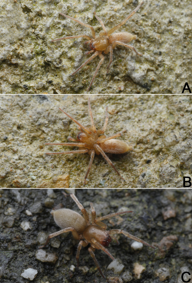
Clubionaxianning sp. nov., male holotype (A, B) and female paratype (C), live specimens. Photographs by Qianle Lu (Shenzhen, Guangdong).
Abdomen (Fig. 4E and F) oval and light brown, dorsally with a wide and more or less oblong scutum extending ca. 2/3 of abdomen length, with two pairs of inconspicuous muscle depressions on either side; venter white with no distinct pattern; spinnerets yellowish-white.
Legs uniformly yellowish-white in ethanol (Fig. 4E and F). Leg length: I 5.85 (1.73, 2.32, 1.24, 0.57), II 6.29 (1.79, 2.44, 1.33, 0.73), III 5.27 (1.69, 1.60, 1.58, 0.40), IV 7.56 (2.19, 2.54, 2.31, 0.52).
Palp (Fig. 3A–E). Femur and patella unmodified. Tibia relatively short, about 2/5 of cymbium length, with two apophyses: a retrolateral one (RTA) that is heavily sclerotised, ca. 1/2 of palpal tibia length, more or less blade-shaped; a partly membranous, laminar apophysis (VTA), ca. 1/3 of palpal tibia length. Bulb nearly pyriform, slightly excavated on prolatero-apical side to accommodate embolus; tegulum oval and slightly expanded, ca. 1.45 longer than wide, sperm duct indistinct in venter view; subtegulum (ST) large, located prolaterally. Embolus (E) wide and heavily sclerotised, about 2/3 of the tegulum length, dagger-shaped, gradually tapering towards its apex, its tip sharp, slightly curved and extending to apex of cymbium. Conductor long and membranous, irregular-shaped in venter view and triangular in retrolateral view, its tip extending above apex of embolus.
Figure 3.

Male left palp of the holotype of Clubionaxianning sp. nov. A Prolateral view; B Rretrolateral view; C Bulb, prolateral view; D Bulb, ventral view; E Bulb, retrolateral view. Abbreviations: C = conductor; E = embolus; EB = embolic base; RTA = retrolateral tibial apophysis; VTA = ventral tibial apophysis. Scale bars: 0.2 mm (equal for A and B, equal for C–E).
Female (Fig. 4G and H). Total length 6.15; carapace 2.64 long, 1.98 wide; abdomen 3.51 long, 1.90 wide. Eye sizes and interdistances: AME 0.16, ALE 0.15, PME 0.14, PLE 0.13; AME–AME 0.07, AME–ALE 0.04, PME–PME 0.22, PME–PLE 0.04. MOQL 0.40, MOQA 0.36, MOQP 0.53. Sternum 1.39 long, 0.96 wide. Measurements of legs: I 5.48 (1.69, 2.12, 1.05, 0.62), II 5.93 (1.90, 2.28, 1.09, 0.66), III 5.29 (1.82, 1.90, 1.14, 0.44), IV 8.07 (2.52, 2.64, 2.32, 0.59). General characters as in female, but slightly larger in size and lighter in colour.
Epigyne (Fig. 4A–D). Epigynal plate slightly wider than long, spermathecae and bursae are indistinctly visible through epigynal plate in ventral view. Atrium (A) large, represented by two symmetrical, spherical, shallow depressions; atrial anterior margin (AAM) distinctly delimited, M-shaped, posterior and lateral anterior margins not rebordered. Two copulatory openings (CO) indistinct, located at medial portion of atrial anterior margin. Spermathecae (SP) with 3 parts: spermathecal head (SH) finger-like and large, ca. 2.7x longer than diameter, the two spermathecal heads separated by 1.58 diameters; spermathecal stalk tubular, running horizontally; (SS) spermathecal base (SB) tubular and convoluted, distinctly thinner than spermathecal head and spermathecal stalk. Fertilisation ducts (FD) short and curved, acicular, located at distal end of spermathecal base.
DNAbarcode:
5'TTCTGGTCAGCTATAGTTGGTACAGCTATAAGAGTTATAATTCGTATAGAATTAGGTCAATCTGGAGCTTTTTTAGGTGATGATCATTTGTATAATGTAGTAGTTACTGCTCATGCTTTTGTTATAATTTTTTTTATAGTAATACCTATTATAATTGGGGGGTTTGGAAATTGATTAGTTCCATTAATATTAGGGGCAGGTGATATAGCTTTTCCTCGTATAAATAATTTAAGTTTTTGACTTTTACCACCTTCATTAATATTATTAGTTATATCATCTATGGCTGAGATGGGAGTTGGGGCTGGATGAACAGTTTATCCCCCTCTTGCTTCTTTAGTAGGTCATACGGGAAGAGCAATGGATTTTGCTATTTTTTCATTACATTTAGCTGGGGCTTCTTCTATTATAGGAGCTGTTAATTTTATTACTACTATTATGAATATACGATCTTTTGGAATAATAATGGAAAAGATTTCATTATTTGTTTGGTCTGTTTTAATTACAGCTATTTTATTATTATTATCTTTGCCAGTTTTAGCCGGGGCTATTACTATATTATTAACTGATCGTAATTTTAATACGTCTTTTTTTGACCCTGCTGGGGGAGGTGATCCTATTTTATTTCAACATTTATTTTGATTTTTTGGTCACCC3' (holotype, YHCLU0272; GenBank: OP675437)
5'TCTGGTCAGCTATAGTTGGTACAGCTATAAGAGTTATAATTCGTATAGAATTAGGTCAATCTGGAGCTTTTTTAGGTGATGATCATTTGTATAATGTAGTAGTTACTGCTCATGCTTTTGTTATAATTTTTTTTATAGTAATACCTATTATAATTGGGGGGTTTGGAAATTGATTAGTTCCATTAATATTAGGGGCAGGTGATATAGCTTTTCCTCGTATAAATAATTTAAGTTTTTGACTTTTACCACCTTCATTAATATTATTAGTTATATCATCTATGGCTGAGATGGGAGTTGGGGCTGGATGAACAGTTTATCCCCCTCTTGCTTCTTTAGTAGGTCATACGGGAAGAGCAATGGATTTTGCTATTTTTTCATTACATTTAGCTGGGGCTTCTTCTATTATAGGAGCTGTTAATTTTATTACTACTATTATGAATATACGATCTTTTGGAATAATAATGGAAAAGATTTCATTATTTGTTTGGTCTGTTTTAATTACAGCTATTTTATTATTATTATCTTTGCCAGTTTTAGCCGGGGCTATTACTATATTATTAACTGATCGTAATTTTAATACGTCTTTTTTTGACCCTGCTGGGGGAGGTGATCCTATTTTATTTCAACATTTATTTTGATTTTTTGGTCACCC3' (paratype, YHCLU0273; GenBank: OP675436).
Diagnosis
Male of the new species resembles that of C.caohai Zhang & Yu, 2020 (Zhang and Yu 2020: 347, figs. 2A–E) in having a blade-shaped RTA and a dagger-shaped embolus, but differs in the following: (1) embolus gradually tapering towards its apex (vs. narrowed in the middle) (cf. Fig. 3D and Zhang and Yu 2020: fig. 2D); (2) conductor nearly triangular, apex sharp and pointing distally (vs. finger-like, apex blunt and pointing prolaterally) (cf. Fig. 3D and E and Zhang and Yu 2020: figs. 2D and E); (3) VTA laminar, relatively large, wider than 1/2 of palpal tibia diameter (vs. papilliform and small, ca. 1/3–1/4 of palpal tibia diameter) (cf. Fig. 3B and Zhang and Yu 2020: fig. 2B). Females of C.xianning sp. nov. can be easily distinguished from other members of the C.corticalis-group, with the exception of C.altissimus Hu, 2001 (Hu 2001: 283, fig. 163.1-3) by the atrium represented by two shallow depressions (atrium absent, or present, but represented by one or two deep cavities in all other corticalis-group species), differ from C.altissimus by: (1) atrium large, nearly as wide as epigynal plate (vs. atrium relatively small, ca. 1/3 of epigyne width) (cf. Fig. 4A and B and Hu 2001: fig. 163.2); (2) spermathecae consisting of head, tubular stalk and base, the two spermathecal heads finger-like, well separated by 1.58 diameters (vs. spermathecae consisting of head and base, the two spermathecal heads reniform, separated by ca. one diameter) (cf. Fig. 4C and D and Hu 2001: fig. 163.3). In addition, C.xianning sp. nov. also can by separated from C.caohai and C.altissimus by their habitus: abdomen without distinct colour pattern in C.xianning sp. nov. (Fig. 4E–H), but with several chevron-shaped bands in C.caohai and C.altissimus (Zhang and Yu 2020: figs. 2 E–H; Hu 2001: fig. 163.1).
Etymology
The specific name refers to the type locality and is a noun in apposition.
Distribution
Known from the Mt. Jiugong, Hubei Province, China (Fig. 1).
Supplementary Material
Acknowledgements
We thank Jie Liu (Wuhan, China), Hirotsugu Ono (Ibaraki-ken, Japan) and a anonymous referee for providing constructive comments on an earlier version of the manuscript. We are especially grateful to Emma McCarroll Shaw (Chiang Mai, Thailand), the subject editor of this manuscript. We are also grateful to Qianle Lu (Shenzhen, China) for his kind help in collecting the specimens and for agreeing to use his picture of live specimens. This work was supported by the National Natural Sciences Foundation of China (NSFC-32060113/32000303), the Natural Science Foundation of Guizhou Province (J [2020] 1Y081), the Natural Sciences Foundation of Xianning City (2022ZRKX063), the Hubei Province Key Laboratory of Conservation Biology for Shennongjia Golden Monkey Foundation (No. SNJKL2021003) and the Special Fund Projects of Hubei Key Laboratory of Radiation Chemistry and Functional Materials (2021ZX12).
Contributor Information
Xusheng Gong, Email: gxs5339@stu.hubu.edu.cn.
Hao Yu, Email: insect1986@126.com.
References
- Folmer Ole, Black Mb, Wr Hoeh, Lutz R, Vrijenhoek Robert C. DNA primers for amplification of mitochondrial Cytochrome C oxidase subunit I from diverse metazoan invertebrates. Molecular Marine Biology and Biotechnology. 1994;3(5):194–299. [PubMed] [Google Scholar]
- Hall T. A. BioEdit: a user-friendly biological sequence alignment editor and analysis program for Windows 95/98/NT. Nucleic Acids Symposium Series. 1999;41:95–98. [Google Scholar]
- Hu JL. Spiders in Qinghai-Tibet Plateau of China. Henan Science and Technology Publishing House; 2001. 658 [Google Scholar]
- Tamura Koichiro, Peterson Daniel, Peterson Nicholas, Stecher Glen, Nei Masatoshi, Kumar Sudhir. MEGA5: Molecular evolutionary genetics analysis using maximum likelihood, evolutioanry distance, and maximum parsimony methods. Molecular Biology and Evolution. 2011;28(10):2731–2739. doi: 10.1093/molbev/msr121. [DOI] [PMC free article] [PubMed] [Google Scholar]
- Thompson J D, Higgins D G, Gibson T J. CLUSTAL W: improving the sensitivity of progressive multiple sequence alignment through sequence weighting, position-specific gap penalties and weight matrix choice. Nucleic acids research. 1994;22(22):4673–4680. doi: 10.1093/nar/22.22.4673. [DOI] [PMC free article] [PubMed] [Google Scholar]
- Wheeler Ward C., Coddington Jonathan A., Crowley Louise M., Dimitrov Dimitar, Goloboff Pablo A., Griswold Charles E., Hormiga Gustavo, Prendini Lorenzo, Ramírez Martín J., Sierwald Petra, Almeida‐Silva Lina, Alvarez‐Padilla Fernando, Arnedo Miquel A., Benavides Silva Ligia R., Benjamin Suresh P., Bond Jason E., Grismado Cristian J., Hasan Emile, Hedin Marshal, Izquierdo Matías A., Labarque Facundo M., Ledford Joel, Lopardo Lara, Maddison Wayne P., Miller Jeremy A., Piacentini Luis N., Platnick Norman I., Polotow Daniele, Silva‐Dávila Diana, Scharff Nikolaj, Szűts Tamás, Ubick Darrell, Vink Cor J., Wood Hannah M., Zhang Junxia. The spider tree of life: phylogeny of Araneae based on target‐gene analyses from an extensive taxon sampling. Cladistics. 2016;33(6):574–616. doi: 10.1111/cla.12182. [DOI] [PubMed] [Google Scholar]
- Catalog World Spider. World Spider Catalog. Version 23.5. http://wsc.nmbe.ch. [2022-09-01T00:00:00+03:00]. http://wsc.nmbe.ch.
- Yu Hao, Li Shuqiang. Eight new species of the genus Clubiona Latreille, 1804 from Xishuangbanna Rainforest, southwestern China (Araneae, Clubionidae) Zootaxa. 2019;4545(2):151–178. doi: 10.11646/zootaxa.4545.2.1. [DOI] [PubMed] [Google Scholar]
- Yu Hao, Li Shuqiang. On further species of the spider genus Clubiona Latreille, 1804 (Araneae, Clubionidae) from Xishuangbanna Rainforest, southwestern China. Zootaxa. 2019;4679(2):201–230. doi: 10.11646/zootaxa.4679.2.1. [DOI] [PubMed] [Google Scholar]
- Zhang Jianshuang, Yu Hao, Zhong Yang. Two new species of the Clubionacorticalis-group from Guizhou Province, China (Araneae: Clubionidae) Zootaxa. 2018;4415(2):393–400. doi: 10.11646/zootaxa.4415.2.10. [DOI] [PubMed] [Google Scholar]
- Zhang Jianshuang, Yu Hao. Three new species of the Clubionacorticalis-group from southern China (Araneae: Clubionidae) Turkish Journal of Zoology. 2020;44(4):346–354. doi: 10.3906/zoo-2003-7. [DOI] [Google Scholar]
- Zhang Jianshuang, Yu Hao, Li Shuqiang. Taxonomic studies on the sac spider genus Clubiona (Araneae, Clubionidae) from Xishuangbanna Rainforest, China. ZooKeys. 2021;1034:1–163. doi: 10.3897/zookeys.1034.59413. [DOI] [PMC free article] [PubMed] [Google Scholar]
Associated Data
This section collects any data citations, data availability statements, or supplementary materials included in this article.