


Abstract
Background
To evaluate the immune checkpoint inhibitors (CPI) for the treatment of patients with advanced cutaneous squamous cell carcinoma (CSCC).
Materials and methods
A meta‐analysis was conducted, and the efficacy and safety of CPI were assessed.
Results
A total of 13 studies with 980 patients were included. The pooled objective response rate (ORR) and disease control rate were 47.2% and 64.4%, separately. In addition, patients with primary tumor located in head and neck (odds ratio [OR]: 0.374, 95% confidence interval [CI]: 0.219–0.640, p < 0.001) and positive expression of programmed death ligand 1 (OR: 0.364, 95% CI: 0.158–0.842, P = 0.018) had superior ORR during CPI treatment. The incidence of progression free survival at 6 and 12 months was 59.3% and 52.8%, and 80.6% and 76.4% for overall survival. As for safety, the overall incidence of adverse events with all grades and 3–4 grade was 76.9% and 20.2%.
Conclusions
Our systematic review confirmed the satisfying efficacy and acceptable toxicity of CPI for advanced CSCC.
Keywords: cemiplimab, cutaneous squamous cell carcinoma, immune checkpoint inhibitors, meta‐analysis, nivolumab, pembrolizumab
1. INTRODUCTION
Cutaneous squamous cell carcinoma (CSCC) is the second most common skin cancer after basal cell carcinoma, 1 with raising incidence worldwide over the decades. 2 Patients with older age, chronic exposure to ultraviolet (UV) radiation, and organ transplant recipients were associated with increased risk of CSCC. 3 About 95% cases of CSCC were localized disease, which could be cured by surgery. 4 However, 15%–28% would experience disease recurrence after surgery resection. 5 , 6 , 7 Besides, in around 5% CSCC cases, the cancer reached locally advanced or metastatic disease at initial diagnosis and could not be managed by surgery or radiation alone. 8 Both locally advanced and metastatic CSCC are referred as advanced CSCC. Mortality rate of advanced CSCC patients exceeded 70%. 5 , 9 However, until recently, there is no consensus recommendation for those patients with advanced disease. 10 , 11
Recently, accumulating evidence indicated that CSCC is a kind of malignant tumor with highly immunogenicity. Long‐term exposure to UV causes increasing DNA damage, thus CSCC has the highest tumor mutation burden among skin cancers, even exceeding to melanoma and head and neck SCC. 12 , 13 , 14 Besides, tumor suppressor genes in CSCC are most frequently altered. 12 In addition, a positive association was also found between the expression of programmed death ligand 1 (PD‐L1) in patients with CSCC and the risk of metastatic disease. 15 These characteristics indicated that CSCC will likely respond to immune checkpoint inhibitors (CPI), including programmed death 1 (PD‐1) inhibitors.
Phase I and II trials of the PD‐1 inhibitor cemiplimab showed an objective response rate (ORR) of 47% in patients with advanced CSCC, 16 which made cemiplimab the first PD‐1 antibody by the Food and Drug Administration (FDA) to treat advanced CSCC in 2018. In addition, two phase II trials (KEYNOTE‐629 and CARSKIN trial) also showed satisfying efficacy of the PD‐1 inhibitor pembrolizumab, with ORR of 34.3% to 41%, and median progression‐free survival of 6.7–6.9 months. The FDA approved pembrolizumab for patients with recurrent or metastatic CSCC in 2020. 17 , 18 In addition, several studies based on real‐world population also showed the superior efficacy of CPI in CSCC patients. 19 , 20 , 21 , 22 , 23 However, the sample sizes of available studies were relatively small, and the reported data of efficacy and safety of CPI varied among different studies. Thus, a review of their findings is needed. A previous systematic review and meta‐analysis confirmed the efficacy and safety of CPI in for advanced and recurrent/metastatic CSCC. 24 However, this study failed to conduct further analysis due to limited references included. In this study, we sought to evaluate the efficacy and safety of CPI in advanced CSCC patients by conducting a systemic review to provide a more evidence‐based evaluation effectiveness of this treatment approach.
2. MATERIALS AND METHODS
2.1. Review protocol
This systematic review and meta‐analysis was conducted and reported according to the recommendations of the Preferred Reporting Items for Systematic Review and Meta‐Analysis guidelines. 25 No published protocol was available for this review.
2.2. Search strategy
PubMed, MEDLINE, Web of Science, EMBASE, and Cochrane Library were searched from inception to November 30, 2021 for available studies. The following Medical Subject Heading and key terms were used during searching: (“cutaneous SCC,” “cutaneous SCC metastatic,” “cSCC,” OR “nonmelanoma skin cancer”) AND (“immunotherapy,” “immune checkpoint inhibitor,” “programmed‐death 1 inhibitor,” “cemiplimab,” “pembrolizumab,” “nivoluimab,” “ipilimumab,” “atezolizumab,” OR “avelumab”). References within searched articles were also reviewed to identify potentially available studies.
2.3. Study selection
All relevant references were manually screened by the title and abstract by two reviewers (Z.H.R and Z.A). The inclusion criteria were as follows: (1) patients with histologically diagnosed with locally advanced or metastatic CSCC; (2) immunotherapy was administrated in one or more interventional arms; (3) studies with or without controlled arms administered with surgery, radiotherapy, or chemotherapy; (4) efficacy (treatment response, survival outcomes, etc.) and/or safety data (adverse events) were reported; (5) both phase I‐III clinical trials and retrospective studies were included. Articles that were not relevant to the purpose of this study were excluded. Exclusive criteria were as follows: (1) studies evaluating the efficacy and safety of immunotherapy in melanoma, basal cell carcinoma, or other non‐CSCC skin cancers; (2) case series, case reports, and review articles; (3) articles that were not written in English were also excluded from this study.
2.4. Data extraction
Two reviewers (Z.H.R and Z.A) independently extracted and summarized the data. All data were checked by the other reviewer for accuracy. Primary outcomes of were the rate of progression‐free survival (PFS) and overall survival (OS) at 6 and 12 months. Secondary outcomes were treatment response including the rate of objective response rate (ORR), disease control rate (DCR), and incidence of treatment‐related adverse events (AEs). The following data were extracted and summarized during review: patient characteristics (number, age, and gender), stage of disease (locally advanced or metastatic CSCC), follow‐up period, rate of PFS and OS at 6 and 12 months, rates of ORR and DCR, as well as the incidence of treatment‐related AEs (all grades and 3–4 grades). All treatment response of included studies was measured using Response Evaluation Criteria in Solid Tumors criteria, and treatment‐related AEs were evaluated using Common Terminology Criteria for Adverse Events.
2.5. Quality assessment
After study selection, we found that all eligible references were single‐armed studies. Thus, The Risk Of Bias In Nonrandomized Studies of Interventions tool was used to assess the risk of bias of included studies. 26 The following seven domains were assessed: bias due to confounding, selection of participants into the study, classification of interventions, deviations from intended interventions, missing data, measurement of the outcome, and selection of the reported results. The risk of bias of each domain was graded as low, moderate, serious, and critical risk of bias. The overall risk of bias across all domains was evaluated according to the guideline of Cochrane Handbook for Systematic Reviews of Interventions. 26
2.6. Statistical analysis
Meta‐analysis was performed using “meta” package on R software (Version 4.0.3 for Mac, R Foundation for Statistical Computing). Pooled measures included PFS and OS rates at 6 and 12 months, ORR, DCR, and incidence of treatment‐related AEs (all grades and 3–4 grades). Odds ratio (OR) was selected as the effect to assess the association of primary tumor location, locally advanced or metastatic disease, programmed death ligand 1 (PD‐L1) expression, and immune status with the efficacy of PD‐1 inhibitors, which was reported along with the 95% confidence interval (95% CI). Values of p < 0.05 were considered statistically significant. Higgins I2 statistic was used to indicative of large heterogeneity. The random‐effects model was used if there was high heterogeneity (I2 > 50%) between the studies; otherwise, the fixed‐effects model was used. Additionally, Egger's test and test were used to evaluate publication bias.
3. RESULTS
A total of 1323 eligible studies were identified after de‐duplication. Then, 1289 studies were excluded after initial screening of titles and abstracts. After reviewing the full text, 13 studies with 980 patients were finally included for final analysis. 16 , 17 , 18 , 19 , 20 , 21 , 22 , 23 , 27 , 28 , 29 , 30 , 31 Figure 1 showed the flowchart of the study screening process. Among all included studies, seven of them were clinical trials, 16 , 17 , 18 , 27 , 28 , 29 , 30 and six were real‐world, retrospective studies. 19 , 20 , 21 , 22 , 23 , 31 Egger's test indicated that there is no significant publication bias within studies (p = 0.282) (Figure S1). Quality assessment also indicated that all included studies were at low risk of bias (Figure S2).
FIGURE 1.
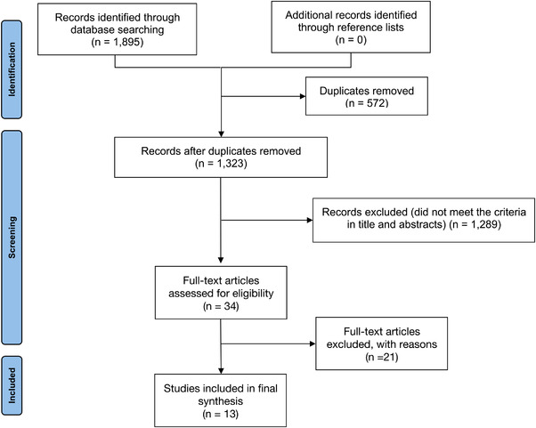
Flowchart of the selection process of eligible studies
3.1. Characteristics of included studies and patients
Characteristics and patient demographic data of all 13 included studies were presented in Table 1. All studies were published between June 2018 to November 2021, and sample sizes ranged from 11 to 159. Among all included patients, 50.8% (498/980) of them had locally advanced disease with no distant metastasis, 37.8% (370/980) had only metastatic lesions, and 11.3% (111/980) had both locally advanced and distant metastatic disease. The most common tumor location was head and neck (61.4%, 427/695), followed by extremities (20.9%, 145/695) and trunk (8.9%, 62/695). Three studies 17 , 18 , 19 reported the status of PD‐L1 expression. With 181 patients included, 142 (78.5%) patients had PD‐L1 positive expression, while the other 39 (21.5%) patients had PD‐L1 negative expression. Details of included patients were also presented in Table 1.
TABLE 1.
Characteristics of included studies and patients
| Primary lesion | ||||||||||||
|---|---|---|---|---|---|---|---|---|---|---|---|---|
| Author (year) | Study design | Number of patients | Age (y), median (range) | Sex, male,n (%) | Locally advanced disease, n (%) | Metastatic disease, n (%) | Both locally advanced and metastatic disea‐ se, n (%) | Head and neck, n (%) | Extremities, n (%) | Trunk, n (%) | Follow‐up (months), median (range) | Treatments |
| Migden et al. 16 2018 | Phase I/II trial | 85 | ‐ | 75 (88.2) | 10 (11.8) | 75 (88.2) | 0 (0) | 38 (64) 56 | 12 (20) 17 | 9 (5) 11 | 11.0 (1.1–17.0) | Cemiplimab |
| Yushak et al. 26 2019 | Phase II trial | 11 | ‐ | 9 (81.8) | 0 (0) | 11 (100) | 0(0) | ‐ | ‐ | ‐ | ‐ | Pembrolizumab |
| Grob et al. 17 2020 | Phase II trial | 105 | 72 (29–95) | 80 (76) | 47 (44.8) | 25 (23.8) | 33 (31.4) | 47 (44.8) | 18 (17.1) | 5 (4.8) | 11.4 (0.4–16.3) | Pembrolizumab |
| Maubec et al. 18 2020 | Phase II trial | 57 | 39 (42–99) | 46 (81) | 7 (12.3) | 36 (63.2) | 14 (24.6) | 36 (63.2) | 16 (28.1) | 4 (7.0) | 11 (0–33) | Pembrolizumab |
| Migden et al. 27 2020 | Phase II trial | 78 | 74 (65–81) | 59 (76) | 78 (100) | 0 (0) | 0 (0) | 62 (79) | 14 (18) | 2 (3) | 9.3 (5.1–15.7) | Cemiplimab |
| Rischin et al. 28 2020 | Phase II trial | 115 | 71 (38–93) | 102 (88.7) | 26 (22.6) | 88 (76.5) | 0 (0) | ‐ | ‐ | ‐ | 8.1 (0.6–14.1) | Cemiplimab |
| Hughes et al. 29 2021 | Phase II trial | 159 | 74 (62–82) | 119 (74.8) | 101 (63.5) | 25 (15.7) | 33 (20.8) | ‐ | ‐ | ‐ | ‐ | Pembrolizumab |
| Salzmann et al. 22 2020 | Real‐world study | 46 | 77 (39–92) | 31 (67) | 6 (13.0) | 40 (87.0) | 0 (0) | 33 (72) | 7 (15) | 4 (9) | 11.8 (0.7–39.7) |
Pembrolizumab; Nivolumab; Cemiplimab |
| Hanna et al. 21 2020 | Real‐world study | 61 | 75 (42–95) | 49 (80) | 14 (23) | 47 (77) | 0 (0) | 17 (28) | 25 (41) | 16 (26) | 8.5 (0–45) |
Pembrolizumab; Nivolumab; Cemiplimab |
| Shalhout et al. 23 2021 | Real‐world study | 76 | 74 (18–98) | 51 (67) | 43 (57) | 33 (43) | 0 (0) | 52 (68) | 19 (25) | 5 (7) | NA |
Pembrolizumab; Nivolumab; Cemiplimab |
| Baggi et al. 20 2021 | Real‐world study | 131 | 79 (19–95) | 90 (68.7) | 91 (69.5) | 18 (13.7) | 22 (16.8) | 91 (69.5) | 18 (13.7) | 8 (6.1) | 5 (5–15+) | Cemiplimab |
| Gino et al. 19 2021 | Real‐world study | 26 | 64.5 (22–92) | 19 (73.1%) | 5 (21.7) | 12 (46.2) | 9 (34.6%) | 10 (38.5) | 6 (23.1) | 5 (19.2) | 5.8 (0.4–31.7) |
Pembrolizumab; Nivolumab; Cemiplimab |
| Strippoli et al. 30 2021 | Real‐world study | 30 | 81 (36–95) | 24 (80) | 25 (83.3) | 5 (16.7) | 0 (0) | 23 (76.7) | 5 (16.7) | 2 (6.7) | ‐ | Cemiplimab |
3.2. Treatment response
3.2.1. ORR
All included studies reported the data of ORR during immunotherapy, which ranged from 31.1% to 76.7% (Table 2). The pooled analysis showed that the overall ORR was 47.2% (95%CI: 40.8%–53.6%) (Table 3). Six studies 16 , 19 , 20 , 28 , 29 , 31 reported the data of ORR in patients treated with cemiplimab, and the pooled ORR for those patients was 52.9% (95%CI: 43.2%–62.6%). As for the five studies reported ORR in pembrolizumab‐treated patients, 17 , 18 , 19 , 27 , 30 the overall ORR was 39.4% (95% CI: 34.3%–44.6%).
TABLE 2.
Summary of outcomes of included patients
| Author (year) | Incidence of PFS1 | Incidence of OS2 | ORR2 | DCR3 | AEs4 | |||
|---|---|---|---|---|---|---|---|---|
| 6 months | 12 months | 6 months | 12 months | All grades | 3‐4 grades | |||
| Migden et al. 16 2018 | 66% | 53% | 83.1% | 80.6% | 47% | 61% | 100% | 42% |
| Yushak et al. 26 2019 | 72% | – | – | – | 64% | 73% | – | – |
| Grob et al. 17 2020 | 50.4% | 32.4% | 79.0% | 60.3% | 34.3% | 52.4% | 66.7% | 5.7% |
| Maubec et al. 18 2020 | 54% | 47% | 93% | 42% | 79% | 71% | 7% | |
| Migden et al. 27 2020 | – | 58.0% | – | 93.0% | 44% | 79% | 99% | 44% |
| Rischin et al. 28 2020 | – | 51.2% | – | 80.7% | 45.2% | 67.8% | 98.3% | 45.2% |
| Hughes et al. 29 2021 | 53.3% | 42.4% | – | 73.6% | 40.3% | 56.6% | 69.2% | 11.9% |
| Salzmann et al. 22 2020 | 67.4% | 58.8% | – | 79.3% | 58.7% | 80.4% | – | 13.0% |
| Hanna et al. 21 2020 | 50.8% | 47.5% | – | – | 31.5% | 41% | – | 20% |
| Shalhout et al. 23 2021 | – | 78.0% | – | 72.0% | 44.7% | 70.0% | – | – |
| Baggi et al. 20 2021 | – | – | – | – | 58.0% | 71.7% | 42.7% | 9.2% |
| Gino et al. 19 2021 | 57.7% | 46.2% | – | – | 42.3% | 65.4% | 73.1% | 19.2% |
| Strippoli et al. 30 2021 | 76.7% | 70.0% | 80.0% | 70.0% | 76.7% | 80.0% | – | 10% |
Abbreviations: AEs, adverse events; DCR, disease control rate; ORR, objective response rate; PFS, progression‐free survival.
TABLE 3.
Meta‐analysis summary results
| Outcomes | Number of observations, n (%) | Number of events, n (%) | Proportion | 95% CI | Heterogeneity | |
|---|---|---|---|---|---|---|
| I 2 | p‐Value | |||||
| Treatment Response | ||||||
| ORR | ||||||
| All | 980 | 448 | 0.472 | 0.408–0.536 | 72.6% | <0.0001 |
| Cemiplimab | 452 | 232 | 0.529 | 0.432–0.626 | 70.9% | 0.0042 |
| Pembrolizumab | 339 | 134 | 0.394 | 0.343–0.446 | 6.2% | 0.3712 |
| DCR | ||||||
| All | 980 | 619 | 0.644 | 0.571–0.718 | 81.3% | <0.0001 |
| Cemiplimab | 439 | 310 | 0.716 | 0.651–0.781 | 55.2% | 0.0627 |
| Pembrolizumab | 332 | 177 | 0.532 | 0.458–0.606 | 47.9% | 0.1238 |
| Survival outcomes | ||||||
| PFS rate at 6 months | 554 | 316 | 0.593 | 0.531–0.655 | 49.6% | 0.0444 |
| PFS rate at 12 months | 812 | 411 | 0.528 | 0.449–0.607 | 83.7% | <0.0001 |
| OS rate at 6 months | 251 | 202 | 0.806 | 0.757–0.855 | 0.0% | 0.9378 |
| OS rate at 12 months | 725 | 547 | 0.764 | 0.701–0.827 | 80.5% | <0.0001 |
| Adverse events | ||||||
| All grades | 730 | 539 | 0.769 | 0.632–0.907 | 97.5% | <0.0001 |
| Cemiplimab | 383 | 300 | 0.835 | 0.571–1.000 | 98.2% | <0.0001 |
| Pembrolizumab | 321 | 220 | 0.686 | 0.635–0.736 | 0.0% | 0.8740 |
| ≥3 grades | 867 | 178 | 0.202 | 0.111–0.293 | 91.4% | <0.0001 |
| Cemiplimab | 413 | 126 | 0.298 | 0.132–0.464 | 95.0% | <0.0001 |
| Pembrolizumab | 321 | 29 | 0.082 | 0.042–0.123 | 42.0% | 0.1782 |
3.2.2. DCR
The data of DCR were available from all included studies, which ranged from 41% to 80.4% (Table 2). The meta‐analysis showed that the pooled DCR was 64.4% (95% CI: 57.1%–71.8%). Five studies 16 , 20 , 28 , 29 , 31 reported the data of DCR in patients with cemiplimab treatment, and the pooled DCR for those patients was 71.6% (95%CI: 65.1%–78.1%). Patients treated with pembrolizumab were available in four studies, 17 , 18 , 27 , 30 and the overall DCR was 53.2% (95% CI: 45.8%–60.6%).
3.2.3. Association between clinicopathological features and treatment response
Some clinicopathological and molecular features of patients were found to be associated with the efficacy of CPI. Patients with CSCC tumor at head and neck had significantly superior ORR (OR: 0.374, 95% CI: 0.219–0.640, p < 0.001) than those with other primary tumor locations (Figure 2A). In addition, patients with locoregional disease had comparable ORR with those with distant metastatic disease (OR: 0.655, 95% CI: 0.391–1.095, p = 0.107) (Figure 2B). Three studies 17 , 18 , 28 assessed the outcomes stratified by PD‐L1 expression status. The meta‐analysis suggested that patients with PD‐L1 positive expression showed superior ORR than those with PD‐L1 negative expression (OR: 0.364, 95% CI: 0.158–0.842, p = 0.018) (Figure 2C). Three studies 21 , 23 , 31 reported the treatment efficacy according to systematic immune status of patients. In the meta‐analysis, the pooled data suggested that patients with different immune status had similar ORR during the treatment of CPI (OR: 1.357, 95% CI: 0.648–2.845, p = 0.419) (Figure 2D).
FIGURE 2.
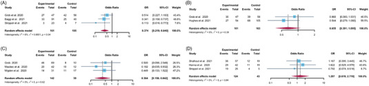
Association between clinicopathological and molecular features of patients and ORR. (A) primary tumor locations; (B) PD‐L1 expression status; (C) immune status
3.3. Survival outcomes
Nine studies 16 , 17 , 18 , 19 , 21 , 22 , 27 , 30 , 31 reported the data of PFS rate at 6 months, which ranged from 50.4% to 76.7%. 16 , 17 , 18 , 19 , 21 , 22 After meta‐analysis, the pooled incidence of PFS rate at 6 months was 59.3% (95% CI: 53.1%–65.5%). Incidence of PFS rate at 12 months was available from eleven studies. 16 , 17 , 18 , 19 , 21 , 22 , 23 , 28 , 29 , 30 , 31 The reported data were varied from 32.4% to 78.0%. After synthesis, the overall incidence of PFS at 12 months was 52.8% (95% CI: 44.9%–60.7%). As for OS, the meta‐analysis suggested that the pooled incidence of OS rate at 6 and 12 months was 80.6% (95% CI: 75.7%–85.5%) and 76.4% (95% CI: 70.1%–82.7%) (Table 3).
3.4. Safety
The pooled analysis of eight studies 16 , 17 , 18 , 19 , 20 , 28 , 29 , 30 suggested that the overall incidence of all grade AEs was 76.9% (95% CI: 63.2%–90.7%). As for the AEs with ≥3 grades, the overall incidence was 20.2% (95% CI: 11.1%–29.3%) after meta‐analysis of eleven studies. 16 , 17 , 18 , 19 , 20 , 21 , 22 , 28 , 29 , 30 , 31 Further analysis indicated that pembrolizumab had better tolerability than cemiplimab. The incidence of all grades and ≥3 grades AEs was 68.6% (95% CI: 63.5%–73.6%) and 8.2% (95% CI: 4.2%–12.3%) for pembrolizumab, and 83.5% (95% CI: 57.1%–100%) and 29.8% (95% CI: 13.2%–46.4%) for cemiplimab (Table 3).
4. DISCUSSION
CSCC is a common skin malignancy with a propensity of locally aggression and distant metastasis. Complete surgical resection could manage CSCC with locally confined disease. 4 However, this treatment approach is not optimal option for advanced CSCC. 6 Despite mortality rate of exceeding 70%, 3 there is no consensus recommendation for those patients with advanced or metastatic disease. 10 , 11 Systematic therapies were considered for the treatment of advanced CSCC. 5‐fluorouracil is the first systematic treatment applied in CSCC, with response rate of around 15% in locally advanced disease. 32 , 33 Capecitabine and IFN are also applied in locally advanced CSCC, but no precise data of efficacy and safety were reported. 32 , 34 , 35 Cetuximab is a monoclonal antibody targeting EGFR, which showed satisfying efficacy in advanced CSCC patients. 36 With the discovery of immunogenicity of CSCC, CPI was applied for the patients with locally advanced or metastatic disease. Phase II trial of cemiplimab showed an ORR of 50%. 16 PD‐1 inhibitor pembrolizumab was also applied in CSCC, with response rate of 34.3%–42%. 17 , 18 Several retrospective studies also suggested the satisfying efficacy of PD‐1 inhibitors in CSCC patients. 19 , 20 , 21 , 22 , 23 However, there is no study based on large population to evaluate the efficacy and safety of PD‐1 inhibitors in locally advanced or metastatic CSCC.
With seven RCTs and six real‐world studies included, our data confirmed the outstanding efficacy of PD‐1 inhibitors for the treatment of CSCC patients, and the pooled ORR and DCR were 47.2% and 71.6%, separately. Furthermore, clinicopathological features of patients showed predictive value for treatment efficacy. Patients with primary head and neck lesions and PD‐L1 positive expression status tend to experience superior ORR during CPI treatment. In contrast, patients with different immune status showed similar ORR during the treatment of CPI. Patients with locoregional disease had comparable ORR with those with distant metastatic disease. For survival outcomes, the overall pooled incidence of PFS at 6 and 12 months were 59.3% and 52.8%, and OS rate at 6 and 12 months were 80.6% and 76.4%. On the contrary, in the phase II trial of cetuximab in treating CSCC patients, the response rate was 27.8%, and the estimated proportion of patients alive at 12 months was 52%, which were lower than those with CPI treatment.
In terms of AEs, the precise data of that in systematic cytokine treatments were not available due to limited data. Sadek et al. suggested that the grade 3–4 AEs was observed in 64.3% (9/14) patients. 32 The phase II trial of cetuximab reported AEs in all included patients, and the incidence of AEs with 3–4 grade was about 64%. 36 In this study, the overall incidence of AEs with all grades was 70.6%, and 8.9% for AEs with 3–4 grades, which were much lower than that of cytokine and cetuximuab treatment, showing the satisfying safety profile for PD‐1 inhibitors.
Mehta et al. conducted a systematic review and meta‐analysis including six prospective clinical trials. The pooled analysis of 392 patients also demonstrated that CPIs conferred ORR of 42.43% (95% CI: 37.53%–47.45%) and DCR of 58.05% (95% CI 53.04%–62.95%), which were similar but numerically lower than that from our study. 24 Consistent with our results, this study also showed that patients with locoregional and distant metastatic disease had comparable ORR during treatment. However, their study failed to assess the relationship between primary lesion, PD‐L1 expression, and immune status and treatment efficacy. Besides, our study involved more studies than Mehta et al. (including one phase II trial and six retrospective studies), and also evaluated the survival outcomes of CPI treatment, which could more comprehensively showed the satisfying efficacy of CPI in treating CSCC.
This systemic review and meta‐analysis has several limitations. Due to the relatively low incidence of this disease, limited data based on clinical trials was reported. Thus, we also collected related retrospective studies and conducted the pooled analysis to estimate the efficacy and safety of PD‐1 inhibitors. In the included retrospective studies, four of them only reported the overall efficacy and safety outcomes with the treatment of cemiplimab, pembrolizumab, and nivolumab, which hindered us for individually analysis of each treatment regiments. Third, the evaluation of PD‐L1 expression differed among included studies. Maubec et al. assessed the PD‐L1 positive expression using tumor proportion score, 18 while Grob et al. conducted this assessment using combined positive score. 17 This difference might reduce the credibility of the result that PD‐L1 expression was a biomarker for PD‐1 inhibitors in CSCC. Forth, only two to three studies were included in the subgroup analysis, which might weaken the credibility of the outcomes. Fifth, the reported AEs were not well defined among included studies. Some of them clearly defined AEs as treatment‐related, while the others did not clarify this issue, which might overestimate the incidence of AEs. Another limitation is that there is no study that directly comparing the efficacy and safety of different PD‐1 inhibitors in CSCC.
5. CONCLUSION
We have provided the first meta‐analysis comprehensively analysis the efficacy and safety of PD‐1 inhibitors for the treatment of advanced CSCC. In this study, we demonstrated the satisfying PFS and treatment response rate of PD‐1 inhibitors, especially cemiplimab and pembrolizumab. Besides, we also indicated that patients with PD‐L1 positive expression and primary head and neck lesion had superior response rate, while patients with different immune status could equally benefit from CPI treatments. On the other hand, PD‐1 inhibitors also showed acceptable toxicity. Future studies are needed to compare the efficacy and safety of available PD‐1 inhibitors for the treatment of locally advanced or metastatic CSCC.
CONFLICT OF INTEREST
The authors have no conflict of interest to declare.
Supporting information
Figure S1. Egger's test
Figure S2. Assessment of risk of bias
ACKNOWLEDGMENTS
This study was financially was supported by Sichuan Science and Technology Program, grant number: 2020YFS0267.
Zhang H, Zhong A, Chen J. Immune checkpoint inhibitors in advanced cutaneous squamous cell carcinoma: A systemic review and meta‐analysis. Skin Res Technol. 2023;29:1–9. 10.1111/srt.13229
DATA AVAILABILITY STATEMENT
All data are available from the corresponding author upon request.
REFERENCES
- 1. Rogers HW, Weinstock MA, Feldman SR, Coldiron BM. Incidence estimate of nonmelanoma skin cancer (keratinocyte carcinomas) in the U.S. population, 2012. AMA Dermatol. 2015;151(10):1081‐1086. [DOI] [PubMed] [Google Scholar]
- 2. Siegel RL, Miller KD, Fuchs HE, Jemal A. Cancer statistics, 2021. CA Cancer J Clin. 2021;71(1):7‐33. [DOI] [PubMed] [Google Scholar]
- 3. Leiter U, Keim U, Eigentler T, et al. Incidence, mortality, and trends of nonmelanoma skin cancer in Germany. J Invest Dermatol. 2017;137(9):1860‐1867. [DOI] [PubMed] [Google Scholar]
- 4. Kauvar ANB, Arpey CJ, Hruza G, Olbricht SM, Bennett R, Mahmoud BH. Consensus for nonmelanoma skin cancer treatment, part II: squamous cell carcinoma, including a cost analysis of treatment methods. Dermatol Surg. 2015;41(11):1214‐1240. [DOI] [PubMed] [Google Scholar]
- 5. Brunner M, Veness Michael J, Ch'ng S, Elliott M, Clark JR. Distant metastases from cutaneous squamous cell carcinoma–analysis of AJCC stage IV. Head Neck. 2013;35(1):72‐75. [DOI] [PubMed] [Google Scholar]
- 6. Veness MJ, Morgan GJ, Palme CE, Gebski V. Surgery and adjuvant radiotherapy in patients with cutaneous head and neck squamous cell carcinoma metastatic to lymph nodes: combined treatment should be considered best practice. Laryngoscope. 2005;115(5):870–875. [DOI] [PubMed] [Google Scholar]
- 7. Veness MJ, Palme CE, Morgan GJ. High‐risk cutaneous squamous cell carcinoma of the head and neck: results from 266 treated patients with metastatic lymph node disease. Cancer. 2006;106(11):2389‐2396. [DOI] [PubMed] [Google Scholar]
- 8. Hillen U, Leiter U, Haase S, et al. Advanced cutaneous squamous cell carcinoma: a retrospective analysis of patient profiles and treatment patterns‐results of a non‐interventional study of the DeCOG. Eur J Cancer. 2018;96:34‐43. [DOI] [PubMed] [Google Scholar]
- 9. Karia PS, Han J, Schmults CD. Cutaneous squamous cell carcinoma: estimated incidence of disease, nodal metastasis, and deaths from disease in the United States, 2012. J Am Acad Dermatol. 2013;68(6):957‐966. [DOI] [PubMed] [Google Scholar]
- 10. Jarkowski A, Hare R, Loud P, et al. Systemic therapy in advanced cutaneous squamous cell carcinoma (CSCC): the Roswell Park experience and a review of the literature. Am J Clin Oncol. 2016;39(6):545‐548. [DOI] [PubMed] [Google Scholar]
- 11. Cranmer LD, Engelhardt C, Morgan SS. Treatment of unresectable and metastatic cutaneous squamous cell carcinoma. Oncologist. 2010;15(2):1320‐1328. [DOI] [PMC free article] [PubMed] [Google Scholar]
- 12. Pickering CR, Zhou JH, Lee JJ, et al. Mutational landscape of aggressive cutaneous squamous cell carcinoma. Clin Cancer Res. 2014;20(24):6582‐6592. [DOI] [PMC free article] [PubMed] [Google Scholar]
- 13. Yarchoan M, Hopkins A, Jaffee EM. Tumor mutational burden and response rate to PD‐1 inhibition. N Engl J Med. 2017;377(25):2500‐2501. [DOI] [PMC free article] [PubMed] [Google Scholar]
- 14. Kim Y, He Y‐Y. Ultraviolet radiation‐induced non‐melanoma skin cancer: regulation of DNA damage repair and inflammation. Genes Dis. 2014;1(2):188‐198. [DOI] [PMC free article] [PubMed] [Google Scholar]
- 15. Madan V, Lear JT, Szeimies R‐M. Non‐melanoma skin cancer. Lancet. 2010;375(9715):673‐685. [DOI] [PubMed] [Google Scholar]
- 16. Migden MR, Rischin D, Schmults CD, et al. PD‐1 blockade with cemiplimab in advanced cutaneous squamous‐cell carcinoma. N Engl J Med. 2018;379(4):341‐351. [DOI] [PubMed] [Google Scholar]
- 17. Grob JJ, Gonzalez R, Basset‐Seguin N, et al. Pembrolizumab monotherapy for recurrent or metastatic cutaneous squamous cell carcinoma: a single‐arm phase II trial (KEYNOTE‐629). J Clin Oncol. 2020;38(25):2916‐2925. [DOI] [PMC free article] [PubMed] [Google Scholar]
- 18. Maubec E, Boubaya M, Petrow P, et al. Phase II study of pembrolizumab as first‐line, single‐drug therapy for patients with unresectable cutaneous squamous cell carcinomas. J Clin Oncol. 2020;38(26):3051‐3061. [DOI] [PubMed] [Google Scholar]
- 19. In GK, Vaidya P, Filkins A, et al. PD‐1 inhibition therapy for advanced cutaneous squamous cell carcinoma: a retrospective analysis from the University of Southern California. J Cancer Res Clin Oncol. 2021;147(6):1803‐1811. [DOI] [PMC free article] [PubMed] [Google Scholar]
- 20. Baggi A, Quaglino P, Rubatto M, et al. Real world data of cemiplimab in locally advanced and metastatic cutaneous squamous cell carcinoma. Eur J Cancer. 2021;157:250‐258. [DOI] [PubMed] [Google Scholar]
- 21. Hanna GJ, Ruiz ES, LeBoeuf NR, et al. Real‐world outcomes treating patients with advanced cutaneous squamous cell carcinoma with immune checkpoint inhibitors (CPI). Br J Cancer. 2020;123(10):1535‐1542. [DOI] [PMC free article] [PubMed] [Google Scholar]
- 22. Salzmann M, Leiter U, Loquai C, et al. Programmed cell death protein 1 inhibitors in advanced cutaneous squamous cell carcinoma: real‐world data of a retrospective, multicenter study. Eur J Cancer. 2020;138:125‐132. [DOI] [PubMed] [Google Scholar]
- 23. Shalhout SZ, Park JC, Emerick KS, Sullivan RJ, Kaufman HL, Miller DM. Real‐world assessment of response to anti‐programmed cell death 1 therapy in advanced cutaneous squamous cell carcinoma. J Am Acad Dermatol. 2021;85(4):1038‐1040. [DOI] [PubMed] [Google Scholar]
- 24. Mehta NK, Li AR, Nguyen SA, Kaczmar JM, Neskey DM, Day TA. Immune checkpoint inhibitors for advanced cutaneous squamous cell carcinoma: a systematic review with meta‐analysis. Targ Oncol. 2021;16(6):743‐752. [DOI] [PubMed] [Google Scholar]
- 25. Hutton B, Salanti G, Caldwell DM, et al. The PRISMA extension statement for reporting of systematic reviews incorporating network meta‐analyses of health care interventions: checklist and explanations. Ann Intern Med. 2015;162(11):777‐784. [DOI] [PubMed] [Google Scholar]
- 26. Higgins J, Thomas J, Chandler J, et al. Cochrane Handbook for Systematic Reviews of Interventions version 6.1. Published online September 7, 2020. www.training.cochrane.org/handbook
- 27. Yushak ML, Lawson DH, Goings M, et al. Phase II trial of pembrolizumab (MK‐3475) in metastatic cutaneous squamous cell carcinoma: an updated analysis. J Clin Oncol. 2019;37(15_suppl):e21015‐e21015. [Google Scholar]
- 28. Migden MR, Khushalani NI, Chang ALS, et al. Cemiplimab in locally advanced cutaneous squamous cell carcinoma: results from an open‐label, phase 2, single‐arm trial. Lancet Oncol. 2020;21(2):294‐305. [DOI] [PMC free article] [PubMed] [Google Scholar]
- 29. Rischin D, Migden MR, Lim AM, et al. Phase 2 study of cemiplimab in patients with metastatic cutaneous squamous cell carcinoma: primary analysis of fixed‐dosing, long‐term outcome of weight‐based dosing. J Immunother Cancer. 2020;8(1):e000775. [DOI] [PMC free article] [PubMed] [Google Scholar]
- 30. Hughes BGM, Munoz‐Couselo E, Mortier L, et al. Pembrolizumab for locally advanced and recurrent/metastatic cutaneous squamous cell carcinoma (KEYNOTE‐629 study): an open‐label, nonrandomized, multicenter, phase II trial. Ann Oncol. 2021;32(10):1276‐1285. [DOI] [PubMed] [Google Scholar]
- 31. Strippoli S, Fanizzi A, Quaresmini D, et al. Cemiplimab in an elderly frail population of patients with locally advanced or metastatic cutaneous squamous cell carcinoma: a single‐center real‐life experience from Italy. Front Oncol. 2021;11:686308. [DOI] [PMC free article] [PubMed] [Google Scholar]
- 32. Sadek H, Azli N, Wendling JL, et al. Treatment of advanced squamous cell carcinoma of the skin with cisplatin, 5‐fluorouracil, and bleomycin. Cancer. 1990;66(8):1692‐1696. [DOI] [PubMed] [Google Scholar]
- 33. Cartei G, Cartei F, Interlandi G, et al. Oral 5‐fluorouracil in squamous cell carcinoma of the skin in the aged. Am J Clin Oncol. 2000;23(2):181‐184. [DOI] [PubMed] [Google Scholar]
- 34. DeConti RC. Chemotherapy of squamous cell carcinoma of the skin. Semin Oncol. 2012;39(2):145‐149. [DOI] [PubMed] [Google Scholar]
- 35. Wollina U, Hansel G, Koch A, Köstler E. Oral capecitabine plus subcutaneous interferon alpha in advanced squamous cell carcinoma of the skin. J Cancer Res Clin Oncol. 2005;131(5):300‐304. [DOI] [PMC free article] [PubMed] [Google Scholar]
- 36. Maubec E, Petrow P, Scheer‐Senyarich I, et al. Phase II study of cetuximab as first‐line single‐drug therapy in patients with unresectable squamous cell carcinoma of the skin. J Clin Oncol. 2011;29(25):3419‐3426. [DOI] [PubMed] [Google Scholar]
Associated Data
This section collects any data citations, data availability statements, or supplementary materials included in this article.
Supplementary Materials
Figure S1. Egger's test
Figure S2. Assessment of risk of bias
Data Availability Statement
All data are available from the corresponding author upon request.