1. CASE PRESENTATION
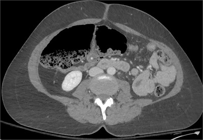
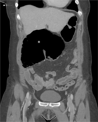
A 52‐year‐old woman presented to the emergency department with severe, acute right‐sided abdominal pain that woke her from sleep 5 hours prior, radiated to the right flank, and had not improved after a bowel movement. She had associated nausea and non‐bilious emesis, but she had no diarrhea or fever. She had no significant medical history or previous abdominal surgeries. She had a screening colonoscopy 1 month prior. Her abdominal examination was soft, non‐distended, and severely tender in the right midabdominal region. Laboratory results were within normal limits. Figures 1 and 2 show an abdomen/pelvis computed tomography (CT), with scrollable CT available in the Appendix.
FIGURE 1.

CT of the abdomen and pelvis with intravenous contrast (axial view) showing dilated large bowel. The terminal ileum is marked with an asterisk. No whirl sign was present. Abbreviation: CT, computed tomography
FIGURE 2.

CT of the abdomen and pelvis with intravenous contrast (coronal view) showing dilated large bowel. The dilated cecum is marked with an asterisk. Abbreviation: CT, computed tomography
2. DIAGNOSIS
Cecal bascule
The CT demonstrated a cecal volvulus, bascule subtype, with cecal closed‐loop obstruction, without bowel ischemia or perforation. There was no whirl sign, a radiographic finding with 73% sensitivity and 100% specificity for axial cecal volvulus. 1 , 2 Bascules are the rarest subtype of cecal volvulus, representing 5%–20% of cecal volvuli. 3 They involve an anterior folding of the cecum rather than the axial twisting found in axial cecal volvulus and thus will not have a whirl sign. 3 , 4 In a literature review, the most common presenting symptom for cecal bascule was abdominal distension (84%), which our patient lacked. 3 Interestingly, colonoscopy can be a precipitating factor for cecal bascule. 5 Abdominal pain was a presenting symptom in only 61%, and vomiting in 30%. 3 Emergency physicians should maintain a nuanced index of suspicion given cecal bascules’ lack of uniform presentation. The patient underwent right hemicolectomy with ileostomy and recovered well.
Supporting information
Supporting Information
Hancock‐Cerutti W, Aliaga L. Woman with right abdominal pain. JACEP Open. 2023;4:e12890. 10.1002/emp2.12890
Meetings: This manuscript has not been presented at an academic meeting.
REFERENCES
- 1. Rosenblat JM, Rozenblit AM, Wolf EL, et al. Findings of cecal volvulus at CT. Radiology. 2010;256(1):169‐175. [DOI] [PubMed] [Google Scholar]
- 2. D'Alessandro AD, Smith AT, Binion TW, et al. Runner with abdominal pain. Ann Emerg Med. 2019;74(4):491‐508. [DOI] [PubMed] [Google Scholar]
- 3. Lung BE, Yelika SB, Murthy AS, et al. Cecal bascule: a systematic review of the literature. Tech Coloproctology. 2018;22(2):75‐80. [DOI] [PubMed] [Google Scholar]
- 4. Kairys N, Skidmore K, Repanshek J, et al. An unlikely cause of abdominal pain. Clin Pract Cases Emerg Med. 2018;2(2):139‐142. [DOI] [PMC free article] [PubMed] [Google Scholar]
- 5. Li T, Myat YM, Nguyen STT, et al. Bowel obstruction: cecal bascule after colonoscopy. ACG Case Rep J. 2022;9(7):e00803. [DOI] [PMC free article] [PubMed] [Google Scholar]
Associated Data
This section collects any data citations, data availability statements, or supplementary materials included in this article.
Supplementary Materials
Supporting Information


