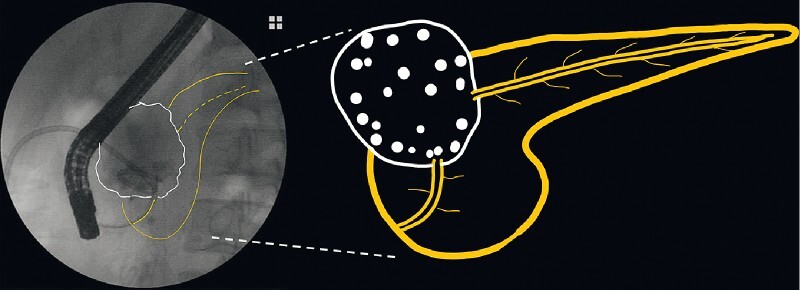
A 48-year-old man with a history of acute necrotizing pancreatitis presented to our institution for management of a persistent external pancreatic fistula. He had required percutaneous catheter drainage for a pseudocyst in the pancreatic neck before this complication. A pancreatogram showed that the pseudocyst was in communication with the main pancreatic duct (MPD) in the head; however, there was no opacification of the upstream MPD, confirming complete MPD disconnection ( Fig. 1 ). It had not been possible to traverse the MPD disconnection with a guidewire during prior attempts.
Fig. 1.

Fluoroscopic image during endoscopic retrograde cholangiopancreatography and accompanying schematic showing the pseudocyst with no opacification of the upstream main pancreatic duct, confirming disconnected pancreatic duct syndrome.
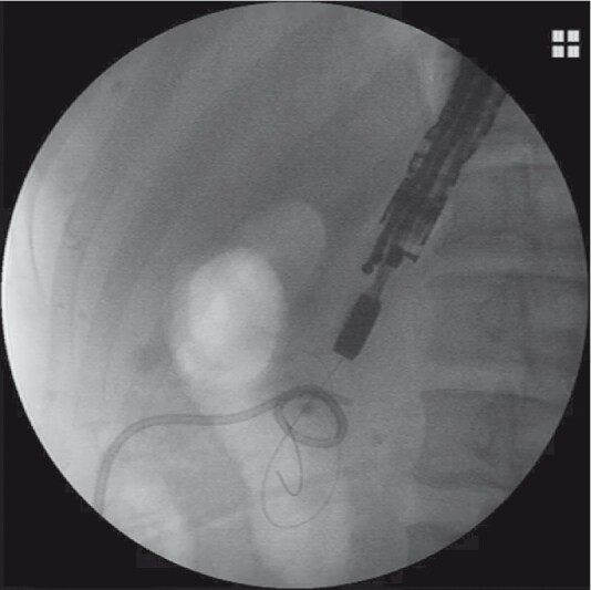
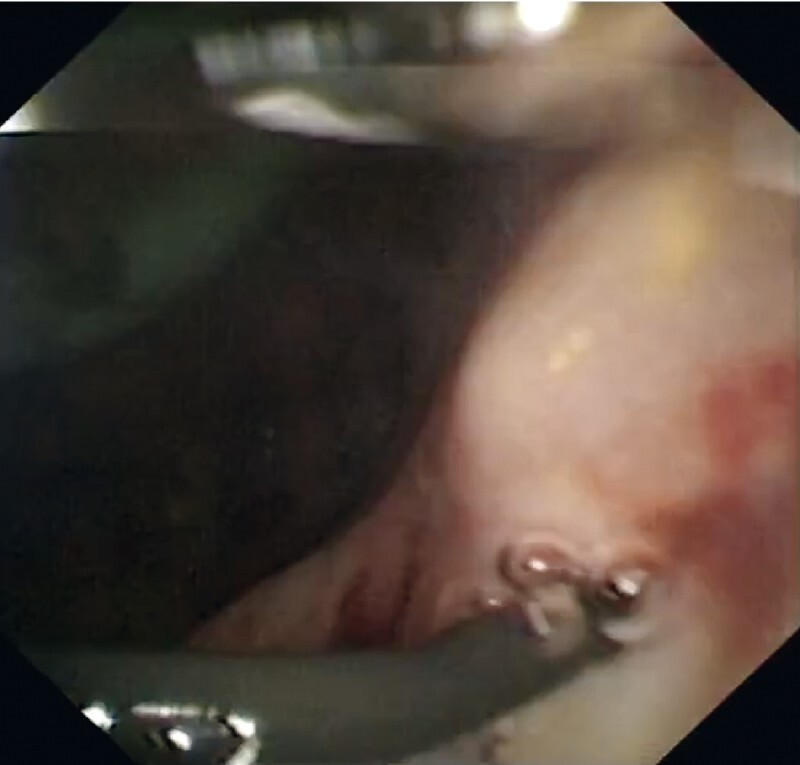
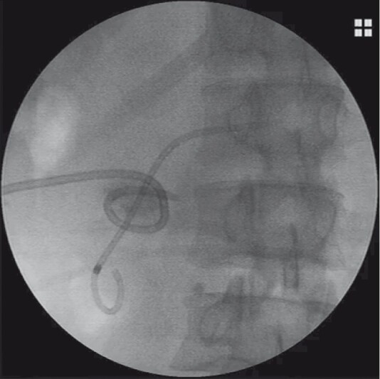
We attempted to advance a novel peroral cholangiopancreatoscope (“eyeMax”, 9 Fr; Micro-Tech Co., Ltd., Nanjing, China) across the papilla into the pseudocyst but were unable to find the opening of the upstream MPD. Therefore, we decided to use the endoscopic ultrasonography-assisted rendezvous technique (EUS-RV) to help locate the opening with the cholangiopancreatoscope. Using a linear echoendoscope, the upstream MPD was recognized and punctured with a 19G fine-needle aspiration (FNA) needle in the stomach. A second guidewire was inserted through the needle into the MPD and further down into the pseudocyst ( Fig. 2 ). Under pancreatoscopic guidance again, we were able to see the EUS guidewire coming out of the disrupted orifice of the upstream MPD ( Fig. 3 ). Following the EUS guidewire, the cholangiopancreatoscope guidewire was smoothly inserted into the upstream MPD ( Video 1 ). An endoscopic retrograde cholangiopancreatography (ERCP) catheter was used to adjust the direction of the guidewire to gain deep access to the MPD and a 7-Fr × 9-cm single-pigtail plastic stent was placed via the papilla across the disconnection ( Fig. 4 ; Video 1 ). The percutaneous drainage catheter was removed, with successful closure of the cutaneous opening of the fistula noted 2 months later.
Fig. 2.

Fluoroscopic image showing endoscopic ultrasonography-guided puncture of the upstream main pancreatic duct, with the guidewire passing into the pseudocyst.
Fig. 3.

Peroral pancreatoscopy showing the endoscopic ultrasonography guidewire in the pseudocyst.
Fig. 4.

Fluoroscopic image showing a single-pigtail plastic stent that was placed via the papilla across the disconnection of the main pancreatic duct.
Video 1 A completely disrupted pancreatic duct is bridged using a novel peroral cholangiopancreatoscope combined with an endoscopic ultrasonography-assisted rendezvous technique.
In conclusion, the combination of ERCP, EUS, and peroral pancreatoscopy offers a novel, accurate, and microinvasive treatment method for pancreatic duct related disorders 1 2 3 .
Endoscopy_UCTN_Code_TTT_1AS_2AD
Footnotes
Competing interests The authors declare that they have no conflict of interest.
Endoscopy E-Videos : https://eref.thieme.de/e-videos .
Endoscopy E-Videos is an open access online section, reporting on interesting cases and new techniques in gastroenterological endoscopy. All papers include a high quality video and all contributions are freely accessible online. Processing charges apply (currently EUR 375), discounts and wavers acc. to HINARI are available. This section has its own submission website at https://mc.manuscriptcentral.com/e-videos
References
- 1.Baron T H, DiMaio C J, Wang A Y et al. American Gastroenterological Association Clinical Practice Update: Management of pancreatic necrosis. Gastroenterology. 2020;158:67–75. doi: 10.1053/j.gastro.2019.07.064. [DOI] [PubMed] [Google Scholar]
- 2.Verma S, Rana S S. Disconnected pancreatic duct syndrome: Updated review on clinical implications and management. Pancreatology. 2020;20:1035–1044. doi: 10.1016/j.pan.2020.07.402. [DOI] [PubMed] [Google Scholar]
- 3.Varadarajulu S, Noone T C, Tutuian R et al. Predictors of outcome in pancreatic duct disruption managed by endoscopic transpapillary stent placement. Gastrointest Endosc. 2005;61:568–575. doi: 10.1016/s0016-5107(04)02832-9. [DOI] [PubMed] [Google Scholar]


