Abstract
Background
Occult breast cancer (OBC) is a rare type of breast cancer, which accounts for 0.3–1.0% of all breast cancers. However, the treatment of OBC remains controversial, especially the local treatment. We aimed to analyze the impact of different treatment and N stage on survival in early-stage OBC patients, and construct a nomogram to predict the prognosis.
Methods
The data of patients with early-stage breast cancer were obtained from 17 registries in the Surveillance, Epidemiology, and End Results (SEER) database. Patient characteristics and breast cancer–specific survival (BCSS) were compared among the groups. Cox proportional risk models were used for both the univariate and multivariate analyses. Variables with a P value <0.07 in the univariate analysis were included in the multivariate analysis. The independent prognostic factors were included in the nomogram and validated internally.
Results
A total of 492 early-stage OBC patients were randomized at a 7:3 ratio into the training cohort (n=348) and the testing cohort (n=144). N2+ stage patients had a worse prognosis than N1 stage patients (P=0.0051). Triple-negative breast cancer (TNBC) patients had the worst prognosis. Early-stage OBC patients benefited from surgery (P=0.0093) and radiotherapy (P=0.0102), but not chemotherapy (P=0.4030). An analysis of OBC patients with different N stages showed that in terms of treatment, N1 stage patients benefited from surgery (P=0.023), but did not benefit significantly from radiotherapy (P=0.0793), whereas N2+ stage patients benefited from radiotherapy (P=0.0098), but the benefit from surgery was not significant (P=0.1005). In the multivariate analysis, N stage, surgery, and radiotherapy remained statistically significant. Based on the results of the multivariate analysis, we constructed a nomogram for estimating the 3- and 5-year BCSS of OBC patients. The concordance index and the calibration plots show that our nomogram had sufficient accuracy and good coordination.
Conclusions
N stage, surgery, and radiotherapy were identified as independent prognostic factors for OBC. We successfully constructed a nomogram using these independent risk factors and demonstrated that it could help predict the 3- and 5-year BCSS of OBC patients. Further data analyses need to be conducted to revise the treatment of early-stage OBC.
Keywords: Occult breast cancer (OBC), breast cancer specific survival (BCSS), treatment, risk factor, nomogram
Highlight box.
Key findings
• We constructed a nomogram to predict the 3- and 5-year BCSS of the OBC patients. And we found that lymph node status in OBC patients should be emphasized.
What is known and what is new?
• Several studies have identified risk factors associated with the prognosis of OBC.
• We developed a prediction model using risk factors for OBC. And we found that N stage had an impact on the benefit of different local treatments for early-stage OBC patients.
What is the implication, and what should change now?
• We believe that more attention should be paid to the N status of OBC patients and local treatment for early-stage OBC patients should be based on N status. Further data analyses need to be conducted to revise the treatment of OBC.
Introduction
Occult breast cancer (OBC) is a rare type of breast cancer, which accounts for 0.3–1.0% of all breast cancers (1-4). OBC mostly presents with regional lymph node metastases in the absence of evidence of a primary lesion, such as axillary lymph node enlargement (1). More than a century has passed since OBC was first reported (5); however, the treatment of OBC remains controversial (6). The current local treatment of OBC is based on surgery and radiotherapy, but which one should be given priority is still unclear (7-9). The National Comprehensive Cancer Network (NCCN) guidelines recommend surgery plus axillary lymph node dissection (ALND) or ALND plus radiotherapy for OBC patients with N1 stage, and systemic therapy may be given to OBC patients according to the recommendations for stage II or III. As in local advanced patients, systemic treatment should be considered before or after surgery for patients with N2–3M0 (10). Although the NCCN guidelines give recommendations for the treatment of early-stage OBC, there are still many studies showing different results in terms of local treatment. Thus, the treatment of OBC needs further exploration, especially the local treatment. In addition, the effect of N stage on treatment is underreported. OBC has a better prognosis than node-positive non-OBC (11-13); several studies have identified risk factors associated with the prognosis of OBC (7,8,11,14,15). However, no study has developed a prediction model using risk factors for OBC. Prospective studies are difficult to conduct because of the low prevalence of OBC. The analysis of large databases plays an important role in gathering evidence for rare diseases, including OBC. Since the treatment of advanced breast cancer is mainly systemic treatment, in this study, we extracted the data of early-stage OBC patients from the Surveillance, Epidemiology, and End Results (SEER) database. We aimed to evaluate the breast cancer-specific survival (BCSS) in early-stage OBC patients, analyze the impact of different treatment and N stage on survival, and construct a nomogram to predict the 3- and 5-year BCSS of the patients, which we also validated internally. We present the following article in accordance with the TRIPOD reporting checklist (available at https://atm.amegroups.com/article/view/10.21037/atm-22-5701/rc).
Methods
Data extraction
The data were obtained from 17 registries in the SEER database, which is an open access resource for epidemiological and survival analyses based on cancer. The data reported in this study represent the latest follow-up data (Nov 2021 Sub). SEER-Stat (version 8.4.0) was used to export the information of the representative patients (http://seer.cancer.gov/).
Because data on human epidermal growth factor receptor 2 (HER2) status have only been available since 2010, we only included eligible patients from the SEER database who met the following criteria: (I) had American Joint Committee on Cancer (AJCC) T0 stage; (II) had been diagnosed between 2010 and 2019; (III) were aged ≥18 years; (IV) were female; (V) had the breast as the first and only and primary cancer site; (VI) had a malignant tumor and a positive histology diagnosis; and (VII) had AJCC M0 stage. Patients with AJCC N0 stage and/or a histological diagnosis of Paget’s or inflammatory breast cancer were excluded from the study.
The following variables were extracted: year of diagnosis, age at diagnosis, race, laterality, AJCC N stage, estrogen receptor (ER) or progesterone receptor (PR) status, HER-2 status, tumor subtypes, records of surgery, radiotherapy and chemotherapy, survival time, vital status, and cause of death. We also excluded patients whose variable contents were unknown. In the SEER database, hormone receptor positive (HR+)/HER2 negative (HER2–), HR+/HER2 positive (HER2+), HR negative (HR–)/HER2+, and HR–/HER2– were defined as the luminal A, luminal B, HER2+, and triple-negative breast cancer (TNBC) subtypes, respectively.
A total of 492 early-stage OBC patients were included in the primary cohort and further randomized at a 7:3 ratio into the training cohort (n=348) and the testing cohort (n=144). We planned to perform survival analysis on all eligible patients. The training cohort was used for the univariate and multivariate analyses to finally obtain independent prognostic factors, and the independent prognostic factors were included in the nomogram, the testing cohort was used to validate it. The study was conducted in accordance with the Declaration of Helsinki (as revised in 2013).
Statistical analysis
The primary endpoint of this study was BCSS. All the survival outcomes were generated using the Kaplan-Meier method, and Kaplan-Meier survival curves for BCSS were plotted. Cox proportional risk models were used for both the univariate and multivariate analysis of all the alternative variables (i.e., year of diagnosis, age at diagnosis, race, laterality, AJCC N stage, ER and PR status, HER-2 status, tumor subtypes, and records of surgery, radiotherapy and chemotherapy). Variables in the univariate analysis with a P value <0.07 were included in the multivariate analysis. The independent prognostic factors were included in the nomogram and validated internally. The concordance index (C-index) was used to evaluate the clinical prediction model; a value of ≥0.60 indicated considerable discriminative ability. Calibration plots were generated to evaluate the accuracy of the nomogram. All the data were analyzed using SPSS 25, GraphPad Prism 9, and R software (4.2.1).
Results
We obtained complete data for 492 early-stage OBC patients with a median follow-up time of 45 months (the training cohort comprised 348 patients, and the testing cohort comprised 144 patients). The clinical characteristics and treatment information of all patients are summarized in Table 1. White was the predominant race of all the OBC patients (386/492, 78.46%). Among the patients with positive lymph nodes, most patients had N1 stage (323/492, 65.65%), but some patients had N2 stage or higher (169/492, 34.35%). Luminal A was the most frequent tumor subtype (229/492, 46.54%), followed by TNBC (117/492, 23.78%), while luminal B (88/492, 17.89%) and HER2+ (58/492, 11.79%) were less common. Less than half of the patients received surgery (233, 47.36%), but more than two thirds of the patients received radiotherapy (335, 68.09%), and most of the patients received chemotherapy (422, 85.77%). By the end of the follow-up period, 421 (85.57%) patients were still alive, 48 (9.76%) had died of breast cancer and 23 (4.67%) had died of other causes. In the training cohort, 38 (10.92%) patients died of breast cancer, while in the testing cohort, only 10 (6.94%) patients died of breast cancer. The Chi-square test results revealed no statistical difference in year of diagnosis, age at diagnosis, race, laterality, AJCC N stage, ER and PR status, HER-2 status, tumor subtypes, and records of surgery, radiotherapy and chemotherapy between the training and testing groups (Table 1).
Table 1. Patient characteristics for the primary cohort.
| Characteristics | Primary cohort (%) | Cohort (%) | P value | |
|---|---|---|---|---|
| Training | Testing | |||
| Year of diagnosis | ||||
| 2010–2014 | 228 (46.34) | 166 (47.70) | 62 (43.06) | 0.347 |
| 2015–2019 | 264 (53.66) | 182 (52.30) | 82 (56.94) | |
| Age at diagnosis, years | ||||
| ≤60 | 249 (50.61) | 173 (49.71) | 76 (52.78) | 0.536 |
| >60 | 243 (49.39) | 175 (50.29) | 68 (47.22) | |
| Race | ||||
| White | 386 (78.46) | 272 (78.16) | 114 (79.17) | 0.964 |
| Asian | 45 (9.15) | 32 (9.20) | 13 (9.03) | |
| Black | 61 (12.40) | 44 (12.64) | 17 (11.81) | |
| Laterality | ||||
| Left | 267 (54.27) | 188 (54.02) | 79 (54.86) | 0.865 |
| Right | 225 (45.73) | 160 (45.98) | 65 (45.14) | |
| AJCC N stage | ||||
| N1 | 323 (65.65) | 231 (66.38) | 92 (63.89) | 0.597 |
| N2+ | 169 (34.35) | 117 (33.62) | 52 (36.11) | |
| ER | ||||
| Negative | 179 (36.38) | 127 (36.49) | 52 (36.11) | 0.936 |
| Positive | 313 (63.62) | 221 (63.51) | 92 (63.89) | |
| PR | ||||
| Negative | 296 (60.16) | 204 (58.62) | 92 (63.89) | 0.277 |
| Positive | 196 (39.84) | 144 (41.38) | 52 (36.11) | |
| HER2 | ||||
| Negative | 346 (70.33) | 244 (70.11) | 102 (70.83) | 0.874 |
| Positive | 146 (29.67) | 104 (29.89) | 42 (29.17) | |
| Tumor subtype | ||||
| Luminal A | 229 (46.54) | 160 (45.98) | 69 (47.92) | 0.940 |
| Luminal B | 88 (17.89) | 64 (18.39) | 24 (16.67) | |
| HER-2 positive | 58 (11.79) | 40 (11.49) | 18 (12.50) | |
| TNBC | 117 (23.78) | 84 (24.14) | 33 (22.92) | |
| Surgery | ||||
| No | 259 (52.64) | 181 (52.01) | 78 (54.17) | 0.663 |
| Yes | 233 (47.36) | 167 (47.99) | 66 (45.83) | |
| Radiotherapy | ||||
| No/unknown | 157 (31.91) | 118 (33.91) | 39 (27.08) | 0.140 |
| Yes | 335 (68.09) | 230 (66.09) | 105 (72.92) | |
| Chemotherapy | ||||
| No/unknown | 70 (14.23) | 50 (14.37) | 20 (13.89) | 0.890 |
| Yes | 422 (85.77) | 298 (85.63) | 124 (86.11) | |
| Cause of death | ||||
| Survive or die of other causes | 444 (90.24) | 310 (89.08) | 134 (93.06) | 0.176 |
| Died of breast cancer | 48 (9.76) | 38 (10.92) | 10 (6.94) | |
| Survival months | ||||
| ≤36 months | 217 (44.11) | 153 (43.97) | 64 (44.44) | 0.286 |
| 37–60 months | 94 (19.11) | 61 (17.53) | 33 (22.92) | |
| ≥61 months | 181 (36.79) | 134 (38.51) | 47 (32.64) | |
| Total | 492 | 348 | 144 | |
AJCC, American Joint Committee on Cancer; ER, estrogen receptor; PR, progesterone receptor; TNBC, triple-negative breast cancer.
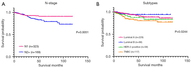
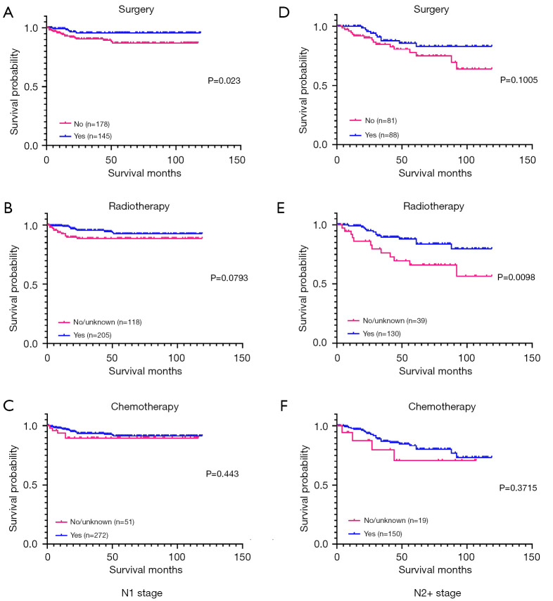
We conducted a Kaplan-Meier analysis to evaluate the effects of the variables on survival. The effects of N stage and the subtypes on survival are shown in Figure 1. Patients with N2+ stage had a worse prognosis than those with N1 stage (P=0.0051). As with other breast cancers, TNBC patients had the worst prognosis, followed by HER-2+ patients and luminal A/B breast cancer (P=0.0244). In relation to treatment, we analyzed the effects of surgery, radiotherapy and chemotherapy on BCSS (Figure 2). Both surgery (P=0.0093) and radiotherapy (P=0.0102) may provide benefits to patients with OBC. However, chemotherapy (P=0.4030) did not significantly affect the prognosis of OBC patients. We conducted a survival analysis of OBC patients with different N stages and found that in terms of treatment (Figure 3), N1 stage patients benefited from surgery (P=0.023), but did not benefit significantly from radiotherapy (P=0.0793). Unlike N1 stage patients, N2+ stage patients benefited from radiotherapy (P=0.0098), but the benefit from surgery was not significant (P=0.1005).
Figure 1.
Kaplan-Meier curves for BCSS stratified by (A) N stage and (B) subtypes. BCSS, breast cancer-specific survival; TNBC, triple-negative breast cancer.
Figure 2.
Kaplan-Meier curves for BCSS stratified by (A) surgery, (B) radiotherapy, and (C) chemotherapy. BCSS, breast cancer-specific survival.
Figure 3.
Kaplan-Meier curves for BCSS stratified by (A) N1 stage surgery, (B) N1 stage radiotherapy, (C) N1 stage chemotherapy, (D) N2+ stage surgery, (E) N2+ stage radiotherapy, and (F) N2+ stage chemotherapy. BCSS, breast cancer-specific survival.
The results of the univariate and multivariate analysis are shown in Table 2. In the univariate analysis, we found that N stage [hazard ratio (HR) 2.441, 95% confidence interval (CI): 1.286–4.631, P<0.07], ER status (HR 0.497, 95% CI: 0.263–0.940, P<0.07), PR status (HR 0.420, 95% CI: 0.199–0.888, P<0.07), surgery (HR 0.386, 95% CI: 0.191–0.778, P<0.07), and radiotherapy (HR 0.543, 95% CI: 0.287–1.026, P<0.07) were significantly associated with BCSS. In the multivariate analysis, N stage (HR 2.650, 95% CI: 1.379–5.090, P<0.05), surgery (HR 0.359, 95% CI: 0.177–0.725, P<0.05), and radiotherapy (HR 0.416, 95% CI: 0.217–0.799, P<0.05) were significantly associated with BCSS, and thus were deemed independent prognostic factors for BCSS.
Table 2. Univariate and multivariate analysis of overall survival for the training cohort.
| Characteristics | Univariate | Multivariate | |||||
|---|---|---|---|---|---|---|---|
| HR | 95% CI | P value | HR | 95% CI | P value | ||
| Year of diagnosis | |||||||
| 2010–2014 | |||||||
| 2015–2019 | 1.490 | 0.735–3.024 | 0.269 | ||||
| Age at diagnosis, years | |||||||
| ≤60 | |||||||
| >60 | 1.483 | 0.774–2.843 | 0.235 | ||||
| Race | |||||||
| White | |||||||
| Asian | 0.654 | 0.156–2.737 | 0.561 | ||||
| Black | 1.042 | 0.405–2.682 | 0.931 | ||||
| Laterality | |||||||
| Left | |||||||
| Right | 0.903 | 0.476–1.711 | 0.754 | ||||
| AJCC N stage | |||||||
| N1 | |||||||
| N2+ | 2.441 | 1.286–4.631 | 0.006* | 2.650 | 1.379–5.090 | 0.003* | |
| ER | |||||||
| Negative | |||||||
| Positive | 0.497 | 0.263–0.940 | 0.031* | ||||
| PR | |||||||
| Negative | |||||||
| Positive | 0.420 | 0.199–0.888 | 0.023* | ||||
| HER–2 | |||||||
| Negative | |||||||
| Positive | 1.027 | 0.509–2.070 | 0.941 | ||||
| Tumor subtype | |||||||
| Luminal A | |||||||
| Luminal B | 0.813 | 0.268–2.472 | 0.716 | ||||
| HER-2 positive | 2.098 | 0.846–5.199 | 0.110 | ||||
| TNBC | 1.911 | 0.898–4.066 | 0.093 | ||||
| Surgery | |||||||
| No | |||||||
| Yes | 0.386 | 0.191–0.778 | 0.008* | 0.359 | 0.177–0.725 | 0.004* | |
| Radiotherapy | |||||||
| No/unknown | |||||||
| Yes | 0.543 | 0.287–1.026 | 0.060* | 0.416 | 0.217–0.799 | 0.008* | |
| Chemotherapy | |||||||
| No/unknown | |||||||
| Yes | 0.882 | 0.369–2.110 | 0.778 | ||||
*, statistically significant. AJCC, American Joint Committee on Cancer; ER, estrogen receptor; PR, progesterone receptor; TNBC, triple-negative breast cancer.
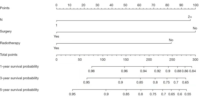
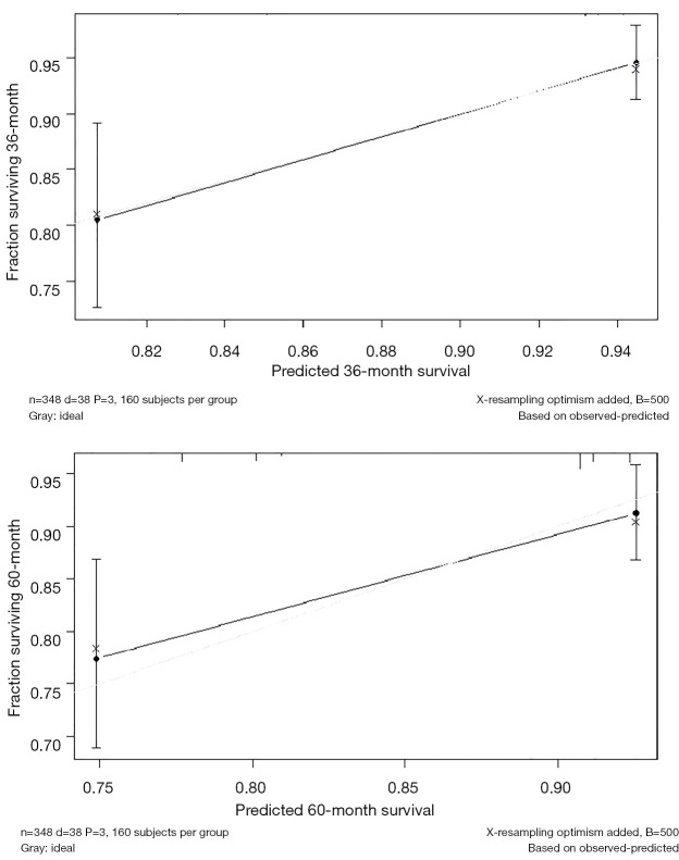
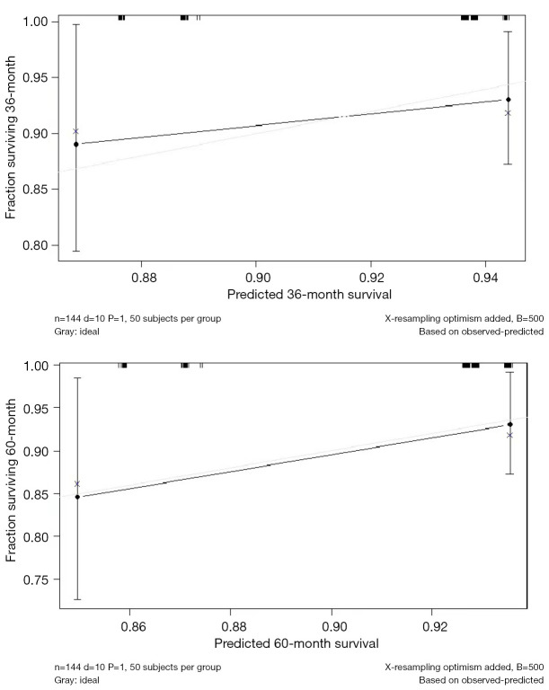
Based on the results obtained from the multivariate analysis, all the independent prognostic factors were included in a nomogram for estimating the 3- and 5-year BCSS of OBC patients (Figure 4). To read the nomogram, a vertical line should be drawn from each variable to the top point row to assign points. Next, the total points for a patient should be added up, and the probability of 3- and 5-year overall survival should be determined by drawing a vertical line from the total points row. The validation results showed that the nomogram had sufficient accuracy in forecasting the prognosis of OBC patients. The C-index values of the training cohort and testing cohort were 0.687 and 0.615, respectively. The calibration plots show good coordination between the predictions of the nomogram model and the observed outcomes of 3- and 5-year BCSS in OBC patients in both the training (Figure 5) and testing (Figure 6) cohorts.
Figure 4.
A nomogram for predicting BCSS. BCSS, breast cancer-specific survival.
Figure 5.
Calibration curves of the training cohort for the nomogram-predicted probability of 3- and 5-year BCSS. BCSS, breast cancer-specific survival.
Figure 6.
Calibration curves of the testing cohort for the nomogram-predicted probability of 3- and 5-year BCSS. BCSS, breast cancer–specific survival.
Discussion
In this retrospective study, we exported the data of 492 early-stage OBC patients from the SEER database, analyzed their clinical characteristics, prognostic factors, and survival, and constructed a nomogram to predict the prognosis of early-stage OBC patients based on the results of our multivariate analysis. No previous study had attempted to construct a nomogram to predict the survival of early-stage OBC patients based on various risk factors. Similar to other studies, we found that axillary lymph node status was an unfavorable prognostic factor for patients with OBC (11,12,14,16,17), and that the higher the N stage, the worse the prognosis of the patients. TNBC was the subtype with the worst prognosis of all OBCs in our results, which is in line with the results of some existing findings (11,12,18).
Our survival analysis of the treatment of early-stage OBC patients showed that both surgery and radiotherapy can be beneficial for early-stage OBC patients, which is in line with the results of previous studies (6,13,18).
The current NCCN Guidelines for Breast Cancer recommend mastectomy plus ALND or ALND plus whole breast irradiation with or without nodal irradiation for OBC patients with stage N1M0, and systemic treatment according to the recommendations for OBC patients with stage II or III. As for local advanced patients, for patients with stage N2–3M0, systemic treatment should be considered before or after ALND and mastectomy (10). However, it should be noted that the evidence supporting the recommendations in the NCCN guidelines is limited.
Previous studies have shown that local therapy provides a survival benefit for OBC patients (8,13,19,20); however, the benefit does not change significantly based on the type of local therapy. In contrast to our results, a retrospective study, which also used data from the SEER database, suggested that there was no difference in the prognosis of all N stage OBC patients who received radiotherapy and surgery (6). Another previous SEER analysis found that the type of local treatment was not associated with an improved prognosis in OBC patients (8). One more study expressed a similar view (13). Similarly, a previous meta-analysis showed that there was no difference in the prognosis of OBC patients who received mastectomy versus radiotherapy, but that radiotherapy improved local recurrence in patients who received ALND (19). Thus, combined ALND and adjuvant radiotherapy should be considered the primary treatment modality for such patients. A recent study based on the SEER database concluded that the type of surgery does not affect the prognosis of OBC patients and additional radiotherapy provides a survival benefit for OBC patients (20).
A slight difference in survival outcomes depending on the type of local therapy has also been observed. Notably, a database study suggests that patients treated with radiotherapy plus ALND had significantly better overall survival than those treated with mastectomy with ALND ± radiotherapy (9). However, as most of the reports did not differentiate between N1 stage and N2+ stage OBC patients, the effect of N stage on treatment is underreported. In our study, we further conducted a survival analysis of early-stage OBC patients with different N stages according to treatment. The results showed that N1 stage patients benefited from surgery, but did not benefit significantly from radiotherapy. Compared to N1 stage patients, N2+ stage patients benefited from radiotherapy, but did not benefit significantly from surgery. This is probably because in patients with fewer lymph node metastases, surgery may be able to remove the metastatic lymph nodes cleanly. Whereas in patients with more lymph node metastases, radiation therapy may be able to more entirely irradiate the area where the lymph node metastases occur. In addition, a Japanese study found that the rate of surgery in OBC patients has decreased in recent years, which may be related to the development of systemic treatment and radiotherapy (21). It has also been suggested that a multimodality treatment based on breast conservation is a good option for OBC patients (22-25). Thus, the local treatment of OBC is still controversial and further data analyses need to be conducted to revise the treatment of OBC.
In our results, no patients with OBC at any N stage received a significant survival benefit from chemotherapy; however, a trend showing the benefit of systemic therapy was observed. A previous study suggested that local treatment after neoadjuvant therapy is a viable option for OBC patients (26). Other studies have also concluded that neoadjuvant therapy might be considered an effective therapy for patients with OBC (22,27). The NCCN also recommends that systemic therapy be administered to OBC patients according to the recommendations for stage II or III (10).
Several studies have identified a number of risk factors that are associated with the clinical outcomes of OBC patients (7,8,11,14,15). The results of our univariate analysis showed that N stage, ER status, PR status, surgery, and radiotherapy were significantly associated with BCSS. We included these 5 factors in our multivariate analysis and ultimately identified 3 independent prognostic factors (i.e., N stage, surgery, and radiotherapy). We then included these 3 independent prognostic factors in a nomogram constructed to predict the 3- and 5-year BCSS rates in OBC patients with lymph node metastasis. Our internal validation results indicated that our nomogram had sufficient accuracy and good coordination.
Our study had a number of limitations. Due to the limitation of the number of cases, we did not compare the prognostic effects of difference treatments. In addition, due to the lack of detailed information on patient treatment, we did not include the treatment of lymph nodes in our analysis, which is an important local treatment for early-stage OBC and may provide benefits in combination with surgery or radiotherapy (15,18,28), and this may have also affected the results of our systemic treatment analysis.
Conclusions
Our results show that N stage, surgery, and radiotherapy were identified as independent prognostic factors for early-stage OBC. We successfully constructed a nomogram using these independent risk factors and demonstrated that it can help predict the 3- and 5-year BCSS of early-stage OBC patients. This nomogram can help doctors intuitively evaluate the prognosis of patients in clinical practice. Furthermore, we found that N stage had an impact on the benefit of different local treatments for early-stage OBC patients. The local treatment of early-stage OBC is still controversial, and prospective studies are difficult to conduct because of the low prevalence of OBC. We believe that more attention should be paid to the N status of OBC patients and local treatment for early-stage OBC patients should be based on N status. Further data analyses need to be conducted to revise the treatment of OBC.
Supplementary
The article’s supplementary files as
Acknowledgments
Funding: We would like to acknowledge the funding support of Chinese anti-cancer association (Grant No. 2021HZ21), the Start-up fund of Shandong Cancer Hospital (Grant No. 2020-PYB10), Wu Jieping Medical Foundation for Clinical Scientific Research (Grant No. 320.6750.2021-14-7), Beijing Science and Technology Innovation Fund (Grant No. KC2021-ZZ-0010-1).
Ethical Statement: The authors are accountable for all aspects of the work in ensuring that questions related to the accuracy or integrity of any part of the work are appropriately investigated and resolved. The study was conducted in accordance with the Declaration of Helsinki (as revised in 2013).
Footnotes
Reporting Checklist: The authors have completed the TRIPOD reporting checklist. Available at https://atm.amegroups.com/article/view/10.21037/atm-22-5701/rc
Conflicts of Interest: All authors have completed the ICMJE uniform disclosure form (available at https://atm.amegroups.com/article/view/10.21037/atm-22-5701/coif). The authors have no conflicts of interest to declare.
References
- 1.Baron PL, Moore MP, Kinne DW, et al. Occult breast cancer presenting with axillary metastases. Updated management. Arch Surg 1990;125:210-4. 10.1001/archsurg.1990.01410140088014 [DOI] [PubMed] [Google Scholar]
- 2.Rosen PP. Axillary lymph node metastases in patients with occult noninvasive breast carcinoma. Cancer 1980;46:1298-306. [DOI] [PubMed] [Google Scholar]
- 3.Galimberti V, Bassani G, Monti S, et al. Clinical experience with axillary presentation breast cancer. Breast Cancer Res Treat 2004;88:43-7. 10.1007/s10549-004-9453-9 [DOI] [PubMed] [Google Scholar]
- 4.Patel J, Nemoto T, Rosner D, et al. Axillary lymph node metastasis from an occult breast cancer. Cancer 1981;47:2923-7. [DOI] [PubMed] [Google Scholar]
- 5.Halsted WS. I. The Results of Radical Operations for the Cure of Carcinoma of the Breast. Ann Surg 1907;46:1-19. 10.1097/00000658-190707000-00001 [DOI] [PMC free article] [PubMed] [Google Scholar]
- 6.Johnson HM, Irish W, Vohra NA, et al. The effect of local therapy on breast cancer-specific mortality of women with occult breast cancer and advanced nodal disease (N2/N3): a population analysis. Breast Cancer Res Treat 2019;177:155-64. 10.1007/s10549-019-05285-x [DOI] [PubMed] [Google Scholar]
- 7.He M, Tang LC, Yu KD, et al. Treatment outcomes and unfavorable prognostic factors in patients with occult breast cancer. Eur J Surg Oncol 2012;38:1022-8. 10.1016/j.ejso.2012.08.022 [DOI] [PubMed] [Google Scholar]
- 8.Walker GV, Smith GL, Perkins GH, et al. Population-based analysis of occult primary breast cancer with axillary lymph node metastasis. Cancer 2010;116:4000-6. 10.1002/cncr.25197 [DOI] [PMC free article] [PubMed] [Google Scholar]
- 9.Hessler LK, Molitoris JK, Rosenblatt PY, et al. Factors Influencing Management and Outcome in Patients with Occult Breast Cancer with Axillary Lymph Node Involvement: Analysis of the National Cancer Database. Ann Surg Oncol 2017;24:2907-14. 10.1245/s10434-017-5928-x [DOI] [PubMed] [Google Scholar]
- 10.National Comprehensive Cancer Network. NCCN Clinical Practice Guidelines in Oncology, Breast Cancer, version 4. 2022.
- 11.Montagna E, Bagnardi V, Rotmensz N, et al. Immunohistochemically defined subtypes and outcome in occult breast carcinoma with axillary presentation. Breast Cancer Res Treat 2011;129:867-75. 10.1007/s10549-011-1697-6 [DOI] [PubMed] [Google Scholar]
- 12.Huang KY, Zhang J, Fu WF, et al. Different Clinicopathological Characteristics and Prognostic Factors for Occult and Non-occult Breast Cancer: Analysis of the SEER Database. Front Oncol 2020;10:1420. 10.3389/fonc.2020.01420 [DOI] [PMC free article] [PubMed] [Google Scholar]
- 13.Ge LP, Liu XY, Xiao Y, et al. Clinicopathological characteristics and treatment outcomes of occult breast cancer: a SEER population-based study. Cancer Manag Res 2018;10:4381-91. 10.2147/CMAR.S169019 [DOI] [PMC free article] [PubMed] [Google Scholar]
- 14.Merson M, Andreola S, Galimberti V, et al. Breast carcinoma presenting as axillary metastases without evidence of a primary tumor. Cancer 1992;70:504-8. [DOI] [PubMed] [Google Scholar]
- 15.Campana F, Fourquet A, Ashby MA, et al. Presentation of axillary lymphadenopathy without detectable breast primary (T0 N1b breast cancer): experience at Institut Curie. Radiother Oncol 1989;15:321-5. 10.1016/0167-8140(89)90077-7 [DOI] [PubMed] [Google Scholar]
- 16.Sohn G, Son BH, Lee SJ, et al. Treatment and survival of patients with occult breast cancer with axillary lymph node metastasis: a nationwide retrospective study. J Surg Oncol 2014;110:270-4. 10.1002/jso.23644 [DOI] [PubMed] [Google Scholar]
- 17.Vlastos G, Jean ME, Mirza AN, et al. Feasibility of breast preservation in the treatment of occult primary carcinoma presenting with axillary metastases. Ann Surg Oncol 2001;8:425-31. 10.1007/s10434-001-0425-6 [DOI] [PubMed] [Google Scholar]
- 18.Kim H, Park W, Kim SS, et al. Prognosis of patients with axillary lymph node metastases from occult breast cancer: analysis of multicenter data. Radiat Oncol J 2021;39:107-12. 10.3857/roj.2021.00241 [DOI] [PMC free article] [PubMed] [Google Scholar]
- 19.Macedo FI, Eid JJ, Flynn J, et al. Optimal Surgical Management for Occult Breast Carcinoma: A Meta-analysis. Ann Surg Oncol 2016;23:1838-44. 10.1245/s10434-016-5104-8 [DOI] [PubMed] [Google Scholar]
- 20.Zhao Z, Zhang T, Yao Y, et al. Clinicopathological characteristics and treatment outcomes of occult breast cancer: a population-based study. BMC Surg 2022;22:143. 10.1186/s12893-022-01472-8 [DOI] [PMC free article] [PubMed] [Google Scholar]
- 21.Terada M, Miyashita M, Kumamaru H, et al. Surgical treatment trends and identification of primary breast tumors after surgery in occult breast cancer: a study based on the Japanese National Clinical Database-Breast Cancer Registry. Breast Cancer 2022;29:698-708. 10.1007/s12282-022-01348-y [DOI] [PubMed] [Google Scholar]
- 22.Rueth NM, Black DM, Limmer AR, et al. Breast conservation in the setting of contemporary multimodality treatment provides excellent outcomes for patients with occult primary breast cancer. Ann Surg Oncol 2015;22:90-5. 10.1245/s10434-014-3991-0 [DOI] [PubMed] [Google Scholar]
- 23.Kim H, Park W, Kim SS, et al. Outcome of breast-conserving treatment for axillary lymph node metastasis from occult breast cancer with negative breast MRI. Breast 2020;49:63-9. 10.1016/j.breast.2019.10.017 [DOI] [PMC free article] [PubMed] [Google Scholar]
- 24.McCartan DP, Zabor EC, Morrow M, et al. Oncologic Outcomes After Treatment for MRI Occult Breast Cancer (pT0N+). Ann Surg Oncol 2017;24:3141-7. 10.1245/s10434-017-5965-5 [DOI] [PMC free article] [PubMed] [Google Scholar]
- 25.Ofri A, Moore K. Occult breast cancer: Where are we at? Breast 2020;54:211-5. 10.1016/j.breast.2020.10.012 [DOI] [PMC free article] [PubMed] [Google Scholar]
- 26.Botty Van den Bruele A, Lavery J, Plitas G, et al. Axillary Downstaging in Occult Primary Breast Cancer After Neoadjuvant Chemotherapy. Ann Surg Oncol 2021;28:968-74. 10.1245/s10434-020-08863-2 [DOI] [PMC free article] [PubMed] [Google Scholar]
- 27.Xu L, Li F, Liu Y, et al. Clinical and pathological portraits of axillary presentation breast cancer and effects of preoperative systemic therapy. Chin J Cancer Res 2017;29:369-73. 10.21147/j.issn.1000-9604.2017.04.10 [DOI] [PMC free article] [PubMed] [Google Scholar]
- 28.Cohen BL, Collier AL, Kelly KN, et al. Surgical Management of the Axilla in Patients with Occult Breast Cancer (cT0 N+) After Neoadjuvant Chemotherapy. Ann Surg Oncol 2020;27:1830-41. 10.1245/s10434-020-08227-w [DOI] [PubMed] [Google Scholar]
Associated Data
This section collects any data citations, data availability statements, or supplementary materials included in this article.
Supplementary Materials
The article’s supplementary files as


