Abstract
Coronavirus disease 2019 (COVID-19) causes ocular manifestations in approximately 11% of patients. Most patients typically develop ocular symptoms within 30 days of the onset of the first COVID-19 symptoms. The most common ocular manifestation is conjunctivitis, which affects nearly 89% of patients with eye problems. Other much less common anterior segment abnormalities caused by severe acute respiratory syndrome coronavirus 2 (SARS-CoV-2) are scleritis, episcleritis, and acute anterior uveitis. Posterior segment abnormalities caused by SARS-CoV-2 are mainly vascular, such as hemorrhages, cotton wool spots, dilated veins, and vasculitis. Herein, we report a rare manifestation of COVID-19 and multiple evanescent white dot syndrome (MEWDS) of the retina. In April 2021, a 40-year-old female patient was admitted to the Eye Clinic of Clinical Center of Montenegro (Podgorica, Montenegro). The patient’s main complaint was sudden vision impairment, which occurred 14 days after a positive polymerase chain reaction (PCR) test result for SARS-CoV-2 infection. A complete eye examination was performed, followed by fundoscopy, optical coherence tomography (OCT), and fluorescein angiography (FA) tests. The results showed retinal changes associated with MEWDS. The patient underwent additional examinations to rule out common causes of multifocal retinitis, all of which were unremarkable. Therefore, it was concluded that retinitis was a complication of COVID-19. Given its non-invasive nature, fundus examination should be used as a standard screening method for retinal changes in patients with COVID-19.
Keywords: COVID-19, Visual acuity, White dot syndromes, Vasculitis, Immunity
What’s Known
Multiple evanescent white dot syndrome (MEWDS) is an inflammation of the retina, more common in healthy myopic women. MEWDS is unilateral and may be the result of a viral infection.
Inflammation seems to involve peripapillary circulation. The inflammatory process develops within the outer retina as a result of choriocapillaris nonperfusion.
What’s New
MEWDS is a rare complication of the coronavirus in the posterior eye segment, following severe acute respiratory syndrome coronavirus 2 (SARS-CoV-2) infection.
SARS CoV-2 disrupts microcirculation, causing capillary congestion, micro thrombosis, and damage to pericytes. These are essential for capillary integrity and barrier function, impairment of which can lead to MEWDS.
Introduction
Coronavirus disease 2019 (COVID-19) has resulted in a wide range of ocular manifestations, the most common are conjunctivitis and ocular irritation. It was reported that 79% of patients develop ocular symptoms within 30 days of the onset of their first COVID-19 symptoms. 1 Anterior segment changes caused by severe acute respiratory syndrome coronavirus 2 (SARS-CoV-2) are common. About 10% of COVID-19 patients are diagnosed with acute follicular conjunctivitis, which is often a primary or sometimes the only symptom of COVID-19. 2 However, posterior segment abnormalities caused by SARS-CoV-2 are less common and mainly vascular, such as hemorrhages (9.25%), cotton wool spots (7.4%), dilated veins (27.7%), and vasculitis. 3 It is still unknown whether retinal abnormalities are caused by the virus or by the activation of the immune response of the host. 3
Case Presentation
We present a case of a 40-year-old female patient who was admitted to the Eye Clinic, Clinical Center of Montenegro (Podgorica, Montenegro) in April 2021. The patient’s main complaint was vision impairment in the left eye, which occurred 14 days after a positive polymerase chain reaction (PCR) test result for SARS-CoV-2 infection. Vision impairment occurred suddenly, four days prior to admission. The only COVID-19 symptoms preceding the vision impairment were an irritating cough and severe headache. These symptoms started a day prior to the diagnosis of COVID-19 and lasted until changes in visual acuity occurred. The patient had hypertension controlled by ramipril (Zdravlje AD, Leskovac, Serbia) with no signs of complications in the kidneys, eyes, or cardiovascular system. In addition, she was diagnosed with Hashimoto thyroiditis 10 years prior to admission, which was successfully treated with levothyroxine (Merck KGaA, Darmstadt, Germany).
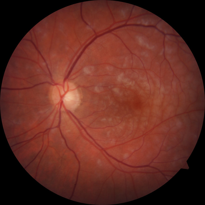
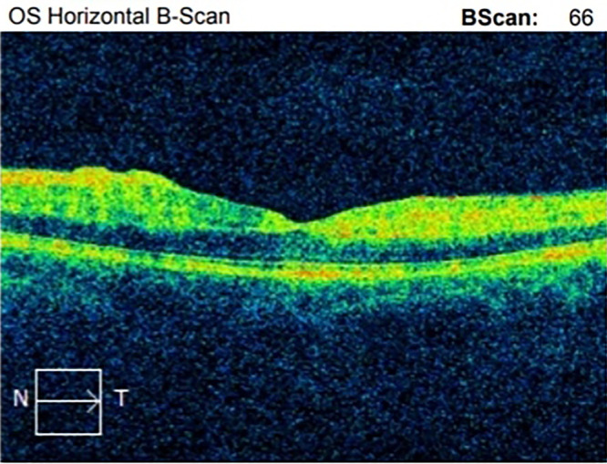
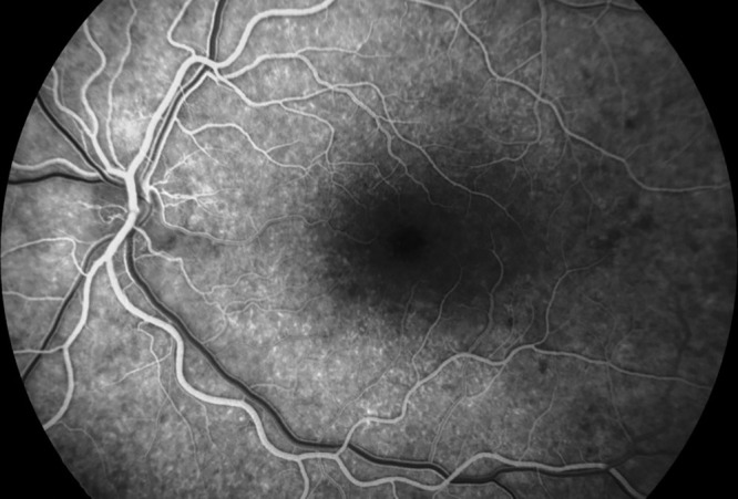
A complete eye examination was performed, followed by optical coherence tomography (OCT), fundus photography (FP), fundus autofluorescence (FAF), and fluorescein angiography (FA) tests. Best corrected visual acuity in the left and right eyes was 0.1 and 1.0, respectively. The affected eye showed a mild relative afferent pupillary defect (RAPD). Biomicroscopic examination showed normal findings. The anterior chamber was transparent with a normal iridocorneal angle. Intraocular pressure was within the normal range. Fundoscopic examination of the right eye was normal, but the fundus of the left eye showed several white dots located around the optic disc and scattered throughout the posterior pole of the retina. The fovea was yellowish with granularity (figure 1). FAF test showed hypofluorescent spots and dots throughout the posterior pole of the retina. The OCT examination of the right eye showed a normal configuration of the macula, but the left eye showed pathological findings – appearing as a multifocal nodular thickening in deep layers of the retina as well as areas of disruption of the ellipsoid zone (EZ) throughout the posterior pole (figure 2). FA test showed early punctate hyperfluorescence in a rim-like pattern and late staining in areas corresponding to the white dots (figure 3).
Figure 1.

The fundus of the left eye shows numerous white dots located around the optic disc and scattered throughout the posterior pole of the retina. The fovea is orange-yellow with a granular appearance.
Figure 2.

Initial optical coherence tomography (OCT) shows increased foveal thickness in the left eye and areas of disruption of the ellipsoid zone.
Figure 3.

Fluorescein angiography (FA) shows subtle early hyperfluorescence of the dots with late staining.
Following an initial diagnosis of multiple evanescent white dot syndrome (MEWDS), we requested additional tests to determine the cause of these abnormalities to provide optimal treatment. Complete blood count (CBC), erythrocyte sedimentation rate (ESR), c-reactive protein (CRP), fibrinogen, blood glucose, urea, creatinine, aspartate aminotransferase (AST), alanine aminotransferase (ALT), lipid panel, and urine analyses were all within normal range. Serum immunoglobulin levels as well as complement components C3 and C4 were also within normal range. Rheumatologic tests for antinuclear antibodies (ANAs), anti-Ro antibodies, antineutrophil cytoplasmic antibodies (ANCA), rheumatoid factor (RF), cyclic citrullinated peptide (CCP) antibodies, anti-smooth muscle antibodies (ASMA), anticardiolipin antibodies, and anti-beta 2 glycoproteins 1 and 2 were all negative. Tests for herpes simplex virus type 1 and 2 (HSV1 and HSV2), herpes zoster virus (HZV), Epstein-Barr virus (EBV), cytomegalovirus (CMV), Borrelia burgdorferi, Treponema pallidum, and Toxoplasma gondii were also negative. The QuantiFERON-TB Gold (QFT) test was false-positive. To rule out tuberculosis, we performed a purified protein derivative (PPD) skin test, which was negative. To exclude pneumonia and related complications, multi-slice computed tomography (MSCT) of the chest was performed, which was normal.
The patient was prescribed nepafenac solution 1 mg/mL (Alcon pharmaceuticals Ltd, Belgrade, Serbia) and acetazolamide (Remedica Ltd, Limassol, Cyprus) to reduce inflammation and edema. After one week of treatment, a follow-up examination showed a decrease in edema and improvement in the corrected visual acuity in the left eye (0.8). A fundus examination of the left eye showed complete resorption of the chorioretinal infiltration. An OCT examination of the eye showed thickening of the choroid, subtle discontinuity of the photoreceptor layer, and scarring of the retinal pigment epithelium.
Written informed consent was obtained from the patient for the publication of this case report.
Discussion
Here we reported one of the first cases of MEWDS associated with SARS-CoV-2 infection. Through extensive testing, we ruled out all common possible causes of retinitis and concluded that changes in the retina of our patient were related to COVID-19 complications. 4 Multimodal imaging at presentation and during follow-up are the standard tools used in the clinical diagnosis of MEWDS. 5
MEWDS is a rare inflammatory eye disease of the outer retina. The condition accounts for 1.24% of uveitis diagnoses. 6 Typically, MEWDS is unilateral and mainly affects healthy women aged 20-50 years, most of whom are moderate myopes. 7 A viral infection precedes MEWDS in 50% of the cases. Our patient thus fully fits the description of the typical patient who develops this condition.
Although the exact pathogenesis of MEWDS is unknown, a viral infection with a possible immune-mediated mechanism and genetic susceptibility is suspected. 8 A predisposition for inflammation seems to be present in the peripapillary circulation. The inflammatory process develops within the outer retina as a result of choriocapillaris nonperfusion. Capillary perfusion is significantly impaired in COVID-19 patients due to endothelial cell damage from both the virus and inflammatory cytokines. SARS-CoV-2 disrupts microcirculation, causing capillary congestion, micro thrombosis, and damage to pericytes. These are essential for capillary integrity and barrier function, impairment of which can presumably lead to MEWDS. 9 Endothelial dysfunction leads to exudation, and inflammatory cells accumulate beneath the retinal pigment epithelium (RPE). Piles of inflammatory cells create a stretching effect beneath the RPE, which leads to its thinning and disruption. It is presumed that the dots visible by multimodal imaging are window defects in areas where the RPE is disrupted. The disease can present with just vision loss, as in our case, or it can manifest with various other signs and symptoms such as shimmering photopsia, dyschromatopsia, paracentral, and often temporal scotomas. 10 While the anterior segment often shows no signs of inflammation, the posterior pole of the fundus displays multiple white dots. 11 Due to the transient nature of white dots, they are not detected in all patients with MEWDS. A more typical finding during fundus examination is a yellowish macula with granularity. In the majority of patients, MEWDS resolves spontaneously without treatment, and vision is restored to baseline level within 7-10 weeks. Recurrences of the syndrome are uncommon. 7
Several studies associated SARS-CoV-2 infection with endothelium dysfunction in various tissues and organs (e.g., lungs, kidneys, and myocardium), leading to various complications. 8 , 9 SARS-CoV-2 targets endothelial cells through the angiotensin-converting enzyme 2 receptor, which leads to significant changes in their morphology resulting in disruption of intercellular junctions, cell swelling, and impaired barrier function. If these abnormalities occur in the peripapillary circulation of the retina, they can lead to an inflammatory process in the outer layer of the retina known as MEWDS. 4 , 9 , 11
As the main limitation of the study, we did not take a sample of vitreous humor to measure its interleukin levels. In the context of COVID-19, it is recommended to further investigate a possible association between changes in the composition of vitreous humor and retinal abnormalities.
Conclusion
Retinal complications due to coronavirus are far more common than it has been reported. Subtle lesions, such as arteriolar or capillary changes in the retinal circulation, can only be detected using OCT, OCT-angiography, FA, or electroretinogram test. However, the required instrumentation for such accurate tests is not available in every hospital/examination site. Consequently, the diagnosis of vision impairment is often attributed to other causes.
Authors’ Contribution
A.AZ: Acquisition, analysis, and interpretation of data, and critical revision. D.V: Conception of the study and drafting of the manuscript. M.D: Acquisition and interpretation of data, and critical revision. L.Z: Acquisition of data and drafting of the manuscript. K.Z: Conception and design of the study, and drafting of the manuscript. All authors have read and approved the final manuscript and agree to be accountable for all aspects of the work in ensuring that questions related to the accuracy or integrity of any part of the work are appropriately investigated and resolved.
Conflict of Interest
None declared.
References
- 1.Feng Y, Park J, Zhou Y, Armenti ST, Musch DC, Mian SI. Ocular Manifestations of Hospitalized COVID-19 Patients in a Tertiary Care Academic Medical Center in the United States: A Cross-Sectional Study. Clin Ophthalmol. 2021;15:1551–6. doi: 10.2147/OPTH.S301040. [ PMC Free Article ] [DOI] [PMC free article] [PubMed] [Google Scholar]
- 2.Guemes-Villahoz N, Burgos-Blasco B, Garcia-Feijoo J, Saenz-Frances F, Arriola-Villalobos P, Martinez-de-la-Casa JM, et al. Conjunctivitis in COVID-19 patients: frequency and clinical presentation. Graefes Arch Clin Exp Ophthalmol. 2020;258:2501–7. doi: 10.1007/s00417-020-04916-0. [ PMC Free Article ] [DOI] [PMC free article] [PubMed] [Google Scholar]
- 3.Hu K, Patel J, Swiston C, Patel BC. Ophthalmic Manifestations Of Coronavirus (COVID-19) Treasure Island (FL): StatPearls; 2022. [PubMed] [Google Scholar]
- 4.Invernizzi A, Torre A, Parrulli S, Zicarelli F, Schiuma M, Colombo V, et al. Retinal findings in patients with COVID-19: Results from the SERPICO-19 study. EClinicalMedicine. 2020;27:100550. doi: 10.1016/j.eclinm.2020.100550. [ PMC Free Article ] [DOI] [PMC free article] [PubMed] [Google Scholar]
- 5.Zicarelli F, Mantovani A, Preziosa C, Staurenghi G. Multimodal Imaging of Multiple Evanescent White Dot Syndrome: A New Interpretation. Ocul Immunol Inflamm. 2020;28:814–20. doi: 10.1080/09273948.2019.1635169. [DOI] [PubMed] [Google Scholar]
- 6.Papasavvas I, Mantovani A, Tugal-Tutkun I, Herbort CP., Jr Multiple evanescent white dot syndrome (MEWDS): update on practical appraisal, diagnosis and clinicopathology; a review and an alternative comprehensive perspective. J Ophthalmic Inflamm Infect. 2021;11:45. doi: 10.1186/s12348-021-00279-7. [ PMC Free Article ] [DOI] [PMC free article] [PubMed] [Google Scholar]
- 7.Abu-Yaghi NE, Hartono SP, Hodge DO, Pulido JS, Bakri SJ. White dot syndromes: a 20-year study of incidence, clinical features, and outcomes. Ocul Immunol Inflamm. 2011;19:426–30. doi: 10.3109/09273948.2011.624287. [ PMC Free Article ] [DOI] [PMC free article] [PubMed] [Google Scholar]
- 8.Rousseau A, Fenolland JR, Labetoulle M. [SARS-CoV-2, COVID-19 and the eye: An update on published data] J Fr Ophtalmol. 2020;43:642–52. doi: 10.1016/j.jfo.2020.05.003. [ PMC Free Article ] [DOI] [PMC free article] [PubMed] [Google Scholar]
- 9.Ostergaard L. SARS CoV-2 related microvascular damage and symptoms during and after COVID-19: Consequences of capillary transit-time changes, tissue hypoxia and inflammation. Physiol Rep. 2021;9:e14726. doi: 10.14814/phy2.14726. [ PMC Free Article ] [DOI] [PMC free article] [PubMed] [Google Scholar]
- 10.Tavallali A, Yannuzzi LA. MEWDS, Common Cold of the Retina. J Ophthalmic Vis Res. 2017;12:132–4. doi: 10.4103/jovr.jovr_241_16. [ PMC Free Article ] [DOI] [PMC free article] [PubMed] [Google Scholar]
- 11.Pellegrini M, Veronese C, Bernabei F, Lupidi M, Cerquaglia A, Invernizzi A, et al. Choroidal Vascular Changes in Multiple Evanescent White Dot Syndrome. Ocul Immunol Inflamm. 2021;29:340–5. doi: 10.1080/09273948.2019. [DOI] [PubMed] [Google Scholar]


