Abstract
Arterial stiffness is a progressive aging process that predicts cardiovascular disease. Pulse wave velocity (PWV) has emerged as a noninvasive, valid, and reliable measure of arterial stiffness and an independent risk predictor for adverse outcomes. However, up to now, PWV measurement has mostly been used as a tool for risk prediction and has not been widely used in clinical practice. This consensus paper aims to discuss multiple PWV measurements currently available in Asia and to provide evidence-based assessment together with recommendations on the clinical use of PWV. For the methodology, PWV measurement including the central elastic artery is essential and measurements including both the central elastic and peripheral muscular arteries, such as brachial-ankle PWV and cardio-ankle vascular index, can be a good alternative. As Asian populations are rapidly aging, timely detection and intervention of “early vascular aging” in terms of abnormally high PWV values are recommended. More evidence is needed to determine if a PWV-guided therapeutic approach will be beneficial to the prevention of cardiovascular diseases beyond current strategies. Large-scale randomized controlled intervention studies are needed to guide clinicians.
Keywords: Arterial stiffness, Pulse wave velocity, Cardiovascular disease, Asia
Introduction
With increasing age, arteries become stiffened (arteriosclerosis) and this increases linearly with the risk of cardiovascular (CV) disease [1]. However, different genetic background, environmental, and lifestyle factors result in large individual variability in the biological age of arteries, even at the same chronological age. This has led to the notion that accelerated arterial aging may be regarded as a failure of the interaction between genetic and environmental factors [2]. The vascular aging CV continuum has similar final outcome to the classic CV continuum, such as end-stage cardiac, cerebral, and renal diseases, and death may occur, but there exists a distinct difference in the underlying pathophysiology [3, 4]. The mechanism of the classic CV continuum is atherosclerosis and cardiac hypertrophy at the beginning. In contrast, that of the vascular CV continuum is fracture of elastic lamellae, aortic stiffening, and dilatation, which induces pulse wave pathology such as pulse wave encephalopathy, pulse wave nephropathy, renal disease, and dementia [3].
Arterial stiffness with aging increases pulse wave velocity (PWV), the measurement of which is a reliable tool to predict CV disease [5]. Although its impact on outcomes has been widely studied and suggested as a clinical aid for primary and secondary CV prevention [6], there are still large hurdles in applying the concept of PWV in clinical use. Hypertension guidelines pay minimal attention to the clinical use of PWV and with many methods available there is no standard method for measuring PWV [7, 8]. Currently, the main tool of measuring vascular aging is PWV, but there are differences in measurement methods. Europeans favor carotid-femoral PWV (cfPWV) [9]. In contrast, in Asia, there is favor toward brachial-ankle PWV (baPWV) [10] or cardio-ankle vascular index (CAVI) [11]. Main differences between methods are the arterial measurement sites that vary from the central elastic aorta to the aorta and peripheral muscular arteries. This review aims to discuss multiple PWV measurement methods and to provide recommendations on the clinical use of PWV in Asia.
Methodologic Aspects
The original concept of “hardening of arteries” was generally understood as a buildup of atheroma and calcification of the arterial lumen, resulting in regional stenosis and leading to obstruction of blood flow [12]. This effect can be readily measured invasively by detecting a pressure drop across the stenotic lesion, or noninvasively using Doppler devices to detect changes in blood flow velocity. However, this concept has now evolved from alterations in the intimal component of the arterial wall to the medial component [12, 13], with altered mechanical properties affecting arterial wall stiffness, which cannot be measured directly noninvasively. The effect of stiffness of large conduit arteries is to increase pulse pressure, an effect largely responsible for isolated systolic hypertension in the elderly [14], but because pulse pressure is a result of stroke volume and the distensibility of the aorta and large arteries, it is not an explicit measure of arterial stiffness. However, from the biophysical relationship of stress and strain in the arterial wall, it has been shown that PWV can be used as a reliable surrogate measure of arterial stiffness, particularly in large conduit arteries [15].
An important consideration in using PWV as a measure of arterial stiffness is that the arterial wall is a type of “hyperelastic” material, i.e., the stiffness depends on blood pressure (BP) − the higher the pressure, the higher the PWV for the same arterial wall. However, the inherent structural material which alters the mechanical properties does not change with increase in pressure immediately, but what does change is the functional arterial stiffness [16]. Increase in structural stiffness is seen as increase in PWV at the same level of BP [17]. These are important methodological aspects to be considered when measuring PWV and interpreting individual patient or population data with respect to contribution of PWV to CV risk beyond BP. An important example of this was shown in two populations in China with distinct geographical separation (north, Beijing; south, Guangzhou). PWV was markedly higher in the Beijing cohort at similar levels of BP in both groups. This implied a difference in inherent structural arterial stiffness, which was most likely related to a lifelong difference in dietary salt consumption, resulting in a marked difference of the prevalence of hypertension in both groups [18, 19].
The sections that follow describe the specific methods used for PWV measurement that will address the use of PWV as a potential clinical measurement that can be performed together with BP in the context of established guidelines for measurement of arterial stiffness [7]. In addition, the innovative application of the pressure dependency of arterial stiffness will be described to obtain a corrected value of PWV, which is independent of BP [20]. The important methodologic advancement with this technique is that the measurement gives information of structural stiffness for an individual patient without the need of statistical information from cohort measurements to correct the measured PWV for the effect of BP.
The measurement of PWV involves a measure of pulse transit time (PTT) obtained from the time delay between two arterial pulses measured simultaneously or sequentially over a given distance. To obtain a reliable measure of PTT, the path length cannot be too short; that is, the longer the distance the smaller the relative error in PTT. In addition, the site of measurement should be convenient and one in which a reliable pulse waveform can be easily recorded. Furthermore, the PTT distance should be applied to cover mainly large conduit arteries. For this, the carotid and femoral sites have been conventionally used as a reasonable compromise which will give meaningful information [7]. However, although these sites have been used extensively in many population cohorts, this measurement method is not favored by investigations among Asian populations, where instead pulses are detected at brachial and ankle sites [21]. Although this methodology offers an unobtrusive form of measurement, the path lengths cover muscular arteries, where the wall stiffness may be modulated by smooth muscle tone and so affect changes of PWV.
Concepts and Clinical Evidence
Brachial-Ankle Pulse Wave Velocity
Concept and Principles
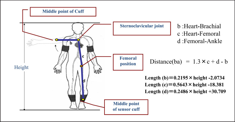
baPWV is calculated by dividing the brachial-ankle distance by the time difference between the brachial and ankle arterial waves. Thus, baPWV is considered as a global measure of arterial stiffness, including the aorto-muscular region. A volume-plethysmographic technique is used to measure baPWV. Pressure cuffs are wrapped at the bilateral brachial and ankle sites to record pulse waves as shown in Figure 1 [22]. The brachial-ankle distance (Distance [ba] in the figure) is determined by the linear equation of height, and the time difference between the brachial and ankle waves is determined by the foot-to-foot method. The height-based path length has been validated by comparing it with the path length determined by magnetic resonance imaging [23]. The unique feature of this equipment is that it simultaneously records the BP at four sites using the oscillometric method, thus also enabling determination of the ankle-brachial pressure index (ABI). This index is critical in confirming the iliotibial circulation and ensuring valid use of baPWV, which becomes invalid if ABI <0.9. The baPWV measurement is easy and reproducible and the generalizability and validity of the methodology have been determined [22]. Thus, this method is suitable for clinical applications.
Fig. 1.
Path length formula for baPWV (source, see ref. 22).
Clinical Evidence
A recent review summarized that baPWV increases in patients with hypertension, diabetes, metabolic syndrome, chronic kidney disease, sleep apnea syndrome, as well as with aging and conditions such as tachycardia and postmenopause [24]. In hypertension and diabetes, higher baPWV is associated with advanced organ damage. The relationship between baPWV and lipid levels remains unclear. Antihypertensive agents, statins, oral diabetic drugs, weight loss, smoking cessation, and continuous positive airway pressure have all been reported to lower baPWV [25].
The risk of CV events increases linearly with an increase in baPWV. Individual participant data meta-analyses of 14,673 individuals, with no previous history of CV events, demonstrated that baPWV can predict both CV events and all-cause mortality Independent of conventional CV risk factors including BP [26]. Every 1-SD increase in baPWV was associated with a 21% increase in the risk of CV disease. Moreover, a 1 m/s increase in baPWV was associated with an increase of 12%, 13%, and 6% in CV events, CV mortality, and all-cause mortality, respectively [27]. Importantly, patients at intermediate risk were reclassified into a higher or lower CV risk category when baPWV was added to a model incorporating the Framingham risk score; net reclassification improved by approximately 25%.
Clinical Use
To implement baPWV measurement in everyday clinical practice, it is necessary to determine reference values. Recently, the expert committee of the Japanese Society of Vascular Failure proposed a physiological diagnostic criterion for various vascular function tests, including baPWV [Table 1] [28]. baPWV was positioned as an arterial stiffness measure to examine the medial layer function of the aorto-muscular region. The measurement of baPWV was categorized as normal (<14 m/s), borderline (14–18 m/s), and abnormal (>18 m/s). These thresholds may be used by health practitioners to individualize non-pharmacological or pharmacological interventions. However, future studies need to verify the clinical significance of these criteria. The inclusion of leg artery stiffness has been long considered a critical limitation of baPWV, but the accumulated data are not fully supportive [28]. This suggests that aorto-muscular artery stiffness is involved more in the modulation of central hemodynamics than initially considered [29, 30]. This topic is the new frontier for future studies.
Table 1.
Reference values and risk associated with PWVs
| Method | Reference values | Risk of CV events |
|---|---|---|
| baPWV | Normal <14 m/s Borderline ≥14 and <18 m/s Abnormal ≥18 m/s |
Every 1-SD increase of baPWV→ 21% increase in the risk of CV disease [26] Every 1 m/s increase of baPWV→ 12%, 13%, and 6% increase in CV events, CV mortality, and all-cause mortality, respectively [27] |
| CAVI | Normal <8 Borderline ≥8 and <9 Abnormal ≥9 |
Every 1.0 index increase of CAVI→ 12.6% increase in the risk of future CV events [128] Cut-off values for CVD events→ 9.0–9.2 in Asian patients [129, 130] 5-year overall net reclassification index→ 16.4% and 33.7% for CVD events in patients with obesity and in patients with ACS, respectively [131] |
| cfPWV | Abnormal ≥10 m/s | Every 1-SD increase of cfPWV→ 30% increase in the risk of CV events after adjustment for traditional risk factors [44] Every 1 m/s increase of cfPWV→ 14%, 15%, and 15% increase in total CV events, CV mortality, and all-cause mortality, respectively [1] 5-year overall net reclassification index→ 14.8% and 19.2% for coronary heart disease and stroke, respectively, in intermediate-risk individuals [44] |
ACS, acute coronary syndrome; baPWV, brachial-ankle PWV; cfPWV, carotid-femoral PWV; CV, cardiovascular; PWV, pulse wave velocity; CAVI, cardio-ankle vascular index; CVD, cardiovascular disease.
Limitations
baPWV measurement becomes unreliable if the circulation is disturbed in any part from iliac to tibial arteries. In patients with ABI <0.9, therefore, baPWV cannot be used as a metric for clinical decisions [25]. In addition, up to now the evidence of baPWV is derived mainly from Asian populations. For global application of baPWV, evidence from non-Asian populations is needed.
Cardio-Ankle Vascular Index
Concept and Principles
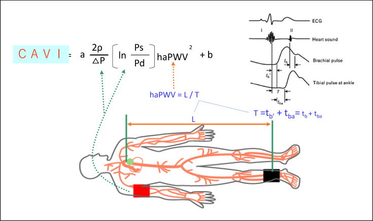
CAVI reflects the arterial stiffness of the arterial tree from the origin of the aorta to the ankle. CAVI is theoretically derived from stiffness parameter β, and the Bramwell-Hill's equation, and is obtained by systolic and diastolic BPs and PWV [20]. The equation and measuring methods are shown in Figure 2. CAVI is measured in the supine position using the VaSera system (Fukuda Denshi, Tokyo, Japan). A feature of CAVI is that it purports to be a measure that is independent of BP at the time of measurement. It is therefore theoretically possible that the effect of antihypertensive drugs on structural arterial stiffness can be evaluated as well as the effect of BP change in chronic phase. The cut-off value of CAVI is proposed to be 9.0 for predicting CV diseases [31].
Fig. 2.
Determination of the cardio-ankle vascular index (CAVI). Ps, systolic blood pressure of brachial artery; Pd, diastolic blood pressure; haPWV, pulse wave velocity from the origin of the aorta to the ankle at mid pressure; ∆P, Ps-Pd; ρ, blood density; a and b, constants to convert the values of CAVI to those of Hasegawa's hfPWV; T, time of the pulse from aortic valve to the ankle; L, length of the arterial tree from the origin of aorta to the ankle.
Clinical Evidence
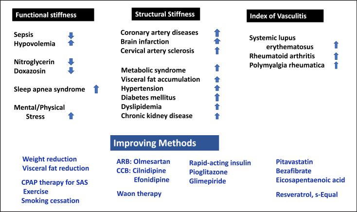
Cross-sectional studies report that CAVI is higher with older age, among men and with arteriosclerotic diseases (coronary artery disease, cerebral infarction, chronic kidney disease) as well as most coronary risk factors such as metabolic syndrome, visceral fat accumulation, hypertension, diabetes mellitus, and dyslipidemia (see Fig. 3) [31]. In vasculitis such as collagen diseases (systemic lupus erythematosus, rheumatoid arthritis) and polymyalgia rheumatica, CAVI is also increased.
Fig. 3.
Clinical implications of CAVI and improving methods. ARB, angiotensin receptor antagonist; CCB, calcium channel blocker; CPAP, continuous positive airway pressure; SAS, sleep apnea syndrome.
CAVI also reflects functional stiffness. CAVI decreases in sepsis [32] and rises in hypovolemia [33]. These facts indicate that CAVI reflects the effect of contraction of arterial smooth muscle cells. Administration of nitroglycerin and the α-blocker, doxazosin acutely decreases CAVI [34, 35]. Patients with sleep apnea syndrome showed high CAVI, probably due to enhanced sympathetic nervous system activation. CAVI was also reported to be elevated after an earthquake [36], indicating a potential influence of mental or physical stress. Prospective studies showed that high CAVI is predictive for mortality and morbidity of coronary artery diseases [31], for the incidence of atrial fibrillation [37] in the general population, and also for deterioration in kidney function [38, 39].
Clinical Use
In routine clinical practice and medical checkup, measuring CAVI gives important information in terms of structural arterial stiffness, functional stiffness, and indices of vasculitis as stated above. When CAVI shows abnormally high value for one's age, the risk factors should be intensively examined, and treatments to address risk factor burden and decrease CAVI are recommended. Reported methods for improving CAVI are listed in Figure 3 [31]. Body weight reduction in metabolic syndrome, especially visceral fat reduction, and continuous positive airway pressure therapy for OSAS decreased CAVI. Exercise, smoking cessation, and Waon therapy also decreased CAVI. BP control with angiotensin receptor II blockers, such as olmesartan, calcium channel blockers such as cilnidipine, efonidipine decreased CAVI. Glucose control with rapid-acting insulin, pioglitazone, or glimepiride improved CAVI. Lipid control with pitavastatin, bezafibrate, and eicosapentaenoic acid also decreased CAVI. Resveratrol improved CAVI. Furthermore, a rapid rise of CAVI in persons with high CAVI might a harbinger of impending CV events. The mechanism is thought to be ischemia of vulnerable plaque due to arterial smooth muscle contraction [36]. Periodic monitoring of CAVI might be useful to predict impending CV events. In summary, measuring CAVI might be useful for a quantitative assessment of vascular aging and the degree of arteriosclerosis, and also for control of risk factors.
Limitations
The CAVI value may not be correct when ABI is less than 0.9 because the pulse at the ankle is too weak to be detected, and PWV which constitutes CAVI cannot be properly obtained with the VaSera system. In patients with aortic valve stenosis, CAVI shows low values [40]. It is suggested that this is due to the PWV decrease associated with the lowered blood flow from the left ventricle to the aorta. Abnormally low age-related CAVI value might indicate the presence of severe aortic valve stenosis.
Carotid-Femoral Pulse Wave Velocity
Concept and Principles
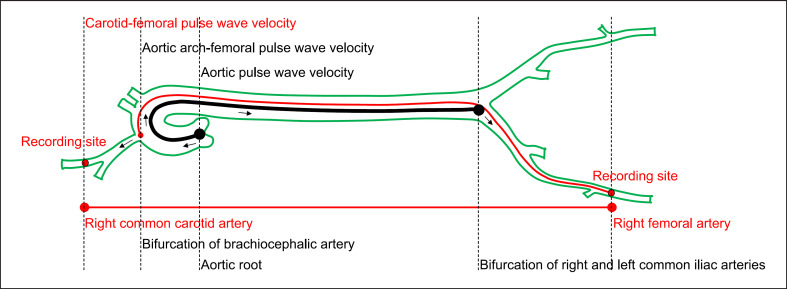
Aortic stiffness can be estimated noninvasively by measuring PWV between the right carotid and right femoral artery (cfPWV) [7, 41]. cfPWV (expressed in m/s) is calculated as the surface distance divided by the PTT between the two arterial sites. The PTT can be measured using the foot-to-foot method from pressure, flow, or volume waveforms recorded with tonometry, Doppler, mechanical sensor, or pulse volume recording device. It should be noted that both the surface distance and the PTT are only crude estimates because physiologically the pulse wave does not actually propagate from the recording site of right carotid artery via aortic arch to the recording site of right femoral artery (Fig. 4) [7].
Fig. 4.
Measurement schematic diagram of carotid-femoral pulse wave velocity, aortic arch-femoral pulse wave velocity, and aortic pulse wave velocity. The surface distance between the recording sites of right common carotid artery and right femoral artery is depicted as the straight red line. The path of the pulse wave travelling from the aortic arch (near the bifurcation of the brachiocephalic artery) to the recording site of right femoral artery is depicted as the red curved line. The path of the pulse wave travelling from the aortic root to the end of abdominal aorta (near the bifurcation of the right and left common iliac arteries) is depicted as the thick black curved line.
Clinical Evidence
It has been recommended that arterial stiffness should be determined noninvasively by measurement of cfPWV [7] and cfPWV is considered as the “gold-standard” measurement of aortic stiffness [41]. However, cfPWV is actually a crude estimate of aortic arch to femoral artery PWV and does not directly measure stiffness of the ascending aorta (Fig. 4) [42].
cfPWV is a sensitive marker of vascular aging [43] and has been validated as an independent, strong marker for future CV events in patients with hypertension, diabetes, and renal failure, and in the general population and apparently healthy subjects [42]. cfPWV is predictive of coronary heart disease, stroke, systolic hypertension, atrial fibrillation, aortic aneurysm formation, heart failure, and CV mortality events [42, 44, 45, 46, 47]. cfPWV improves model fit and reclassifies risk for future CV disease events in models that include standard CV disease risk factors [44]. cfPWV may enable earlier identification of high-risk populations that might benefit from earlier CV disease risk factor management [44]. Therefore, it is reasonable to measure cfPWV to provide incremental information beyond standard CV disease risk factors in the prediction of future CV disease events [7].
Clinical Use
Age- and sex-specific normal reference values and thresholds have been established in a European population, and this may facilitate the clinical application of cfPWV for this demographic [48]. The generalizability of these cfPWV reference values to Asian populations is yet to be known. cfPWV may be clinically useful to identify medium and high-risk CV disease groups, but the prognostic value of arterial stiffness in older adults may be limited [46]. cfPWV is considered as a surrogate target for prevention and intervention [42]. However, large-scale randomized controlled intervention studies are needed to guide clinicians [42].
Limitations
Regarding cerebral structure and function, increased cfPWV has been associated with an increased risk of brain structural abnormalities and worse performance in various subdomains of cognitive function but negative results were also observed [49]. Since cfPWV does not cover the ascending aorta, which may play a critical role in generating aortic pressure/flow pulsatility, it is probably not an ideal parameter to evaluate the association between vascular aging and cognitive function [49].
Measures of PWV vary with age, sex, BP, ethnicity, and measurement techniques and devices [10]. Standardization of the techniques, the validation of devices, and arterial stiffness studies have been recommended [7, 50].
Estimated Pulse Wave Velocity
Concept and Principles
cfPWV by applanation tonometry is one of the widely used methods for the assessment of aortic stiffness. Despite its popularity as a well-standardized and noninvasive measure of aortic stiffness, the routine assessment of cfPWV still requires sophisticated technical skills and specialized equipment, which may limit its widespread incorporation into routine clinical practice. As a strategy to overcome the restraints concerning the assessment of aortic stiffness using cfPWV, researchers have developed the concept of estimated PWV (ePWV) that can be calculated from age and mean BP (MBP) using a regression equation generated from the Reference Values for Arterial Stiffness Collaboration: ePWV = 9.587 − 0.402 × age + 4.560 × 10⁻³ × age2 − 2.621 × 10⁻⁵ × age2 × MBP + 3.176 × 10⁻³ × age × MBP − 1.832 × 10⁻² × MBP [51].
Clinical Evidence
In the high-risk patients from the SPRINT trial, ePWV predicted all-cause mortality and CVD outcomes beyond traditional risk factors, and improved C-statistics beyond the Framingham Risk Score (from 0.65 to 0.69) [52]. Additionally, ePWV was associated with CV mortality and morbidity independently of the Systematic Coronary Risk Evaluation (SCORE) and Framingham Risk Score, but not independently of traditional CV risk in the MORGAM Project with 38 cohorts from 11 countries [53]. ePWV is associated with all-cause and CVD mortality and slightly improves the C-statistics for the primary outcome in the Chinese Study [54].
By contrast, an addition of ePWV to contemporary CVD risk scores does not improve discrimination of all-cause and CVD mortality risk [55]. Similarly, while ePWV is associated with all-cause mortality and MI, independent of traditional risk factors, discrimination is not improved to a clinically meaningful extent in patients with angina pectoris [56]. Given uncertainty related to the usefulness of ePWV, additional research examining the role of ePWV as a predictor of CVD outcomes is clearly warranted, especially in differing outcomes, populations, and racial/ethnic backgrounds.
Clinical Use
Emerging evidence suggests that ePWV is associated with CVD outcomes and mortality, independent of traditional CVD risk factors in USA and European cohorts [52, 53, 55, 56, 57, 58, 59]. The role of ePWV as an independent predictor of CVD outcomes may also extend to Asian populations [54, 60]. Non-device-based estimation of aortic stiffness is rapid, easy, and inexpensive and may be used in clinical practice settings to aid in CVD risk prediction. However, there remains a substantial unexplained variance. Furthermore, the question remains as to whether ePWV can become an alternative for assessment of cfPWV as a marker of vascular aging remains to be determined, and whether ePWV improves CVD risk prediction beyond contemporary CVD risk scores such as Systematic Coronary Risk Evaluation (SCORE) and the Framingham Risk Score, as findings have been sparse and equivocal.
Limitations
To date, only few studies have sought to examine the correlation between ePWV and cfPWV and have reported this correlation to be weak (r ranges: 0.31–0.36) or moderate (r range: 0.52–0.67) [61, 62], but this correlation still remains unexplored in Asian population. It is also unclear whether the current ePWV equation from European cohort data is appropriate to be applied to other populations and racial/ethnic backgrounds. Thus, further validation studies comparing ePWV to other measures of vascular aging are needed in Asian reference populations.
While ePWV is somewhat correlated with cfPWV and an increased risk of CVD outcomes beyond risk scores, it still remains uncertain as to whether ePWV is a sensitive assessment of aortic stiffness and can serve as a substitute for cfPWV. Few studies to date have simultaneously compared the ePWV prediction equation to directly noninvasive or invasive measured aortic stiffness in predicting CVD outcomes. Compared to cfPWV or baPWV, ePWV has been shown to also predict major CV events independently of SCORE and Framingham Risk Score in patients [60, 61]. Interestingly, both ePWV and invasive PWV independently predict CV events and mortality and that ePWV has a similar predictive value for mortality as that of invasive PWV in patients with undergoing coronary angiography [59]. Despite these findings, whether ePWV can become a substitute for cfPWV as a marker of vascular aging remains to be determined. However, as ePWV reflects an interaction between age and MBP [61], this variable may not be viewed as synonymous with cfPWV [61] and may capture different risk information than cfPWV [56, 61], thereby providing another important prognostic value to the prediction of CV health.
Other Pulse Wave Velocities
Regional PWVs
Devices which measure the cfPWV can also be used to measure other regional PWVs, such as the carotid-radial PWV and femoral-ankle PWV. While the usefulness of these regional PWVs for CV risk assessment may be limited, aortic-brachial arterial stiffness mismatch has been reported to be associated with increased mortality in the dialysis population [63].
Apart from the aforementioned regional PWVs, heart-to-brachium PWV (hbPWV) includes a segment of the proximal aorta, and this measurement can be obtained in the baPWV measurement. PTT for hbPWV can be evaluated fairly easily by simultaneous recordings of the heart sounds or electrocardiography and brachial arterial pulse waves are recorded with a high-fidelity sensor (e.g., air-plethysmography) embedded in the BP cuff. The path length is obtained using an equation derived from the gender and height. hbPWV has been shown to be correlated with the aortic systolic BP and augmentation index. Therefore, the hbPWV may be a marker of proximal aortic stiffness [64].
Finger-toe PWV is a simple noninvasive method for measuring regional arterial stiffness. The finger-toe PWV is determined on the basis of a patented height chart for the distance and the PTT between the finger and the toe pulpar artery signals (ft-PTT). Acceptable correlation has been reported between the finger and the toe pulpar artery signals and carotid-femoral PTT [65].
Recently, based on the pulse waveform recorded at the radial and digital arteries, the radial-digital PWV has become available as a measure of the regional stiffness of small conduit arteries [66]. The clinical implication of this measurement has not yet been clarified.
Local PWVs and Ambulatory PWVs
The use of cuff-based oscillometric devices provides an estimated (local) PWV based on pulse wave analysis and wave separation analysis at a single site such as the carotid, brachial, radial, or femoral arteries [67, 68]. These are simple and relatively operator-independent, and enable ambulatory measurements, and the PWV values are associated with aortic stiffness. Sarafidis et al. [69] reported that ambulatory PWV is a useful marker to predict future CV events. Ambulatory PWV was estimated with an oscillometric ambulatory BP monitoring device. In the future, it may be possible for both ambulatory and home monitoring of stiffness parameters and related hemodynamic abnormalities. It is still not known how these may be applied in clinical practice, and robust validations for such devices are needed.
Aortic PWV by Magnetic Resonance
For measurement of the aortic PWV by magnetic resonance, which is the most reliable noninvasive method to measure the aortic PWV, a fully automatic method has become available [70], although this measurement is limited to research use because of its cost and availability in limited institutions. In patients with hypertrophic cardiomyopathy, the aortic strain of the descending aorta assessed by magnetic resonance was significantly decreased as compared with that in control subjects and correlated with the native T1 values. Aortic strain may be a marker of myocardial fibrosis in patients with hypertrophic cardiomyopathy [71].
Standardization of PWV Measurement
Validation is a crucial step for standardization. For the validation of devices, performance in terms of precision (i.e., repeatability/reproducibility) and accuracy (i.e., closeness to real value) must be assessed [72] because these features determine the reliability and validity of a device in clinical practice. Validation is a fundamental prerequisite for a device to be clinically useful. For this reason, structured standardized protocols providing evidence of the performance of a system need to be implemented in the development of any medical device. Currently, available guidelines provided by the Artery Society in 2010 [73] have been used in the last 10 years for validation of devices measuring carotid-femoral cfPWV. However, since 2010, many devices measuring PWV on arterial paths other than carotid-femoral have been developed, raising many methodological and clinical questions. A detailed evaluation of these issues is beyond the scope of this article: an international working group is now working to provide an updated document including those cases. However, it is worthwhile to mention some open issues posed as questions and answers below:
Q1: Are PWV values from different arterial segments directly comparable in a single individual?
A1: baPWV is systematically higher than cfPWV [74], even when correction formulas are used [23]. This is a consequence of different arterial segments containing different distributions of elastic and muscular arteries. One possible standardization could be to make reference to normal percentiles, or vascular age calculation, for each technique, noting that validation of such approach is still needed.
Q2: Are PWVs from different arterial segments to be validated against the same reference standard (i.e., invasive aortic PWV)?
A2: As elaborated in Q1, PWVs differ quantitatively between arterial beds. As far as baPWV is concerned, the brachial-ankle arterial bed cannot realistically be assessed invasively using catheters. As an alternative, new baPWV devices can be validated against existing (noninvasive) baPWV devices that have shown prognostic relevance beyond classical risk factor assessment [26]. Thus, new baPWV devices can be validated against these proven devices. For arterial beds other than brachial-ankle, the answer is less clear.
Q3: Do PWVs from different arterial segments provide similar prognostic information/risk stratification in a single individual?
A3: cfPWV and baPWV predict CV events beyond classical CV risk assessment and improve risk reclassification [26, 44]. To the authors' knowledge, a formal head-to-head comparison between cfPWV and baPWV in terms of risk prediction is not available at present. International collaborations are encouraged to address this important gap. However, in 2005, Pannier et al. [75] directly compared the ability of cfPWV, carotid-radial PWV, and femoral-tibial PWV to predict CV events in end-stage renal disease and found only cfPWV to significantly predict CV events.
Other devices, measuring finger-to-toe PWV by finger plethysmography or heart-to-foot PWV by a combination of ballistocardiography and foot impedance measurement [65, 76], have demonstrated agreement with noninvasive cfPWV, but only in small samples; no information on prognostic value is available. Furthermore, an increasing number of approaches claim to evaluate arterial stiffness by use of machine learning algorithms applied mostly to arterial waveforms, but sometimes also using clinical variables such as age, sex, or BP [77]. Though promising given their ease of use and with the potential of evaluating beat-to-beat PWV for some methods, these techniques cannot be recommended for clinical use to date.
The standardization of PWV measures must also consider the standardization of how these measures should be recorded in clinical practice. This is crucial given the dependence of PWV measurements on the hemodynamic status of the patient. We recommend standard operating procedures which are similar to those of BP measurements [78] or ECG, and that are summarized in the 2012 paper about measurement of cfPWV in daily practice [9]. Most importantly, we recommend that the measurement is taken in a quiet and stable-temperature environment after 10 min rest, avoiding smoking, caffeine, alcohol, and eating in the hours preceding the measurement, and not speaking during the measurement. Noteworthy, each technique may have specific contraindications (e.g., carotid stenosis for cfPWV, peripheral arterial disease for baPWV). With an increasing number of devices allowing out-of-office, self-measurement of arterial stiffness [79], the issue of measure standardization will become even more critical.
Clinical Implication of PWV Measurement
Arterial stiffness is the primary parameter to detect age-related CV risk before CV risk factors and organ damage become clinically overt. In addition, even after clinically overt risk factors, organ damage, and/or CV disease developed, arterial stiffness is closely associated with these risks and is useful for the management [80]. Thus, arterial stiffness is a useful parameter across the broad scope of healthcare and medicine [28, 81].
CV Risk Stratification for Medical Use
A clinical implication of arterial stiffness is the risk stratification for CV events from the community-dwelling population to outpatients with CV risk factors and/or CV diseases. The hypertension guidelines or expert consensus documentation include measures of arterial stiffness for risk stratification and better management of hypertension [82].
There is ample evidence which demonstrates that increased arterial stiffness, assessed by different measures such as cfPWV, baPWV, and CAVI, is associated with organ damage and CV events [28]. Although arterial stiffness is closely associated with high BP, all arterial stiffness measures are associated with CV event risk even after controlling for BP.
First, cfPWV is a well-established measure of arterial stiffness, independently associated with organ damage and CV event risk. Theoretically, it is the measure of stiffness of large arteries, however, to detect the pulse at the femoral site limits its clinical use. Second, compared with cfPWV, baPWV included the arterial properties of muscular peripheral artery distal to the femoral site. Thus, this indicator is more closely affected by BP. baPWV is associated with CV risk factors and a risk predictor of CV events, and it is useful for the risk stratification [83, 84, 85]. Third, CAVI is a new measure of arterial stiffness that reflects the stiffness from the ascending aorta to the ankle arteries and demonstrates less dependence on BP during the evaluation [20]. The systematic review to assess the association between CAVI and CV diseases (9 prospective studies (n = 5,214) and 17 cross-sectional eligible studies (n = 7,309), with most enrolling high CVD risk populations in Asia), demonstrated a modest association between CAVI and incident CVD risk [86]. A recent prospective study, CAVI-J, demonstrated that CAVI is predictive of stroke and heart failure in outpatients with CV risk [87].
The challenges of arterial stiffness are to demonstrate the benefit of arterial stiffness-guided management of risk factors on future CV prognosis. To address this issue, the Coupling study, a nationwide-prospective study, is now ongoing to demonstrate the association of serial change of CAVI and CV risk [88, 89]. A recent prospective study on the serial cfPWV change in patients with resistant hypertension, reducing, or preventing progression of aortic stiffness was associated with significant CV protection in patients with resistant hypertension [90].
Prediction of Hypertension in Healthcare
In healthy subjects, arterial stiffness may predict the future development of hypertension and hypertension-related organ damage. In subjects without hypertension, increase in CAVI or baPWV predicted hypertension [91]. Stress and obesity per se could accelerate the age-related arterial stiffening [92, 93, 94]. Other age-related diseases, such as cognitive dysfunction-associated small artery disease and atrial fibrillation are also associated with increased arterial stiffness [95, 96].
Challenges and Concept of Systemic Hemodynamic Atherothrombotic Syndrome
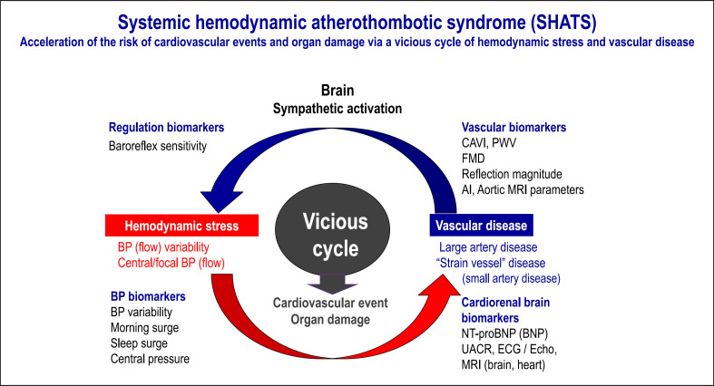
There is the vicious cycle between arterial stiffness and BP variability to facilitate the organ damage and CV events. The concept of systemic hemodynamic atherothrombotic syndrome (SHATS) has been proposed, describing an age-related and synergistic vicious cycle of hemodynamic stress and vascular disease [97]. This was presented and discussed at the 2018 Scientific meeting of Pulse of Asia, Kyoto (Fig. 5) [98]. The importance of SHATS is based on the assumption that the assessment of BP variability and arterial disease is likely to provide an effective opportunity to intervene early to reduce progression to hypertension in younger patients or to CV disease events and organ damage in older patients. We propose a new SHATS score to diagnose and assess the severity of SHATS. The score includes two components − a BP score and a vascular score − which are multiplied to generate the SHATS score [98]. This reflects the synergistic, rather than additive, effects of BP and vascular disease on target organ damage and CV disease events. Recently, we demonstrate the interaction of arterial stiffness with the association between home BP variability and CV risk in the prospective J-HOP study [99, 100]. First, the regression line of levels of BNP, a measure of cardiac overload, against home BP variability was steeper in those with baPWV >1,800 cm/s than those with baPWV <1,800 cm/s in the cross-sectional analysis [100]. Second, in the prospective analysis, the regression lines of CV events against the home BP variability were also steeper in those with baPWV >1,800 cm/s than those with that <1,800 cm/s [100]. Although it requires refinement and validation in future studies and in other populations, early detection of SHATS using tools such as the proposed score, combined with population-based stratification and technology-based anticipation medicine incorporating real-time individual data, has the potential to contribute to meaningful reductions in rates of CV disease events and target organ damage.
Fig. 5.
Concept and biomarkers of the systemic hemodynamic atherothrombotic syndrome. AI, augmentation index; BNP, B-type natriuretic peptide; CAVI, cardio-ankle vascular index; FMD, flow-mediated dilation; UACR, urinary albumin/creatinine ratio. (Source: Kario. Nat Rev Nephrol. 2013;9:726–738 and Kario. Prog Cardiovasc Dis. 2016;59:262–281.)
Perspective of PWV Measurement
Methodology Perspectives
There is no methodologic consensus for PWV in risk prediction models of CV disease. Japan Brachial-Ankle Pulse Wave Velocity Individual Participant Data Meta-Analysis of Prospective Studies (J-BAVELs) suggested the steno-stiffness approach improved CV risk assessment in primary prevention using the inter-arm BP difference, the ABI, and baPWV [83]. Asian population might have different responses in PWV profiles to vascular aging or BP lowering treatment. Vascular aging or hypertension-related medial degeneration is the dominant factor associated with increased arterial stiffness (arteriosclerosis) more than the narrowing of the artery (atherosclerosis) in Asian populations [18]. Asian populations also have a particular predisposition to increased central aortic pulse pressure because of the relatively larger diameter and thinner media at the proximal aorta that modulates the interaction between ventricular ejection and arterial load than other ethnicities [101]. Still, there are no specific indications for the vascular markers in most Asian CV prevention guidelines. However, recently the Japanese Society of Hypertension Guidelines introduced the implication of PWV analysis [102]. Both cfPWV and baPWV could improve the prediction of existing risk models; however, with the improvement of the prognostic ability being larger when baPWV was used in the low-risk group [26], cfPWV may perform better when used in cases of moderate or higher risk [44]. Also, the guideline recommended to conduct vascular evaluation upon stabilization of BP after the initiation of antihypertensive treatment rather than before treatment. One of the fundamental limitations of PWV measurement is that BP change substantially influences its value, namely “functional stiffness.” Therefore, after BP lowering, the PWV values are reduced rapidly within weeks before the vascular rigidity is actually reversed. There is little possibility that any advances in PWV methodology might completely remove functional components from the PWV values. However, the standard method of PWV evaluation after BP lowering treatment, e.g., taking stable doses of medication for 2–3 months to minimize the effect of BP lowering or antihypertensive medications, will be necessary to remove the functional component, thus solely evaluating the vascular structural changes.
Clinical Perspectives
Modern clinical practice stresses the importance of basing healthcare practices and health policy on the best available clinical evidence. However, it is a long journey to translate research evidence into routine clinical practice through closing fundamental translational gaps [103]. There have been numerous studies and meta-analyses demonstrating the prognostic value of arterial stiffness beyond established CV disease risk factors, including age and BP [1, 44]. Nevertheless, clinical practice guidelines rarely recommend the routine use of arterial stiffness in daily care [104, 105]. To facilitate the routine use of arterial stiffness measurements, the corresponding translational gaps in the clinical application should be analyzed and addressed with appropriately designed clinical trials.
To justify the routine clinical use of any biomarkers, several criteria must be met. For the many methodologies to estimate arterial stiffness, their reliability, and reproducibility in a standard clinical setting should be firstly demonstrated [106]. Moreover, in a busy clinical environment, a less complicated measurement procedure with operator independence is more welcomed [105]. Second, these techniques must carry the ability to add additional risk discrimination beyond the conventional risk prediction systems such as Framingham risk score or SCORE and should be cost-effective. Not only the independent prognostic value but also the significant reclassification ability should be shown [44]. Lastly, clear indication of the implementation of arterial stiffness techniques should be provided including but not limited to being used for risk stratification, treatment monitoring, or as a therapeutic target.
Previous studies pertaining to arterial stiffness have endeavored to address most but not all the above requirements. The independent value of arterial stiffness in predicting CV events have been presented [1, 44, 107]. Moreover, its ability to predict incident hypertension [108] and target organ damage, including brain [109, 110], heart [111], and kidney [112] have also been confirmed. In an individual patient, data meta-analysis of cfPWV for subjects at intermediate risk [44] adding cfPWV into standard risk factors rendered a net reclassification of 15% and 27% for coronary heart disease events and CVD death, respectively. Such analysis provided justification to apply arterial stiffness measurements in routine clinical practice. They can make risk prediction more accurate by reclassifying subjects with intermediate CV risk into a higher risk level, in which treatment is indicated, or into a lower risk level, in which further therapy could be safely avoided. In subjects with intermediate risk such as white coat hypertension [113], isolated diastolic hypertension [114], or borderline hypertension, the uncertainty in risk prediction may be further reduced by measuring arterial stiffness.
Of the techniques for estimating arterial stiffness, advantages and disadvantages of each technique are observed. Although cfPWV has the strongest evidence base supporting clinical value [7], the operator dependence limits its widely use in routine practice [105]. Cuff-based devices require less training but may be less accurate [106]. Further research is required to address this important unmet need. Besides, recommendation of the routine use of arterial stiffness techniques could be further supported by conducting randomized controlled trials adopting arterial stiffness as a treatment target, a treatment monitoring instrument, or as a tool incorporated in the intervention strategies such as risk stratification for subjects with intermediate risk profiles. To obtain health insurance reimbursement, cost-effectiveness analysis accompanied with the prospective studies for arterial stiffness is also warranted to support its routine clinical implement.
The distinctive clinical value of arterial stiffness techniques relies on the ability to provide important information regarding BP progression and susceptibility to end organ damage. The most useful scenario seems to be the more accurate risk classification for subjects at intermediate CVD risk, in which condition the need for treatment is uncertain, such as patients with masked or white coat hypertension. Therefore, arterial stiffness can be seen as complimentary to current BP measurement, and both should be considered when risk assessment is required for making timely and relevant treatment decisions. The role of arterial stiffness as a treatment target, treatment monitoring strategy in response to therapy, or a tool in the intervention strategy should be further uncovered in prospective studies or randomized controlled trials.
Research Perspectives
Comparison between Various PWVs and Exploration on Structural Stiffness
Irrespective of the cfPWV or baPWV, the PWV measurements are highly dependent on BP, which often makes the interpretations of separate interventional effects on structural and functional stiffness difficult [16]. CAVI is less dependent on BP, but it reflects not only aortic stiffness but also stiffness of the femoral and tibial arteries, similar to baPWV [20]. cfPWV, baPWV, and CAVI were all associated with target organ damage and predictive of adverse clinical outcomes. However, comparisons between these parameters in their associations with target organ damage are inconsistent [115, 116]. It remains to be determined whether the predictive value would be different between various PWVs, and what values are added on each other. Furthermore, there is still a need to develop new methods for the calculation of PWV-derived values independent of BP, especially for interventional research.
Asian-Specific Reference Values
In addition to age and BP, the two major determinants of PWV, other factors include sex, body height, body weight, heart rate, blood glucose, and lipids. Populations of different ethnicity or environmental background may share similar major risk profiles of PWV but may be different in risk factors and strengths of associations between these risk factors and PWV [117]. Indeed, Europeans and Asians are different in body height, heart rate, and metabolic risk profiles [117]. It is therefore necessary to explore whether the currently proposed reference values of PWV fit the Asian population.
New Devices and Parameters
Recent advanced techniques make it possible to monitor the diurnal PWV changes during ambulatory BP monitoring [118]. In normotensive volunteers, the 24-hour PWV follows a similar circadian pattern as BP [119]. However, the clinical significance of 24-hour, daytime, and nighttime PWVs remains to be elucidated. It is expected that the “white-coat” effect and the “masked” phenomenon also exist for the PWV measurement because of its BP-dependent nature. Indeed, in patients with chronic kidney disease, 24-hour PWV increased only in patients with masked uncontrolled hypertension and sustained uncontrolled hypertension, but not those with controlled hypertension [120].
Emerging newly developed smart wearable devices can estimate PWV via signals of electrocardiography and photoplethysmography in a tracing of 30 s [121]. Such a device (Huawei Watch GT2/3 Pro) has been recently commercialized in China. According to the results of a validation study in adults, the mean difference in comparison with the estimation by the Complior device (France) was within the acceptable pass criteria [121]. It remains to be determined what this new and convenient measurement of PWV can add on the management of arterial stiffness in populations.
During PWV measurement, PTT varies in a beat-to-beat manner. In the Chinese and Swedish elderly populations, the beat-to-beat variation of PTT within a 10-s measurement period independently predicted all-cause and CV mortality outcomes and improved risk prediction beyond PWV and other conventional risk factors [122]. Further studies are warranted to address whether the longer term variation of PWV or PTT could add to risk stratification and would be modifiable by lifestyle modifications and drug treatment.
New Strategies and Drugs
In most of the previous research, PWV has been used as a risk predictor. The SPARTE trial was the first to investigate whether a PWV-guided therapeutical strategy would be better than conventional antihypertensive treatment in improving CV outcomes [123]. Although the PWV normalization driven strategy, compared with usual BP driven therapeutic strategy, did not result in a statistically significant reduction in the risk of CV events because of inadequate power, it resulted in significant treatment intensification, reduction in office and ambulatory BP, and prevention of vascular aging. The research hypothesis is warranted to be tested in future adequately powered trials.
The lack of effective interventions to improve arterial stiffness is a major barrier for the clinical application of PWV measurement. Conventional lifestyle improvement and RAS inhibitors, especially in high doses, such as 40 and 80 mg olmesartan, were able to significantly remodel or destiffen the arterial wall material during long-term treatment, partly independent of BP lowering effects [124, 125]. Selective sodium-glucose cotransporter inhibitors were recently demonstrated to reduce PWV independent of change in systolic BP and CV risk factors [126], although not CAVI [127]. However, it is important to look for new therapeutic targets. The promising effect of several new agents on the pathways of caloric restriction, inflammation, and fibrosis, such as the mTOR inhibitors, AMPK, sirtuin and PPAR-γ activators, and TNFα antagonism, is anticipated to be tested in future clinical research [125].
Conclusion
Arterial stiffness, as a marker of subclinical target organ damage, is an important and independent predictor for CV mortality and morbidity. PWV is a noninvasive and reliable tool for the assessment of arterial stiffness. Various methods of PWV measurement, depending on which arterial sites are involved, are now available. PWV measurement including the central elastic artery is essential and measurements including both the central elastic and peripheral muscular arteries can be a good alternative, as PWVs, with either measurement methodology, are predictive of outcomes. As Asian populations are rapidly aging, timely detection and intervention of “early vascular aging” are recommended. Convenient and wearable devices may facilitate the application of PWV measurement in diverse settings; nonetheless, the validation of devices is also necessary pending the development of consensus on the validation protocol. More evidence is urgently needed to prove a PWV-guided therapeutic approach will be beneficial to the prevention of CV diseases beyond current strategies.
Conflict of Interest Statement
Dr. Yan Li reports having received research grants from A&D, Bayer, Omron, Salubris, and Shyndec and lecture fees from A&D, Omron, Servier, Salubris, and Shyndec. Dr Hao-min Cheng served as an advisor or consultant for LeadTek Technology, Inc.; Inovise Medical, Inc., served as a speaker or member of speakers bureau AstraZeneca; Pfizer Inc.; Bayer AG; Boehringer Ingelheim Pharmaceuticals, Inc.; Daiichi Sankyo, Novartis Pharmaceuticals, Inc.; SERVIER; Co., Pharmaceuticals Corporation; Sanofi; TAKEDA Pharmaceuticals International; Eli Lilly received grants for clinical research from Microlife Co., Ltd and LeadTek Technology, Inc. No other author has conflicts of interest.
Funding Sources
The authors received no financial support for the research, authorship, and/or publication of this article.
Author Contributions
Jeong Bae Park, James E Sharman, and Yan Li contribute to the design of this review. Jeong Bae Park contributes to the writing of the Introduction and Conclusion. Alberto P Avolio contributes to the writing of Methodologic aspect. Masanori Munakata, Kohji Shirai, Chen-Huan Chen, Sae Young Jae, Hirofumi Tomiyama, and Hisanori Kosuge contribute to the writing of baPWV, CAVI, cfPWV, ePWV, and others, respectively. Rosa Maria Bruno, Bart Spronck RM, and James E Sharman contribute to the writing of how to standardize the methods. Kazuomi Kario contributes to the writing of clinical implication. Hae Young Lee, Hao-Min Cheng, Jiguang Wang, and Yan Li contribute to the writing of perspectives on methodologic, clinical, and research aspects. Matthew Budoff and Raymond Townsend contribute to the final version of the manuscript as external reviewers. All authors discussed and commented on the manuscript.
Acknowledgments
Special thanks to Drs Atsuhito Saiki, Daiji Nagayama, and Kazuhiro Shimizu for co-writing a section of CAVI.
Funding Statement
The authors received no financial support for the research, authorship, and/or publication of this article.
References
- 1.Vlachopoulos C, Aznaouridis K, Stefanadis C. Prediction of cardiovascular events and all-cause mortality with arterial stiffness: a systematic review and meta-analysis. J Am Coll Cardiol. 2010;55((13)):1318–27. doi: 10.1016/j.jacc.2009.10.061. [DOI] [PubMed] [Google Scholar]
- 2.Lakatta EG. So! What's aging? Is cardiovascular aging a disease? J Mol Cell Cardiol. 2015;83:1–13. doi: 10.1016/j.yjmcc.2015.04.005. [DOI] [PMC free article] [PubMed] [Google Scholar]
- 3.O'Rourke MF, Safar ME, Dzau V. The Cardiovascular Continuum extended: aging effects on the aorta and microvasculature. Vasc Med. 2010;15((6)):461–8. doi: 10.1177/1358863X10382946. [DOI] [PubMed] [Google Scholar]
- 4.Kim SA, Park JB, O'Rourke MF. Vasculopathy of aging and the revised cardiovascular continuum. Pulse. 2015;3((2)):141–7. doi: 10.1159/000435901. [DOI] [PMC free article] [PubMed] [Google Scholar]
- 5.Avolio AP, Kuznetsova T, Heyndrickx GR, Kerkhof PLM, Li JKJ. Arterial flow, pulse pressure and pulse wave velocity in men and women at various ages. Adv Exp Med Biol. 2018;1065:153–68. doi: 10.1007/978-3-319-77932-4_10. [DOI] [PubMed] [Google Scholar]
- 6.Vlachopoulos C, Xaplanteris P, Aboyans V, Brodmann M, Cifkova R, Cosentino F, et al. The role of vascular biomarkers for primary and secondary prevention. A position paper from the European society of cardiology working group on peripheral circulation: endorsed by the association for research into arterial structure and physiology (ARTERY) society. Atherosclerosis. 2015;241((2)):507–32. doi: 10.1016/j.atherosclerosis.2015.05.007. [DOI] [PubMed] [Google Scholar]
- 7.Townsend RR, Wilkinson IB, Schiffrin EL, Avolio AP, Chirinos JA, Cockcroft JR, et al. Recommendations for improving and standardizing vascular research on arterial stiffness: a scientific statement from the American heart association. Hypertension. 2015;66((3)):698–722. doi: 10.1161/HYP.0000000000000033. [DOI] [PMC free article] [PubMed] [Google Scholar]
- 8.Rey-García J, Townsend RR. Large artery stiffness: a companion to the 2015 AHA science statement on arterial stiffness. Pulse. 2021;9((1–2)):1–10. doi: 10.1159/000518613. [DOI] [PMC free article] [PubMed] [Google Scholar]
- 9.Van Bortel LM, Laurent S, Boutouyrie P, Chowienczyk P, Cruickshank JK, De Backer T, et al. Expert consensus document on the measurement of aortic stiffness in daily practice using carotid-femoral pulse wave velocity. J Hypertens. 2012;30((3)):445–8. doi: 10.1097/HJH.0b013e32834fa8b0. [DOI] [PubMed] [Google Scholar]
- 10.Tomiyama H, Matsumoto C, Shiina K, Yamashina A. Brachial-ankle PWV: current status and future directions as a useful marker in the management of cardiovascular disease and/or cardiovascular risk factors. J Atheroscler Thromb. 2016;23((2)):128–46. doi: 10.5551/jat.32979. [DOI] [PubMed] [Google Scholar]
- 11.Miyoshi T, Ito H. Arterial stiffness in health and disease: the role of cardio-ankle vascular index. J Cardiol. 2021;78((6)):493–501. doi: 10.1016/j.jjcc.2021.07.011. [DOI] [PubMed] [Google Scholar]
- 12.Wilkinson IB, McEniery CM, Cockcroft JR. Arteriosclerosis and atherosclerosis: guilty by association. Hypertension. 2009;54((6)):1213–5. doi: 10.1161/HYPERTENSIONAHA.109.142612. [DOI] [PubMed] [Google Scholar]
- 13.O'Rourke M. Mechanical principles in arterial disease. Hypertension. 1995;26((1)):2–9. doi: 10.1161/01.hyp.26.1.2. [DOI] [PubMed] [Google Scholar]
- 14.Laurent S, Boutouyrie P. Arterial stiffness and hypertension in the elderly. Front Cardiovasc Med. 2020;7:544302. doi: 10.3389/fcvm.2020.544302. [DOI] [PMC free article] [PubMed] [Google Scholar]
- 15.Avolio A. Arterial stiffness. Pulse. 2013;1((1)):14–28. doi: 10.1159/000348620. [DOI] [PMC free article] [PubMed] [Google Scholar]
- 16.van der Bruggen M, Spronck B, Bos S, Heusinkveld MHG, Taddei S, Ghiadoni L, et al. Pressure-corrected carotid stiffness and young's modulus: evaluation in an outpatient clinic setting. Am J Hypertens. 2021;34((7)):737–43. doi: 10.1093/ajh/hpab028. [DOI] [PMC free article] [PubMed] [Google Scholar]
- 17.Spronck B, Heusinkveld MHG, Vanmolkot FH, Roodt JO't, Hermeling E, Delhaas T, et al. Pressure-dependence of arterial stiffness: potential clinical implications. J Hypertens. 2015;33((2)):330–8. doi: 10.1097/HJH.0000000000000407. [DOI] [PubMed] [Google Scholar]
- 18.Avolio AP, Chen SG, Wang RP, Zhang CL, Li MF, O'Rourke MF. Effects of aging on changing arterial compliance and left ventricular load in a northern Chinese urban community. Circulation. 1983;68((1)):50–8. doi: 10.1161/01.cir.68.1.50. [DOI] [PubMed] [Google Scholar]
- 19.Avolio AP, Deng FQ, Li WQ, Luo YF, Huang ZD, Xing LF, et al. Effects of aging on arterial distensibility in populations with high and low prevalence of hypertension: comparison between urban and rural communities in China. Circulation. 1985;71((2)):202–10. doi: 10.1161/01.cir.71.2.202. [DOI] [PubMed] [Google Scholar]
- 20.Shirai K, Utino J, Otsuka K, Takata M. A novel blood pressure-independent arterial wall stiffness parameter: cardio-ankle vascular index (CAVI) J Atheroscler Thromb. 2006;13((2)):101–7. doi: 10.5551/jat.13.101. [DOI] [PubMed] [Google Scholar]
- 21.Yamashina A, Tomiyama H, Takeda K, Tsuda H, Arai T, Hirose K, et al. Validity, reproducibility, and clinical significance of noninvasive brachial-ankle pulse wave velocity measurement. Hypertens Res. 2002;25((3)):359–64. doi: 10.1291/hypres.25.359. [DOI] [PubMed] [Google Scholar]
- 22.Munakata M. Brachial-ankle pulse wave velocity: background, method, and clinical evidence. Pulse. 2016;3((3–4)):195–204. doi: 10.1159/000443740. [DOI] [PMC free article] [PubMed] [Google Scholar]
- 23.Sugawara J, Hayashi K, Tanaka H. Arterial path length estimation on brachial-ankle pulse wave velocity: validity of height-based formulas. J Hypertens. 2014;32((4)):881–9. doi: 10.1097/HJH.0000000000000114. [DOI] [PubMed] [Google Scholar]
- 24.Tomiyama H, Shiina K. State of the art review: brachial-ankle PWV. J Atheroscler Thromb. 2020;27((7)):621–36. doi: 10.5551/jat.RV17041. [DOI] [PMC free article] [PubMed] [Google Scholar]
- 25.Munakata M. Brachial-ankle pulse wave velocity in the measurement of arterial stiffness: recent evidence and clinical applications. Curr Hypertens Rev. 2014;10((1)):49–57. doi: 10.2174/157340211001141111160957. [DOI] [PubMed] [Google Scholar]
- 26.Ohkuma T, Ninomiya T, Tomiyama H, Kario K, Hoshide S, Kita Y, et al. Brachial-ankle pulse wave velocity and the risk prediction of cardiovascular disease: an individual participant data meta-analysis. Hypertension. 2017;69((6)):1045–52. doi: 10.1161/HYPERTENSIONAHA.117.09097. [DOI] [PubMed] [Google Scholar]
- 27.Vlachopoulos C, Aznaouridis K, Terentes-Printzios D, Ioakeimidis N, Stefanadis C. Prediction of cardiovascular events and all-cause mortality with brachial-ankle elasticity index: a systematic review and meta-analysis. Hypertension. 2012;60((2)):556–62. doi: 10.1161/HYPERTENSIONAHA.112.194779. [DOI] [PubMed] [Google Scholar]
- 28.Tanaka A, Tomiyama H, Maruhashi T, Matsuzawa Y, Miyoshi T, Kabutoya T, et al. Physiological diagnostic criteria for vascular failure. Hypertension. 2018;72((5)):1060–71. doi: 10.1161/HYPERTENSIONAHA.118.11554. [DOI] [PubMed] [Google Scholar]
- 29.Beck DT, Martin JS, Casey DP, Braith RW. Exercise training reduces peripheral arterial stiffness and myocardial oxygen demand in young prehypertensive subjects. Am J Hypertens. 2013;26((9)):1093–102. doi: 10.1093/ajh/hpt080. [DOI] [PMC free article] [PubMed] [Google Scholar]
- 30.Alvarez-Alvarado S, Jaime SJ, Ormsbee MJ, Campbell JC, Post J, Pacilio J, et al. Benefits of whole-body vibration training on arterial function and muscle strength in young overweight/obese women. Hypertens Res. 2017;40((5)):487–92. doi: 10.1038/hr.2016.178. [DOI] [PubMed] [Google Scholar]
- 31.Saiki A, Ohira M, Yamaguchi T, Nagayama D, Shimizu N, Shirai K, et al. New horizons of arterial stiffness developed using cardio-ankle vascular index (CAVI) J Atheroscler Thromb. 2020;27((8)):732–48. doi: 10.5551/jat.RV17043. [DOI] [PMC free article] [PubMed] [Google Scholar]
- 32.Nagayama D, Imamura H, Endo K, Saiki A, Sato Y, Yamaguchi T, et al. Marker of sepsis severity is associated with the variation in cardio-ankle vascular index (CAVI) during sepsis treatment. Vasc Health Risk Manag. 2019;15:509–16. doi: 10.2147/VHRM.S228506. [DOI] [PMC free article] [PubMed] [Google Scholar]
- 33.Nagasawa Y, Shimoda A, Shiratori H, Morishita T, Sakuma K, Chiba T, et al. Analysis of effects of acute hypovolemia on arterial stiffness in rabbits monitored with cardio-ankle vascular index. J Pharmacol Sci. 2022;148((3)):331–6. doi: 10.1016/j.jphs.2022.01.008. [DOI] [PubMed] [Google Scholar]
- 34.Yamamoto T, Shimizu K, Takahashi M, Tatsuno I, Shirai K. The effect of nitroglycerin on arterial stiffness of the aorta and the femoral-tibial arteries. J Atheroscler Thromb. 2017;24((10)):1048–57. doi: 10.5551/jat.38646. [DOI] [PMC free article] [PubMed] [Google Scholar]
- 35.Shirai K, Song M, Suzuki J, Kurosu T, Oyama T, Nagayama D, et al. Contradictory effects of β1- and α1- aderenergic receptor blockers on cardio-ankle vascular stiffness index (CAVI): CAVI independent of blood pressure. J Atheroscler Thromb. 2011;18((1)):49–55. doi: 10.5551/jat.3582. [DOI] [PubMed] [Google Scholar]
- 36.Shimizu K, Takahashi M, Sato S, Saiki A, Nagayama D, Harada M, et al. Rapid rise of cardio-ankle vascular index may be a trigger of cerebro-cardiovascular events: proposal of smooth muscle cell contraction theory for plaque rupture. Vasc Health Risk Manag. 2021;17:37–47. doi: 10.2147/VHRM.S290841. [DOI] [PMC free article] [PubMed] [Google Scholar]
- 37.Nagayama D. Psychological stress-induced increase in the cardio-ankle vascular index (CAVI) may be a predictor of cardiovascular events. Hypertens Res. 2022;45((10)):1672–4. doi: 10.1038/s41440-022-00996-z. [DOI] [PubMed] [Google Scholar]
- 38.Itano S, Yano Y, Nagasu H, Tomiyama H, Kanegae H, Makino H, et al. Association of arterial stiffness with kidney function among adults without chronic kidney disease. Am J Hypertens. 2020;33((11)):1003–10. doi: 10.1093/ajh/hpaa097. [DOI] [PMC free article] [PubMed] [Google Scholar]
- 39.Nagayama D, Fujishiro K, Miyoshi T, Horinaka S, Suzuki K, Shimizu K, et al. Predictive ability of arterial stiffness parameters for renal function decline: a retrospective cohort study comparing cardio-ankle vascular index, pulse wave velocity and cardio-ankle vascular index. J Hypertens. 2022;40((7)):1294–302. doi: 10.1097/HJH.0000000000003137. [DOI] [PMC free article] [PubMed] [Google Scholar]
- 40.Plunde O, Franco-Cereceda A, Bäck M. Cardiovascular risk factors and hemodynamic measures as determinants of increased arterial stiffness following surgical aortic valve replacement. Front Cardiovasc Med. 2021;8:754371. doi: 10.3389/fcvm.2021.754371. [DOI] [PMC free article] [PubMed] [Google Scholar]
- 41.Laurent S, Cockcroft J, Van Bortel L, Boutouyrie P, Giannattasio C, Hayoz D, et al. Expert consensus document on arterial stiffness: methodological issues and clinical applications. Eur Heart J. 2006;27((21)):2588–605. doi: 10.1093/eurheartj/ehl254. [DOI] [PubMed] [Google Scholar]
- 42.Chirinos JA, Segers P, Hughes T, Townsend R. Large-artery stiffness in health and disease: JACC state-of-the-art review. J Am Coll Cardiol. 2019;74((9)):1237–63. doi: 10.1016/j.jacc.2019.07.012. [DOI] [PMC free article] [PubMed] [Google Scholar]
- 43.Schellinger IN, Mattern K, Raaz U. The hardest part. Arterioscler Thromb Vasc Biol. 2019;39((7)):1301–6. doi: 10.1161/ATVBAHA.118.311578. [DOI] [PubMed] [Google Scholar]
- 44.Ben-Shlomo Y, Spears M, Boustred C, May M, Anderson SG, Benjamin EJ, et al. Aortic pulse wave velocity improves cardiovascular event prediction: an individual participant metaanalysis of prospective observational data from 17, 635 subjects. J Am Coll Cardiol. 2014;63((7)):636–46. doi: 10.1016/j.jacc.2013.09.063. [DOI] [PMC free article] [PubMed] [Google Scholar]
- 45.Shaikh AY, Wang N, Yin X, Larson MG, Vasan RS, Hamburg NM, et al. Relations of arterial stiffness and brachial flow-mediated dilation with new-onset atrial fibrillation: the Framingham heart study. Hypertension. 2016;68((3)):590–6. doi: 10.1161/HYPERTENSIONAHA.116.07650. [DOI] [PubMed] [Google Scholar]
- 46.Kim ED, Ballew SH, Tanaka H, Heiss G, Coresh J, Matsushita K. Short-term prognostic impact of arterial stiffness in older adults without prevalent cardiovascular disease. Hypertension. 2019;74((6)):1373–82. doi: 10.1161/HYPERTENSIONAHA.119.13496. [DOI] [PMC free article] [PubMed] [Google Scholar]
- 47.Boczar KE, Boodhwani M, Beauchesne L, Dennie C, Chan KL, Wells GA, et al. Aortic stiffness, central blood pressure, and pulsatile arterial load predict future thoracic aortic aneurysm expansion. Hypertension. 2021;77((1)):126–34. doi: 10.1161/HYPERTENSIONAHA.120.16249. [DOI] [PubMed] [Google Scholar]
- 48.Reference Values for Arterial Stiffness' Collaboration Determinants of pulse wave velocity in healthy people and in the presence of cardiovascular risk factors: “establishing normal and reference values.”. Eur Heart J. 2010;31((19)):2338–50. doi: 10.1093/eurheartj/ehq165. [DOI] [PMC free article] [PubMed] [Google Scholar]
- 49.Lin CH, Cheng HM, Wang JJ, Peng LN, Chen LK, Wang PN, et al. Excess pressure but not pulse wave velocity is associated with cognitive function impairment: a community-based study. J Hypertens. 2022;40((9)):1776–85. doi: 10.1097/HJH.0000000000003217. [DOI] [PubMed] [Google Scholar]
- 50.Townsend RR. Arterial stiffness: recommendations and standardization. Pulse. 2017;4((Suppl 1)):3–7. doi: 10.1159/000448454. [DOI] [PMC free article] [PubMed] [Google Scholar]
- 51.Greve SV, Blicher MK, Kruger R, Sehestedt T, Gram Kampmann E, Rasmussen S, et al. Estimated carotid femoral pulse wave velocity has similar predictive value as measured carotid femoral pulse wave velocity. J Hypertens. 2016;34((7)):1279–89. doi: 10.1097/HJH.0000000000000935. [DOI] [PubMed] [Google Scholar]
- 52.Vlachopoulos C, Terentes Printzios D, Laurent S, Nilsson PM, Protogerou AD, Aznaouridis K, et al. Association of estimated pulse wave velocity with survival: a secondary analysis of sprint. JAMA Netw Open. 2019;2((10)):e1912831. doi: 10.1001/jamanetworkopen.2019.12831. [DOI] [PMC free article] [PubMed] [Google Scholar]
- 53.Vishram Nielsen JKK, Laurent S, Nilsson PM, Linneberg A, Sehested TSG, Greve SV, et al. Does estimated pulse wave velocity add prognostic information: morgam prospective cohort project. Hypertension. 2020;75((6)):1420–8. doi: 10.1161/HYPERTENSIONAHA.119.14088. [DOI] [PubMed] [Google Scholar]
- 54.Ji C, Gao J, Huang Z, Chen S, Wang G, Wu S, et al. Estimated pulse wave velocity and cardiovascular events in Chinese. Int J Cardiol Hypertens. 2020;7:100063. doi: 10.1016/j.ijchy.2020.100063. [DOI] [PMC free article] [PubMed] [Google Scholar]
- 55.Jae SY, Heffernan KS, Park JB, Kurl S, Kunutsor SK, Kim JY, et al. Association between estimated pulse wave velocity and the risk of cardiovascular outcomes in men. Eur J Prev Cardiol. 2021;28((7)):e25–e27. doi: 10.1177/2047487320920767. [DOI] [PubMed] [Google Scholar]
- 56.Laugesen E, Olesen KKW, Peters CD, Buus NH, Maeng M, Botker HE, et al. Estimated pulse wave velocity is associated with all-cause mortality during 8.5 Years follow-up in patients undergoing elective coronary angiography. J Am Heart Assoc. 2022;11((10)):e025173. doi: 10.1161/JAHA.121.025173. [DOI] [PMC free article] [PubMed] [Google Scholar]
- 57.Heffernan KS, Jae SY, Loprinzi PD. Association between estimated pulse wave velocity and mortality in U.S. Adults. J Am Coll Cardiol. 2020;75((15)):1862–4. doi: 10.1016/j.jacc.2020.02.035. [DOI] [PubMed] [Google Scholar]
- 58.Jae SY, Heffernan KS, Kurl S, Kunutsor SK, Laukkanen JA. Association between estimated pulse wave velocity and the risk of stroke in middle aged men. Int J Stroke. 2021;16((5)):551–5. doi: 10.1177/1747493020963762. [DOI] [PubMed] [Google Scholar]
- 59.Eber B, Wassertheurer S, Rammer M, Haiden A, Hametner B, Weber T. Aortic pulse wave velocity predicts cardiovascular events and mortality in patients undergoing coronary angiography: a comparison of invasive measurements and noninvasive estimates. Hypertension. 2012;60((2)):534–41. doi: 10.1161/HYPERTENSIONAHA.120.15336. [DOI] [PubMed] [Google Scholar]
- 60.Hsu PC, Lee WH, Tsai WC, Chen YC, Chu CY, Yen HW, et al. Comparison between estimated and brachial-ankle pulse wave velocity for cardiovascular and overall mortality prediction. J Clin Hypertens. 2021;23((1)):106–13. doi: 10.1111/jch.14124. [DOI] [PMC free article] [PubMed] [Google Scholar]
- 61.Greve SV, Laurent S, Olsen MH. Estimated pulse wave velocity calculated from age and mean arterial blood pressure. Pulse. 2017;4:175–9. doi: 10.1159/000453073. [DOI] [PMC free article] [PubMed] [Google Scholar]
- 62.Heffernan KS, Stoner L, Meyer ML, Keifer A, Bates L, Lassalle PP, et al. Associations between estimated and measured carotid-femoral pulse wave velocity in older Black and White adults: the atherosclerosis risk in communities (ARIC) study. J Cardiovasc Aging. 2022;2:7. doi: 10.20517/jca.2021.22. [DOI] [PMC free article] [PubMed] [Google Scholar]
- 63.Fortier C, Mac-Way F, Desmeules S, Marquis K, De Serres SA, Lebel M, et al. Aortic-brachial stiffness mismatch and mortality in dialysis population. Hypertension. 2015;65((2)):378–84. doi: 10.1161/HYPERTENSIONAHA.114.04587. [DOI] [PubMed] [Google Scholar]
- 64.Sugawara J, Tomoto T, Tanaka H. Heart-to-Brachium pulse wave velocity as a measure of proximal aortic stiffness: MRI and longitudinal studies. Am J Hypertens. 2019;32((2)):146–54. doi: 10.1093/ajh/hpy166. [DOI] [PMC free article] [PubMed] [Google Scholar]
- 65.Obeid H, Khettab H, Marais L, Hallab M, Laurent S, Boutouyrie P. Evaluation of arterial stiffness by finger-toe pulse wave velocity: optimization of signal processing and clinical validation. J Hypertens. 2017;35((8)):1618–25. doi: 10.1097/HJH.0000000000001371. [DOI] [PubMed] [Google Scholar]
- 66.Obeid H, Fortier C, Garneau CA, Pare M, Boutouyrie P, Bruno RM, et al. Radial-digital pulse wave velocity: a noninvasive method for assessing stiffness of small conduit arteries. Am J Physiol Heart Circ Physiol. 2021;320((4)):H1361–9. doi: 10.1152/ajpheart.00551.2020. [DOI] [PubMed] [Google Scholar]
- 67.Salvi P, Scalise F, Rovina M, Moretti F, Salvi L, Grillo A, et al. Noninvasive estimation of aortic stiffness through different approaches. Hypertension. 2019;74((1)):117–29. doi: 10.1161/HYPERTENSIONAHA.119.12853. [DOI] [PubMed] [Google Scholar]
- 68.Bia D, Zócalo Y. Physiological age- and sex-related profiles for local (aortic) and regional (Carotid-Femoral, carotid-radial) pulse wave velocity and center-to-periphery stiffness gradient, with and without blood pressure adjustments: reference intervals and agreement between methods in healthy subjects (3–84 Years) J Cardiovasc Dev Dis. 2021;8((1)):3. doi: 10.3390/jcdd8010003. [DOI] [PMC free article] [PubMed] [Google Scholar]
- 69.Sarafidis PA, Loutradis C, Karpetas A, Tzanis G, Piperidou A, Koutroumpas G, et al. Ambulatory pulse wave velocity is a stronger predictor of cardiovascular events and all-cause mortality than office and ambulatory blood pressure in hemodialysis patients. Hypertension. 2017;70((1)):148–57. doi: 10.1161/HYPERTENSIONAHA.117.09023. [DOI] [PubMed] [Google Scholar]
- 70.Shahzad R, Shankar A, Amier R, Nijveldt R, Westenberg JJM, de Roos A, et al. Quantification of aortic pulse wave velocity from a population based cohort: a fully automatic method. J Cardiovasc Magn Reson. 2019;21((1)):27. doi: 10.1186/s12968-019-0530-y. [DOI] [PMC free article] [PubMed] [Google Scholar]
- 71.Hachiya S, Kosuge H, Fujita Y, Hida S, Tomiyama H, Chikamori T. American Heart Association Scientific Session 2022. Abstract 12273: Correlation Between Aortic Strain and Native T1 Value in Hypertrophic Cardiomyopathy.
- 72.Streiner DL, Norman GR. “Precision” and “accuracy”: two terms that are neither. J Clin Epidemiol. 2006;59((4)):327–30. doi: 10.1016/j.jclinepi.2005.09.005. [DOI] [PubMed] [Google Scholar]
- 73.Wilkinson IB, McEniery CM, Schillaci G, Boutouyrie P, Segers P, Donald A, et al. ARTERY Society guidelines for validation of non-invasive haemodynamic measurement devices: Part 1, arterial pulse wave velocity. Artery Res. 2010;4((2)):34–40. [Google Scholar]
- 74.Tanaka H, Munakata M, Kawano Y, Ohishi M, Shoji T, Sugawara J, et al. Comparison between carotid-femoral and brachial-ankle pulse wave velocity as measures of arterial stiffness. J Hypertens. 2009;27((10)):2022–7. doi: 10.1097/HJH.0b013e32832e94e7. [DOI] [PubMed] [Google Scholar]
- 75.Pannier B, Guérin AP, Marchais SJ, Safar ME, London GM. Stiffness of capacitive and conduit arteries: prognostic significance for end-stage renal disease patients. Hypertension. 2005;45((4)):592–6. doi: 10.1161/01.HYP.0000159190.71253.c3. [DOI] [PubMed] [Google Scholar]
- 76.Campo D, Khettab H, Yu R, Genain N, Edouard P, Buard N, et al. Measurement of aortic pulse wave velocity with a connected bathroom scale. Am J Hypertens. 2017;30((9)):876–83. doi: 10.1093/ajh/hpx059. [DOI] [PMC free article] [PubMed] [Google Scholar]
- 77.Bikia V, Fong T, Climie RE, Bruno RM, Hametner B, Mayer C, et al. Leveraging the potential of machine learning for assessing vascular ageing: state- of-the-art and future research. Eur Heart J Digit Health. 2021;2((4)):676–90. doi: 10.1093/ehjdh/ztab089. [DOI] [PMC free article] [PubMed] [Google Scholar]
- 78.Muntner P, Shimbo D, Carey RM, Charleston JB, Gaillard T, Misra S, et al. Measurement of blood pressure in humans: a scientific statement from the American heart association. Hypertension. 2019;73((5)):e35–66. doi: 10.1161/HYP.0000000000000087. [DOI] [PMC free article] [PubMed] [Google Scholar]
- 79.Bruno RM, Pépin JL, Empana JP, Yang RY, Vercamer V, Jouhaud P, et al. Home monitoring of arterial pulse-wave velocity during COVID-19 total or partial lockdown using connected smart scales. Eur Heart J Digital Health. 2022;3((3)):362–72. doi: 10.1093/ehjdh/ztac027. [DOI] [PMC free article] [PubMed] [Google Scholar]
- 80.Budoff MJ, Alpert B, Chirinos JA, Fernhall B, Hamburg N, Kario K, et al. Clinical applications measuring arterial stiffness: an expert consensus for the application of cardio-ankle vascular index. Am J Hypertens. 2022;35((5)):441–53. doi: 10.1093/ajh/hpab178. [DOI] [PMC free article] [PubMed] [Google Scholar]
- 81.Kario K. Essential manual of perfect 24 hour blood pressure management from morning to nocturnal hypertension. 2nd ed. London: Wiley; 2022. pp. p. 1–374. [Google Scholar]
- 82.Nemcsik J, Cseprekál O, Tislér A. Measurement of arterial stiffness: a novel tool of risk stratification in hypertension. Adv Exp Med Biol. 2017;956:475–88. doi: 10.1007/5584_2016_78. [DOI] [PubMed] [Google Scholar]
- 83.Tomiyama H, Ohkuma T, Ninomiya T, Mastumoto C, Kario K, Hoshide S, et al. Steno-stiffness approach for cardiovascular disease risk assessment in primary prevention. Hypertension. 2019;73((3)):508–13. doi: 10.1161/HYPERTENSIONAHA.118.12110. [DOI] [PubMed] [Google Scholar]
- 84.Tomiyama H, Vlachopoulos C, Xaplanteris P, Nakano H, Shiina K, Ishizu T, et al. Usefulness of the SAGE score to predict elevated values of brachial-ankle pulse wave velocity in Japanese subjects with hypertension. Hypertens Res. 2020;43((11)):1284–92. doi: 10.1038/s41440-020-0472-7. [DOI] [PubMed] [Google Scholar]
- 85.Fujii M, Tomiyama H, Nakano H, Iwasaki Y, Matsumoto C, Shiina K, et al. Differences in longitudinal associations of cardiovascular risk factors with arterial stiffness and pressure wave reflection in middle-aged Japanese men. Hypertens Res. 2021;44((1)):98–106. doi: 10.1038/s41440-020-0523-0. [DOI] [PubMed] [Google Scholar]
- 86.Matsushita K, Ding N, Kim ED, Budoff M, Chirinos JA, Fernhall B, et al. Cardio-ankle vascular index and cardiovascular disease: systematic review and meta-analysis of prospective and cross-sectional studies. J Clin Hypertens. 2019;21((1)):16–24. doi: 10.1111/jch.13425. [DOI] [PMC free article] [PubMed] [Google Scholar]
- 87.Miyoshi T, Ito H, Shirai K, Horinaka S, Higaki J, Yamamura S, et al. Predictive value of the cardio-ankle vascular index for cardiovascular events in patients at cardiovascular risk. J Am Heart Assoc. 2021;10((16)):e020103. doi: 10.1161/JAHA.120.020103. [DOI] [PMC free article] [PubMed] [Google Scholar]
- 88.Kabutoya T, Hoshide S, Fujiwara T, Negishi K, Nishizawa M, Yamamoto M, et al. Age-related difference of the association of cardiovascular risk factors with the cardio-ankle vascular index in the Cardiovascular Prognostic Coupling Study in Japan (the Coupling Registry) J Clin Hypertens. 2020;22((7)):1208–15. doi: 10.1111/jch.13896. [DOI] [PMC free article] [PubMed] [Google Scholar]
- 89.Kario K, Kabutoya T, Fujiwara T, Negishi K, Nishizawa M, Yamamoto M, et al. Rationale, design, and baseline characteristics of the cardiovascular prognostic COUPLING study in Japan (the COUPLING registry) J Clin Hypertens. 2020;22((3)):465–74. doi: 10.1111/jch.13764. [DOI] [PMC free article] [PubMed] [Google Scholar]
- 90.Cardoso CRL, Salles GF. Prognostic value of changes in aortic stiffness for cardiovascular outcomes and mortality in resistant hypertension: a cohort study. Hypertension. 2022;79((2)):447–56. doi: 10.1161/HYPERTENSIONAHA.121.18498. [DOI] [PubMed] [Google Scholar]
- 91.Kario K, Kanegae H, Oikawa T, Suzuki K. Hypertension is predicted by both large and small artery disease. Hypertension. 2019;73((1)):75–83. doi: 10.1161/HYPERTENSIONAHA.118.11800. [DOI] [PubMed] [Google Scholar]
- 92.Tajima T, Ikeda A, Steptoe A, Takahashi K, Maruyama K, Tomooka K, et al. The independent association between salivary alpha-amylase activity and arterial stiffness in Japanese men and women: the Toon Health Study. Hypertens Res. 2022;45((8)):1249–62. doi: 10.1038/s41440-022-00963-8. [DOI] [PubMed] [Google Scholar]
- 93.Nagayama D, Fujishiro K, Nakamura K, Watanabe Y, Yamaguchi T, Suzuki K, et al. Cardio-ankle vascular index is associated with prevalence and new-appearance of atrial fibrillation in Japanese urban residents: a retrospective cross-sectional and cohort study. Vasc Health Risk Manag. 2022;18:5–15. doi: 10.2147/VHRM.S351602. [DOI] [PMC free article] [PubMed] [Google Scholar]
- 94.Cobos-Palacios L, Ruiz-Moreno MI, Muñoz-Ubeda M, López-Sampalo A, Vilches-Perez A, Vargas-Candela A, et al. A healthy lifestyle is associated with lower arterial stiffness in a metabolically healthy elderly population with overweight or obesity. J Hypertens. 2022;40((9)):1808–14. doi: 10.1097/HJH.0000000000003227. [DOI] [PubMed] [Google Scholar]
- 95.Coffin C, Suerken CK, Bateman JR, Whitlow CT, Williams BJ, Espeland MA, et al. Vascular and microstructural markers of cognitive pathology. Alzheimers Dement. 2022;14((1)):e12332. doi: 10.1002/dad2.12332. [DOI] [PMC free article] [PubMed] [Google Scholar]
- 96.Stone K, Fryer S, Faulkner J, Meyer ML, Heffernan K, Kucharska-Newton A, et al. The aortic-femoral arterial stiffness gradient is blood pressure independent in older adults: the atherosclerosis risk in communities (ARIC) study. J Hypertens. 2021;39((12)):2361–9. doi: 10.1097/HJH.0000000000002937. [DOI] [PMC free article] [PubMed] [Google Scholar]
- 97.Kario K. Orthostatic hypertension-a new haemodynamic cardiovascular risk factor. Nat Rev Nephrol. 2013;9((12)):726–38. doi: 10.1038/nrneph.2013.224. [DOI] [PubMed] [Google Scholar]
- 98.Kario K, Chirinos JA, Townsend RR, Weber MA, Scuteri A, Avolio A, et al. Systemic hemodynamic atherothrombotic syndrome (SHATS): coupling vascular disease and blood pressure variability: proposed concept from pulse of Asia. Prog Cardiovasc Dis. 2020;63((1)):22–32. doi: 10.1016/j.pcad.2019.11.002. [DOI] [PubMed] [Google Scholar]
- 99.Ishiyama Y, Hoshide S, Kanegae H, Kario K. Increased ArterialStiffness amplifies the association between home blood pressure variability and cardiac overload: the J-HOP study. Hypertension. 2020;75((6)):1600–6. doi: 10.1161/HYPERTENSIONAHA.119.14246. [DOI] [PubMed] [Google Scholar]
- 100.Ishiyama Y, Hoshide S, Kanegae H, Kario K. Impact of home blood pressure variability on cardiovascular outcome in patients with arterial stiffness: results of the J-HOP study. J Clin Hypertens. 2021;23((8)):1529–37. doi: 10.1111/jch.14327. [DOI] [PMC free article] [PubMed] [Google Scholar]
- 101.Avolio A. Genetic and environmental factors in the function and structure of the arterial wall. Hypertension. 1995;26((1)):34–7. doi: 10.1161/01.hyp.26.1.34. [DOI] [PubMed] [Google Scholar]
- 102.Umemura S, Arima H, Arima S, Asayama K, Dohi Y, Hirooka Y, et al. The Japanese society of hypertension guidelines for the management of hypertension (JSH 2019) Hypertens Res. 2019;42((9)):1235–481. doi: 10.1038/s41440-019-0284-9. [DOI] [PubMed] [Google Scholar]
- 103.Califf RM. The future of cardiovascular medicine from the regulatory perspective. J Am Coll Cardiol. 2016;68((7)):766–9. doi: 10.1016/j.jacc.2016.07.001. [DOI] [PubMed] [Google Scholar]
- 104.Whelton PK, Carey RM, Aronow WS, Casey DE, Jr, Collins KJ, Dennison Himmelfarb C, et al. 2017 ACC/AHA/AAPA/ABC/ACPM/AGS/APhA/ASH/ASPC/NMA/PCNA guideline for the prevention, detection, evaluation, and management of high blood pressure in adults: a report of the American college of cardiology/American heart association task force on clinical practice guidelines. Hypertension. 2018;71((6)):e13–15. doi: 10.1161/HYP.0000000000000065. [DOI] [PubMed] [Google Scholar]
- 105.Williams B, Mancia G, Spiering W, Agabiti Rosei E, Azizi M, Burnier M, et al. 2018 ESC/ESH Guidelines for the management of arterial hypertension. Eur Heart J. 2018;39((33)):3021–104. doi: 10.1093/eurheartj/ehy339. [DOI] [PubMed] [Google Scholar]
- 106.Wilkinson IB, Maki-Petaja KM, Mitchell GF. Uses of arterial stiffness in clinical practice. Arterioscler Thromb Vasc Biol. 2020;40((5)):1063–7. doi: 10.1161/ATVBAHA.120.313130. [DOI] [PMC free article] [PubMed] [Google Scholar]
- 107.Guerin AP, Blacher J, Pannier B, Marchais SJ, Safar ME, London GM. Impact of aortic stiffness attenuation on survival of patients in end-stage renal failure. Circulation. 2001;103((7)):987–92. doi: 10.1161/01.cir.103.7.987. [DOI] [PubMed] [Google Scholar]
- 108.Kaess BM, Rong J, Larson MG, Hamburg NM, Vita JA, Levy D, et al. Aortic stiffness, blood pressure progression, and incident hypertension. JAMA. 2012;308((9)):875–81. doi: 10.1001/2012.jama.10503. [DOI] [PMC free article] [PubMed] [Google Scholar]
- 109.Cooper LL, Woodard T, Sigurdsson S, van Buchem MA, Torjesen AA, Inker LA, et al. Cerebrovascular damage mediates relations between aortic stiffness and memory. Hypertension. 2016;67((1)):176–82. doi: 10.1161/HYPERTENSIONAHA.115.06398. [DOI] [PMC free article] [PubMed] [Google Scholar]
- 110.Mitchell GF, van Buchem MA, Sigurdsson S, Gotal JD, Jonsdottir MK, Kjartansson O, et al. Arterial stiffness, pressure and flow pulsatility and brain structure and function: the age, gene/environment susceptibility–Reykjavik study. Brain. 2011;134((Pt 11)):3398–407. doi: 10.1093/brain/awr253. [DOI] [PMC free article] [PubMed] [Google Scholar]
- 111.Borlaug BA, Melenovsky V, Redfield MM, Kessler K, Chang HJ, Abraham TP, et al. Impact of arterial load and loading sequence on left ventricular tissue velocities in humans. J Am Coll Cardiol. 2007;50((16)):1570–7. doi: 10.1016/j.jacc.2007.07.032. [DOI] [PubMed] [Google Scholar]
- 112.Singer J, Trollor JN, Baune BT, Sachdev PS, Smith E. Arterial stiffness, the brain and cognition: a systematic review. Ageing Res Rev. 2014;15:16–27. doi: 10.1016/j.arr.2014.02.002. [DOI] [PubMed] [Google Scholar]
- 113.Cohen JB, Lotito MJ, Trivedi UK, Denker MG, Cohen DL, Townsend RR. Cardiovascular events and mortality in white coat hypertension: a systematic review and meta-analysis. Ann Intern Med. 2019;170((12)):853–62. doi: 10.7326/M19-0223. [DOI] [PMC free article] [PubMed] [Google Scholar]
- 114.Huang M, Long L, Tan L, Shen A, Deng M, Peng Y, et al. Isolated diastolic hypertension and risk of cardiovascular events: a systematic review and meta-analysis of cohort studies with 489, 814 participants. Front Cardiovasc Med. 2021;8:810105. doi: 10.3389/fcvm.2021.810105. [DOI] [PMC free article] [PubMed] [Google Scholar]
- 115.Lu Y, Zhu M, Bai B, Chi C, Yu S, Teliewubai J, et al. Comparison of carotid-femoral and brachial-ankle pulse-wave velocity in association with target organ damage in the community-dwelling elderly Chinese: the Northern Shanghai Study. J Am Heart Assoc. 2017;6((2)):e004168. doi: 10.1161/JAHA.116.004168. [DOI] [PMC free article] [PubMed] [Google Scholar]
- 116.Wang T, Fan F, Gong Y, Gao L, Liu Z, Jia J, et al. Comparison of brachial-ankle pulse wave velocity and carotid-femoral pulse wave velocity in association with albuminuria in a community of Beijing: a cross-sectional study. J Hum Hypertens. 2022 doi: 10.1038/s41371-022-00697-7. [DOI] [PubMed] [Google Scholar]
- 117.Guo QH, Muhammad IF, Borné Y, Sheng CS, Persson M, Wang JG, et al. Difference in the risk profiles of carotid-femoral pulse wave velocity: results from two community-based studies in China and Sweden. J Hum Hypertens. 2020;34((3)):207–13. doi: 10.1038/s41371-018-0160-0. [DOI] [PubMed] [Google Scholar]
- 118.Papaioannou TG, Argyris A, Protogerou AD, Vrachatis D, Nasothimiou EG, Sfikakis PP, et al. Non-invasive 24 hour ambulatory monitoring of aortic wave reflection and arterial stiffness by a novel oscillometric device: the first feasibility and reproducibility study. Int J Cardiol. 2013;169((1)):57–61. doi: 10.1016/j.ijcard.2013.08.079. [DOI] [PubMed] [Google Scholar]
- 119.Kuznetsova TY, Korneva VA, Bryantseva EN, Barkan VS, Orlov AV, Posokhov IN, et al. The 24-hour pulse wave velocity, aortic augmentation index, and central blood pressure in normotensive volunteers. Vasc Health Risk Manag. 2014;10:247–51. doi: 10.2147/VHRM.S61978. [DOI] [PMC free article] [PubMed] [Google Scholar]
- 120.Scheppach JB, Raff U, Toncar S, Ritter C, Klink T, Störk S, et al. Blood pressure pattern and target organ damage in patients with chronic kidney disease. Hypertension. 2018;72((4)):929–36. doi: 10.1161/HYPERTENSIONAHA.118.11608. [DOI] [PubMed] [Google Scholar]
- 121.Sun NL, Wang LY, Xi Y, Yang F, Chen YY, Wang HY, et al. Assessment of arterial stiffness using smart watches. Chin Cir J. 2022;37((8)):836–42. [Google Scholar]
- 122.An DW, Muhammad IF, Li MX, Borné Y, Sheng CS, Persson M, et al. Carotid-femoral pulse transit time variability predicted mortality and improved risk stratification in the elderly. Hypertension. 2021;78((5)):1287–95. doi: 10.1161/HYPERTENSIONAHA.121.17891. [DOI] [PubMed] [Google Scholar]
- 123.Laurent S, Chatellier G, Azizi M, Calvet D, Choukroun G, Danchin N, et al. SPARTE study: normalization of arterial stiffness and cardiovascular events in patients with hypertension at medium to very high risk. Hypertension. 2021;78((4)):983–95. doi: 10.1161/HYPERTENSIONAHA.121.17579. [DOI] [PMC free article] [PubMed] [Google Scholar]
- 124.Laurent S, Boutouyrie P, Vascular Mechanism Collaboration Dose-dependent arterial destiffening and inward remodeling after olmesartan in hypertensives with metabolic syndrome. Hypertension. 2014;64((4)):709–16. doi: 10.1161/HYPERTENSIONAHA.114.03282. [DOI] [PubMed] [Google Scholar]
- 125.Nowak KL, Rossman MJ, Chonchol M, Seals DR. Strategies for achieving healthy vascular aging. Hypertension. 2018;71((3)):389–402. doi: 10.1161/HYPERTENSIONAHA.117.10439. [DOI] [PMC free article] [PubMed] [Google Scholar]
- 126.Katakami N, Mita T, Yoshii H, Shiraiwa T, Yasuda T, Okada Y, et al. Effect of tofogliflozin on arterial stiffness in patients with type 2 diabetes: prespecified sub-analysis of the prospective, randomized, open-label, parallel-group comparative UTOPIA trial. Cardiovasc Diabetol. 2021;20((1)):4. doi: 10.1186/s12933-020-01206-1. [DOI] [PMC free article] [PubMed] [Google Scholar]
- 127.Kario K, Okada K, Murata M, Suzuki D, Yamagiwa K, Abe Y, et al. Effects of luseogliflozin on arterial properties in patients with type 2 diabetes mellitus: the multicenter, exploratory LUSCAR study. J Clin Hypertens. 2020;22((9)):1585–93. doi: 10.1111/jch.13988. [DOI] [PMC free article] [PubMed] [Google Scholar]
- 128.Sato Y, Nagayama D, Saiki A, Watanabe R, Watanabe Y, Imamura H, et al. Cardio-ankle vascular index is independently associated with future cardiovascular events in outpatients with metabolic disorders. J Atheroscler Thromb. 2016;23((5)):596–605. doi: 10.5551/jat.31385. [DOI] [PubMed] [Google Scholar]
- 129.Kusunose K, Sato M, Yamada H, Saijo Y, Bando M, Hirata Y, et al. Prognostic implications of non-invasive vascular function tests in highrisk atherosclerosis patients. Circ J. 2016;80((4)):1034–40. doi: 10.1253/circj.CJ-15-1356. [DOI] [PubMed] [Google Scholar]
- 130.Chung SL, Yang CC, Chen CC, Hsu YC, Lei MH. Coronary artery calcium score compared with cardio-ankle vascular index in the prediction of cardiovascular events in asymptomatic patients with type 2 diabetes. J Atheroscler Thromb. 2015;22((12)):1255–65. doi: 10.5551/jat.29926. [DOI] [PubMed] [Google Scholar]
- 131.Kirigaya J, Iwahashi N, Tahakashi H, Minamimoto Y, Gohbara M, Abe T, et al. Impact of cardio-ankle vascular index on longterm outcome in patients with acute coronary syndrome. J Atheroscler Thromb. 2020;27((7)):657–68. doi: 10.5551/jat.51409. [DOI] [PMC free article] [PubMed] [Google Scholar]


