Abstract
Background
Prostate cancer (PCa) screening is not routinely conducted in men aged 55 and younger, although this age group accounts for more than 10% of cases. Polygenic risk scores (PRSs) and patient data applied toward early prediction of PCa may lead to earlier interventions and increased survival. We have developed machine learning (ML) models to predict PCa risk in men 55 and under using PRSs combined with patient data.
Methods
We conducted a retrospective study on 91,106 male patients aged 35–55 using the UK Biobank database. Five gradient boosting models were developed and validated utilizing routine screening data, PRSs, additional clinical data, or combinations of the three.
Results
Combinations of PRSs and patient data outperformed models that utilized PRS or patient data only, and the highest performing models achieved an area under the receiver operating characteristic curve of 0.788. Our models demonstrated a substantially lower false positive rate (35.4%) in comparison to standard screening using prostate‐specific antigen (60%–67%).
Conclusion
This study provides the first preliminary evidence for the use of PRSs with patient data in a ML algorithm for PCa risk prediction in men aged 55 and under for whom screening is not standard practice.
Keywords: algorithms, decision support, machine learning, polygenic risk score, prostate cancer
Prostate cancer screening is not routinely conducted in men aged 55 and younger, despite the fact that more than 10% of cases are in this age group. We developed machine learning models to predict prostate cancer risk in men 55 and under using polygenic risk scores and other patient data.

1. INTRODUCTION
Prostate cancer (PCa) is the second most common cancer in men and responsible for 375,000 deaths worldwide. 1 Although it presents an indolent clinical course, PCa still remains a major health burden with mortality rates expected to rise 1.05% by 2040. 2 PCa is generally asymptomatic in the early and later stages. 3 , 4 Routine cancer screening can prevent future health complications by facilitating early detection and allowing for timely intervention. The most common screening methods for PCa are the digital rectal examination (DRE) and prostate‐specific antigen (PSA) test. The largest conducted trial of DRE and PSA screening demonstrated the usefulness of screening with a subsequent risk reduction in PCa‐related deaths of up to 49%. 5 However, there is controversy surrounding the effectiveness of PSA screening as false positive results, overdiagnosis and overtreatment are associated with the use of this screening tool. 6 In 2012, the United States Preventive Services Task Force issued a recommendation discouraging routine PCa screening in men regardless of risk factors, causing high‐grade cases to increase by 11.3%. 4 Further efforts are warranted to improve current PCa initial screening approaches and methods.
Screening is generally recommended for men aged 55 and older, as the majority of PCa cases are diagnosed in older men. Although the average age of PCa diagnosis is 66, with the highest incidence seen in those older than 65, 7 more than 10% of cases occur in men aged 55 and younger 8 and current research indicates that younger men diagnosed with high‐grade PCa have an overall poorer prognosis. 9 Developing an accurate screening tool to predict the risk of PCa for patients younger than the standard screening age would therefore allow for earlier identification of those younger patients at risk and potentially reduce the public health burden.
The high heritability of PCa 10 demonstrates that genetic factors play a considerable role in its development. Several genome‐wide association studies have identified over 200 single nucleotide polymorphisms (SNPs) that are associated with an increased risk of PCa. 11 , 12 , 13 These genetic variants can be combined to determine an individual's polygenic risk score (PRS), and PRSs have been demonstrated to have a large clinical utility potential for numerous diseases, including PCa. 14
Prostate cancer is also associated with additional known risk factors, such as age and ethnicity, 15 that can be routinely entered into electronic health records. PRSs along with patient data may be used for earlier and more accurate predictions of PCa, leading to earlier interventions, increased survival, and reduced healthcare costs. We have developed and validated machine learning (ML) models to predict PCa diagnosis specifically in younger men (age ≤ 55) based on PRS and relevant patient data. This risk assessment screening method is not contingent on the use of PSA or DRE results.
2. METHODS
2.1. Data source
Data from 502,460 participants in the UK Biobank (UKBB) were analyzed in this retrospective study. UKBB is a longitudinal electronic health record repository that incorporates clinical and genetic data. Patient data from hospitals and UKBB assessment centers between January 2007 and June 2020 were used in this study. Prior to use, passive patient data gathering and de‐identification were conducted in compliance with the Health Insurance Portability and Accountability Act. The use of de‐identified retrospective data is classified as a non‐human subject study and exempt from Institutional Review Board approval.
2.2. Cohort definition and gold standard
We included all male UKBB participants aged 35–55 who had genotypic data. The gold standard labels for a positive PCa diagnosis were defined using data from two fields: International Classification of Diseases, Tenth Revision, Clinical Modification (ICD‐10) diagnoses, and self‐reported cancer code. The ICD‐10 code used to define PCa was C61—malignant neoplasm of prostate, and the self‐reported cancer code was 1044—prostate cancer. Any patient fitting either of these two criteria were labeled as positive. Those that had a PCa diagnosis prior to this visit were excluded. All other patients were considered negative cases.
2.3. Genetic data and PRS
The PRSs were created using the PRSice tool (https://www.prsice.info/quick_start/). 16 The polygenic score (PGS) weights found on the PGS Catalog website (https://www.pgscatalog.org/score/PGS000333/) 17 were used to generate the PRSs for every participant. The genome‐wide association study for this set of weights was performed on a cohort of European ancestry for PCa among other traits, and included more than 170 validated SNPs associated with PCa. 11 This set of weights were then trained and validated on the FINRISK biobank cohort. 17 The study reported weights and summary statistics of 6,606,785 SNPs. The UKBB cohort had a total of 784,256 variants out of which 541,268 variants overlapped with the variants reported by the study, and included more than 200 validated PCa SNPs. 12
2.4. Machine learning algorithm, input features, and prediction models
We used XGBoost, a gradient boosting algorithm, 18 implemented in Python. This algorithm was chosen because it allows the analysis of contributions of individual features to the algorithm results. Five models were developed: (1) PRSs only, (2) Features I: utilizing only age, father's history, sibling history, and ethnicity, (3) PRSs + Features I, (4) Minimal features: age, father's history, and body mass index (BMI), and (5) PRSs + minimal features. Four additional models were investigated: (1) Features II: Features I + BMI, smoking status, glycated hemoglobin, C‐reactive protein, and insulin‐like growth factor 1, (2) PRS + Features II, (3) Features III: Features II + number of sex partners, diabetes diagnosis, and diabetes medication and (4) PRS + Features III. The selection of these additional variables is based on previous UKBB studies that developed PCa prediction models and identified modified risk due to smoking status, 19 glycated hemoglobin, 20 C‐reactive protein, 20 insulin‐like growth factor, 20 number of sex partners, 19 diabetes diagnosis, 21 and diabetes medication 21 as important risk factors and predictors of PCa. We partitioned the dataset into training (60%), validation (20%), and hold‐out testing (20%) splits prior to training of the model. The validation set was used during training to validate the model performance. The hold‐out test set was not seen by the model during the training or validation phase. Results are reported for the hold‐out test set. The model was trained to predict PCa up to 11 years, the maximum time between the patient's visit to the health facility used for training and the first diagnosis of PCa in the data set. Missing values in the continuous features were filled as null, and missing values in categorical features were filled with the appropriate data code for “Unknown” or “No response”. Family history features are binary features, created by checking the presence of the code for PCa in the appropriate columns for father and sibling history. The hyperparameters of the model were tuned on the validation set using a threefold grid search cross‐validation approach. The hyperparameters that were tuned were eta (learning rate), gamma (minimum loss reduction to split), and lambda (L2 regularization term). The number of estimators were fixed to be 100, and the maximum depth to be 6.
2.5. Statistical analysis
The performance of each model was evaluated on the 20% hold‐out test set with respect to the area under the receiver operating characteristic (AUROC), sensitivity, specificity, diagnostic odds ratio, and positive and negative likelihood ratios. The threshold for predicting labels was calculated by setting the minimum sensitivity value to 0.800. 95% confidence intervals for these metrics were constructed using 1000 bootstrapped samples. We conducted a SHapely Additive ExPlanations (SHAP) plot 22 to evaluate feature importance.
3. RESULTS
3.1. Subject characteristics
There were 502,460 UKBB participants before exclusion of any patients. The number of patients were 229,106 after excluding female patients. After exclusion of male patients over the age of 55 and those with a prior PCa diagnosis, a total of 91,106 men were included in the study: 90,419 control participants and 687 participants with a PCa diagnosis. Figure 1 represents the attrition chart. Table 1 summarizes the patient characteristics for positive cases (participants with PCa diagnosis) and controls (participants with no PCa diagnosis). Group differences were calculated with Fisher's exact test. Age (p < 0.0001) and black ethnicity (p < 0.0001), showed significant group differences. Table 2 provides the input features for the five ML models: PRS only, Features I (age, father's history, sibling history, and ethnicity), PRS + Features I, Minimal Features (age, father's history, and BMI), PRS + Minimal Features. Table S1 lists the input variables for the four additional ML models: Features II, PRS+ Features II, Features III, and PRS + Features III.
FIGURE 1.

Attrition chart for inclusion criteria of UK Biobank participants. PCa, Prostate cancer.
TABLE 1.
Demographic data and other patient characteristics for individuals with and without prostate cancer (PCa) included in the analysis
| Demographics | With PCa (n = 687) | Without PCa (n = 90,419) | p‐value |
|---|---|---|---|
| Age | |||
| 35–45 | 29 (4.2%) | 23,334 (25.8%) | <0.0001 |
| 45–55 | 658 (95.8%) | 67,085 (74.2%) | <0.0001 |
| Ethnicity | |||
| White | 624 (90.8%) | 82,384 (91.1%) | 0.978 |
| Black | 37 (5.4%) | 2239 (2.5%) | <0.0001 |
| Asian | 13 (1.9%) | 3377 (3.7%) | 0.010 |
| Mixed | 7 (1.0%) | 683 (0.8%) | 0.375 |
| Other | 6 (0.9%) | 1736 (1.9%) | 0.048 |
| BMI | |||
| Mean | 27.53 | 27.76 | 0.186 |
| Range (Min–Max) | 16.75–43.16 | 14.87–63.44 | – |
| Smoking status | |||
| Current smoker | 95 (13.8%) | 13,886 (15.4%) | 0.369 |
| Previous smoker | 185 (26.9%) | 24,721 (27.3%) | 0.901 |
| Never smoker | 406 (59.1%) | 51,413 (56.9%) | 0.549 |
| Did not answer | 1 (0.1%) | 399 (0.4%) | 0.381 |
Abbreviations: BMI, body mass index.
TABLE 2.
Input variables for the five machine learning models: PRS only, Features I, PRS + Features I, Minimal Features, and PRS + Minimal Features
| Machine learning model | |||||
|---|---|---|---|---|---|
| Input variables | (1) PRS only | (2) Features I | (3) PRS + Features I | (4) Minimal Features | (5) PRS + Minimal Features |
| Genetic | PRS | PRS | PRS | ||
| Demographics | Age | Age | Age | Age | |
| Father's history | Father's history | Father's history | Father's history | ||
| Sibling history | Sibling history | ||||
| Ethnicity | Ethnicity | ||||
| Clinical measurements | BMI | BMI | |||
Abbreviations: BMI, body mass index; PRS, polygenic risk score.
3.2. ML algorithm performance
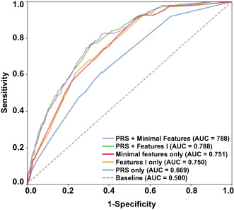
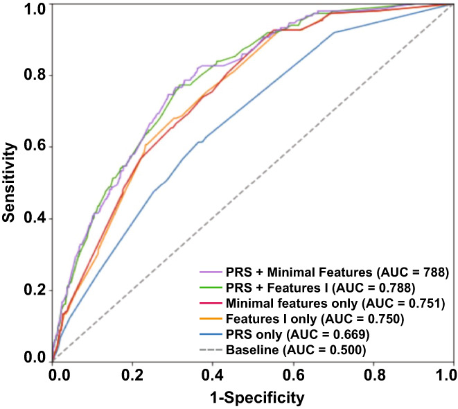
Figure 2 shows the ROC curves for the five models. Table 3 summarizes the performance metrics of all five models and includes the AUROC, sensitivity, specificity, diagnostic odds ratio, false positive rates, and positive and negative likelihood ratios. The PRS + Features I and PRS + Minimal Features models' performance were comparable and demonstrated the highest AUROCs, 0.788 (95% CI = 0.758–0.819) and 0.788 (95% CI = 0.757–0.820), respectively. At a sensitivity of 0.800, the PRS + Features I model demonstrated a specificity of 0.629, and the PRS + Minimal Features model a specificity of 0.646. The PRS + Minimal Features model had a false positive rate of 35.4%. The performance metrics of the four additional models (Features II, PRS + Features II, Features III, and PRS + Features III) are presented in Table S2.
FIGURE 2.

Receiver operating characteristic (ROC) curves of the five machine learning algorithm models for risk prediction of prostate cancer (PCa): PRSs only, Features I, PRSs + Features I, Minimal Features, and PRSs + Minimal Features. AUC, area under the curve; PRS, polygenic risk score.
TABLE 3.
Performance metrics of all five machine learning algorithm models
| Only PRS scores | Features I only | PRS + Features I | Minimal Features only | PRS + Minimal Features | |
|---|---|---|---|---|---|
| AUROC | 0.669 (0.634, 0.708) | 0.750 (0.714, 0.781) | 0.788 (0.758, 0.819) | 0.751 (0.714, 0.784) | 0.788 (0.757, 0.820) |
| Sensitivity | 0.920 (0.877, 0.963) | 0.807 (0.743, 0.870) | 0.800 (0.736, 0.864) | 0.807 (0.743, 0.870) | 0.800 (0.736, 0.864) |
| Specificity | 0.295 (0.289, 0.302) | 0.552 (0.545, 0.560) | 0.629 (0.622, 0.636) | 0.563 (0.556, 0.570) | 0.646 (0.639, 0.653) |
| DOR | 4.819 (4.229, 5.410) | 5.151 (4.744, 5.557) | 6.783 (6.382, 7.184) | 5.377 (4.971, 5.784) | 7.299 (6.897, 7.700) |
| LR+ | 1.306 (1.244, 1.370) | 1.802 (1.664, 1.953) | 2.157 (1.986, 2.341) | 1.846 (1.704, 2.000) | 2.260 (2.081, 2.454) |
| LR‐ | 0.271 (0.157, 0.466) | 0.350 (0.252, 0.485) | 0.318 (0.231, 0.438) | 0.343 (0.248, 0.476) | 0.310 (0.225, 0.426) |
| FPR | 0.705 (0.698, 0.711) | 0.448 (0.440, 0.455) | 0.371 (0.364, 0.378) | 0.437 (0.430, 0.444) | 0.354 (0.347, 0.361) |
Abbreviations: AUROC, area under the receiver operating characteristic; DOR, diagnostic odds ratio; FPR, false positive rate; LR+, likelihood ratio positive; LR−, likelihood ratio negative; PRS, polygenic risk score.
3.3. Feature importance
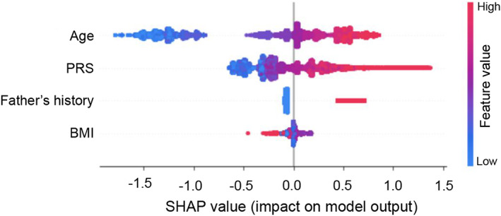
The SHAP plot (Figure 3) shows the features with the highest contribution to the XGB results for the PRS + Minimal Features model. Age, PRS, and father's PCa history were identified as the top features having a positive association with PCa risk, whereas higher BMI was associated with lower risk. Sibling history and ethnicity were also identified as high‐importance predictors in the Features I model; however, inclusion of PRS resulted in a sharp decrease in their feature importance. Figure S1 presents the SHAP plots of the Features I, PRSs + Features I, Features II, PRS + Features II model, and Minimal Features ML models. While the risk factors featured in our additional models are not typically used in routine clinical examinations for PCa, investigation in previously published ML‐based studies prompted analysis in our study. Apart from glycated hemoglobin, the supplementary features did not improve model performance and thus are not the main features of focus in our study.
FIGURE 3.

SHapely Additive exPlanations (SHAP) plot of the PRS + Minimal Features model. Display of the top predictor correlations and distribution of feature importance. PRS, polygenic risk score; BMI, body mass index.
4. DISCUSSION
4.1. Summary of the study
This is the first study demonstrating the utility of ML algorithms for PCa risk assessment in younger men who have not reached the recommended age for routine PCa screening. We achieved the same accuracy as the PRS + Features I model with fewer inputs (PRS + Minimal Features) and successfully created a risk assessment tool for identifying high‐risk individuals among men aged 55 and younger. PCa incidence in men of this age group has been steadily increasing over the last few decades and is expected to continue rising. Younger men with high grade PCa have a significantly diminished overall survival and disease‐specific survival compared to older men. 23 , 24 Our ML‐based prediction model may aid in the early detection of PCa in young at‐risk individuals to prompt further examination and provide an opportunity for early treatment and prevention options.
4.2. Significance and impact of PRS in PCa prediction models
There are notable biological differences between early and late onset PCa, which can have significant clinical implications. 25 The early onset of PCa in younger men is thought to be largely attributed to genetic factors. 26 This is consistent with the presence of PRS and paternal PCa history among the top features of the ML prediction models. Although familial history is a known PCa risk factor, using genetic data in the form of a PRS can provide a more objective risk profile that is not contingent upon accurate information from an individual's family members. 27 Previous studies have shown the clinical utility of using PRS data by accurately predicting PCa risk and demonstrating that this method may also aid in reducing overdiagnosis. 28 , 29 As research continues to identify new PCa susceptibility loci, 11 we expect that future ML models will incorporate improved PRSs in parallel with new genetic discoveries.
4.3. Importance of other features
When PRS was not included in our model, ethnicity was among the significant features. PCa is known to be disproportionately higher in Black men, 30 , 31 which is consistent with the significant overrepresentation of Black men with PCa cases in comparison to other racial groups in our study. Sibling history was an additional model feature determined to be of high importance, although the addition of PRS to the ML model substantially reduced its importance as a predictor. Incorporation of PRS diminished the need for these additional features and maintained high accuracy in our Minimal Features model. Interaction and overlapping effects between PRS and race/ethnicity, sibling history and glycated hemoglobin were not explored in this study and warrant further investigation.
Age was identified as the most important feature in predicting PCa, in agreement with established literature. 7 Addition of other clinical data (BMI) also slightly improved performance metrics. BMI has been reported to influence PCa aggressiveness, however, the mechanisms are not yet known. 32 , 33 , 34 We observed a negative association between BMI and PCa risk. Similar findings were reported by Giovannuci et al. 35 who found a lower risk of PCa in men with higher BMI only if they were younger (<60 years old) or had a family history of PCa, and attributed their findings to the complex relationship between obesity and various hormones.
4.4. Comparison with other models
Previous studies in older men (ranging from 55 to 80 years) have reported that the inclusion of PRS data improves the performance of different PCa prediction models. 36 , 37 , 38 , 39 Oh et al. 38 identified several ethnicity‐specific SNPs with moderate predictive performance (an AUROC of 0.637) in men 60 years and older. Aly et al. 39 developed a baseline model in men 80 years and under based on age, PSA levels and familial history and determined that the inclusion of PRS improved performance metrics (AUROC of 0.64–0.67) as well as reduced the number of required diagnostic biopsies. Our models did not rely on the use of PSA tests, which can be associated with false positive results leading to biopsy complications, 40 particularly in younger men who have a distinctive phenotype characterized by early‐onset PCa where poorly differentiated adenocarcinoma may impact the accuracy of PSA as a PCa risk predictor. 9 The distribution of baseline PSA levels in younger men has also been reported to vary across ethnic groups, 41 suggesting that screening guidelines involving PSA values in younger men may not be applicable to all population groups. Our model demonstrated a substantial reduction in false positive rates for ML PCa screening compared to using PSA serum tests: 35.4% for our PRS + Minimal Features model in men aged 35–55, versus 60% to 67% for PSA screening in men aged 55–71. 42 This was also a considerable improvement from the 70.5% false positive rate of our PRS only model, which is comparable to current false positive rates of PSA screening in older men. Our ML models' combination use of genetic and patient data demonstrated increased accuracy in the identification of PCa risk in a younger cohort of men.
4.5. Study limitations
There are several limitations to this study. First, this work was conducted retrospectively, therefore we cannot determine how this model would perform in prospective clinical practice. Additionally, PRS studies that use cohorts of mainly European descent, as is the case with our dataset, may not be generalizable to other populations and may affect risk prediction accuracy for individuals of non‐European ancestry. 43 Information on Gleason grades was not available in our database. Our presented model aims to prompt evaluation and thorough testing in patients at high‐risk for PCa. Consideration of Gleason grades and genetic variants is important for management and treatment, and are an area for future research. Other PCa risk assessment tools 44 , 45 require PSA or DRE measures, which were not available for direct comparison of risk assessment in this population. The clinical applicability and statistical power of these findings are limited by a relatively small number of cases, particularly among the population groups with the youngest ages and nonwhite race categories, as well as a limited number of race‐agnostic SNPs. Adjustments for PRSs were not made using race‐specific SNPs; race should be accounted for (or considered) in calculating PRS in future studies. This work provides promising preliminary evidence for PCa risk evaluation in younger men and warrants future studies that should include validation of our ML algorithm in a prospective clinical setting and assessing how patient care and outcomes are affected.
5. CONCLUSION
Machine learning algorithms which include PRS information and basic patient data can provide risk assessment for PCa in a young population not routinely screened. Efforts to identify men at risk in earlier age groups can help decrease the burden of PCa. Future work to support implementation of ML algorithms for PCa risk assessment of younger men in clinical practice is needed.
AUTHOR CONTRIBUTIONS
Amita Varma: Formal analysis, software, visualization, and writing—original draft. Jenish Maharjan: Formal analysis and software. Myrna Hurtado: writing—original draft and writing—review and editing. Sepideh Shokouhi: writing—original draft and writing—review and editing. Anurag Garikipati: Conceptualization. Qingqing Mao: Conceptualization.
FUNDING INFORMATION
The authors received no public funding for this work.
CONFLICT OF INTERESTS
The authors are or have been employees or contractors of Dascena, Inc.
ETHICS APPROVAL AND CONSENT TO PARTICIPATE
Data were collected passively and de‐identified in compliance with the Health Insurance Portability and Accountability Act, thus, this study was considered non‐human subjects research and did not require Institutional Review Board approval.
Supporting information
Appendix S1 Supporting Information
ACKNOWLEDGMENTS
The authors thank Misty M Attwood, PhD for her contribution to the research and development of this project.
Varma A, Maharjan J, Garikipati A, Hurtado M, Shokouhi S, Mao Q. Early prediction of prostate cancer risk in younger men using polygenic risk scores and electronic health records. Cancer Med. 2023;12:379‐386. doi: 10.1002/cam4.4934
Contributor Information
Myrna Hurtado, Email: lhurtado@dascena.com.
Sepideh Shokouhi, Email: shokous@gmail.com.
DATA AVAILABILITY STATEMENT
The data used in this study are available from UKBB and may be accessed by completing an application via https://www.ukbiobank.ac.uk/register‐apply/. The ML algorithm code developed in this study is proprietary and not publicly available.
REFERENCES
- 1. Sung H, Ferlay J, Siegel RL, et al. Global cancer statistics 2020: GLOBOCAN estimates of incidence and mortality worldwide for 36 cancers in 185 countries. CA Cancer J Clin. 2021;71(3):209‐249. doi: 10.3322/caac.21660 [DOI] [PubMed] [Google Scholar]
- 2. Ferlay J, Colombet M, Soerjomataram I, et al. Estimating the global cancer incidence and mortality in 2018: GLOBOCAN sources and methods. Int J Cancer. 2019;144(8):1941‐1953. doi: 10.1002/ijc.31937 [DOI] [PubMed] [Google Scholar]
- 3. Merriel SWD, Funston G, Hamilton W. Prostate cancer in primary care. Adv Ther. 2018;35(9):1285‐1294. doi: 10.1007/s12325-018-0766-1 [DOI] [PMC free article] [PubMed] [Google Scholar]
- 4. Leslie SW, Soon‐Sutton TL, Sajjad H, Siref LE. Prostate Cancer. StatPearls Publishing; 2021. Accessed September 9, 2021. http://www.ncbi.nlm.nih.gov/books/NBK470550/ [Google Scholar]
- 5. van den Bergh RCN, Loeb S, Roobol MJ. Impact of early diagnosis of prostate cancer on survival outcomes. Eur Urol Focus. 2015;1(2):137‐146. doi: 10.1016/j.euf.2015.01.002 [DOI] [PubMed] [Google Scholar]
- 6. Palsdottir T, Nordstrom T, Karlsson A, Grönberg H, Clements M, Eklund M. The impact of different prostate‐specific antigen (PSA) testing intervals on Gleason score at diagnosis and the risk of experiencing false‐positive biopsy recommendations: a population‐based cohort study. BMJ Open. 2019;9(3):e027958. doi: 10.1136/bmjopen-2018-027958 [DOI] [PMC free article] [PubMed] [Google Scholar]
- 7. Rawla P. Epidemiology of prostate cancer. World J Oncol. 2019;10(2):63‐89. doi: 10.14740/wjon1191 [DOI] [PMC free article] [PubMed] [Google Scholar]
- 8. Siegel R, Naishadham D, Jemal A. Cancer statistics, 2012. CA Cancer J Clin. 2012;62(1):10‐29. doi: 10.3322/caac.20138 [DOI] [PubMed] [Google Scholar]
- 9. Gupta S, Gupta A, Saini AK, Majumder K, Sinha K, Chahal A. Prostate cancer: how young is too young? Curr Urol. 2017;9(4):212‐215. doi: 10.1159/000447143 [DOI] [PMC free article] [PubMed] [Google Scholar]
- 10. Mucci LA, Hjelmborg JB, Harris JR, et al. Familial risk and heritability of cancer among twins in Nordic countries. JAMA. 2016;315(1):68‐76. doi: 10.1001/jama.2015.17703 [DOI] [PMC free article] [PubMed] [Google Scholar]
- 11. Schumacher FR, Al Olama AA, Berndt SI, et al. Association analyses of more than 140,000 men identify 63 new prostate cancer susceptibility loci. Nat Genet. 2018;50(7):928‐936. doi: 10.1038/s41588-018-0142-8 [DOI] [PMC free article] [PubMed] [Google Scholar]
- 12. Conti DV, Darst BF, Moss LC, et al. Trans‐ancestry genome‐wide association meta‐analysis of prostate cancer identifies new susceptibility loci and informs genetic risk prediction. Nat Genet. 2021;53(1):65‐75. doi: 10.1038/s41588-020-00748-0 [DOI] [PMC free article] [PubMed] [Google Scholar]
- 13. Huynh‐Le MP, Karunamuni R, Fan CC, et al. Prostate cancer risk stratification improvement across multiple ancestries with new polygenic hazard score. Prostate Cancer Prostatic Dis. 2022;12:1‐7. doi: 10.1038/s41391-022-00497-7 [DOI] [PMC free article] [PubMed] [Google Scholar]
- 14. Lambert SA, Abraham G, Inouye M. Towards clinical utility of polygenic risk scores. Hum Mol Genet. 2019;28(R2):R133‐R142. doi: 10.1093/hmg/ddz187 [DOI] [PubMed] [Google Scholar]
- 15. Leitzmann MF, Rohrmann S. Risk factors for the onset of prostatic cancer: age, location, and behavioral correlates. Clin Epidemiol. 2012;4:1‐11. doi: 10.2147/CLEP.S16747 [DOI] [PMC free article] [PubMed] [Google Scholar]
- 16. Choi SW, O'Reilly PF. PRSice‐2: polygenic risk score software for biobank‐scale data. GigaScience. 2019;8(7):1‐6. doi: 10.1093/gigascience/giz082 [DOI] [PMC free article] [PubMed] [Google Scholar]
- 17. Mars N, Koskela JT, Ripatti P, et al. Polygenic and clinical risk scores and their impact on age at onset and prediction of cardiometabolic diseases and common cancers. Nat Med. 2020;26(4):549‐557. doi: 10.1038/s41591-020-0800-0 [DOI] [PubMed] [Google Scholar]
- 18. Chen T, Guestrin C. XGBoost: a scalable tree boosting system. Proceedings of the 22nd ACM SIGKDD International Conference on Knowledge Discovery and Data mining. Published online August 13, 2016:785‐794. doi: 10.1145/2939672.2939785 [DOI]
- 19. Perez‐Cornago A, Key TJ, Allen NE, et al. Prospective investigation of risk factors for prostate cancer in the UKbiobank cohort study. Br J Cancer. 2017;117(10):1562‐1571. doi: 10.1038/bjc.2017.312 [DOI] [PMC free article] [PubMed] [Google Scholar]
- 20. Monroy‐Iglesias MJ, Russell B, Crawley D, et al. Metabolic syndrome biomarkers and prostate cancer risk in the UKbiobank. Int J Cancer. 2021;148(4):825‐834. doi: 10.1002/ijc.33255 [DOI] [PubMed] [Google Scholar]
- 21. Peila R, Rohan TE. Diabetes, glycated hemoglobin, and risk of cancer in the UKbiobank study. Cancer Epidemiol Biomarkers Prev. 2020;29(6):1107‐1119. doi: 10.1158/1055-9965.EPI-19-1623 [DOI] [PubMed] [Google Scholar]
- 22. Rodríguez‐Pérez R, Bajorath J. Interpretation of compound activity predictions from complex machine learning models using local approximations and Shapley values. J Med Chem. 2020;63(16):8761‐8777. doi: 10.1021/acs.jmedchem.9b01101 [DOI] [PubMed] [Google Scholar]
- 23. Lin DW, Porter M, Montgomery B. Treatment and survival outcomes in young men diagnosed with prostate cancer: a population based cohort study. Cancer. 2009;115(13):2863‐2871. doi: 10.1002/cncr.24324 [DOI] [PMC free article] [PubMed] [Google Scholar]
- 24. Merrill RM, Bird JS. Effect of young age on prostate cancer survival: a population‐based assessment (United States). Cancer Causes Control CCC. 2002;13(5):435‐443. doi: 10.1023/a:1015764507609 [DOI] [PubMed] [Google Scholar]
- 25. Salinas CA, Tsodikov A, Ishak‐Howard M, Cooney KA. Prostate cancer in young men: an important clinical entity. Nat Rev Urol. 2014;11(6):317‐323. doi: 10.1038/nrurol.2014.91 [DOI] [PMC free article] [PubMed] [Google Scholar]
- 26. Lange EM, Salinas CA, Zuhlke KA, et al. Early onset prostate cancer has a significant genetic component. The Prostate. 2012;72(2):147‐156. doi: 10.1002/pros.21414 [DOI] [PMC free article] [PubMed] [Google Scholar]
- 27. Sun J, Na R, Hsu FC, et al. Genetic score is an objective and better measurement of inherited risk of prostate cancer than family history. Eur Urol. 2013;63(3):585‐587. doi: 10.1016/j.eururo.2012.11.047 [DOI] [PMC free article] [PubMed] [Google Scholar]
- 28. Pashayan N, Pharoah PD, Schleutker J, et al. Reducing overdiagnosis by polygenic risk‐stratified screening: findings from the Finnish section of the ERSPC. Br J Cancer. 2015;113(7):1086‐1093. doi: 10.1038/bjc.2015.289 [DOI] [PMC free article] [PubMed] [Google Scholar]
- 29. Seibert TM, Fan CC, Wang Y, et al. Polygenic hazard score to guide screening for aggressive prostate cancer: development and validation in large scale cohorts. BMJ. 2018;360:j5757. doi: 10.1136/bmj.j5757 [DOI] [PMC free article] [PubMed] [Google Scholar]
- 30. Edwards BK, Howe HL, Ries LAG, et al. Annual report to the nation on the status of cancer, 1973‐1999, featuring implications of age and aging on U.S. cancer burden. Cancer. 2002;94(10):2766‐2792. doi: 10.1002/cncr.10593 [DOI] [PubMed] [Google Scholar]
- 31. Tsodikov A, Gulati R, de Carvalho TM, et al. Is prostate cancer different in black men? Answers from 3 natural history models. Cancer. 2017;123(12):2312‐2319. doi: 10.1002/cncr.30687 [DOI] [PMC free article] [PubMed] [Google Scholar]
- 32. Haque R, Van Den Eeden SK, Wallner L, et al. Association of body mass index and prostate cancer mortality. Obes Res Clin Pract. 2014;8(4):e374‐e381. doi: 10.1016/j.orcp.2013.06.002 [DOI] [PMC free article] [PubMed] [Google Scholar]
- 33. Rodriguez C, Freedland SJ, Deka A, et al. Body mass index, weight change, and risk of prostate cancer in the cancer prevention study II nutrition cohort. Cancer Epidemiol Prev Biomark. 2007;16(1):63‐69. doi: 10.1158/1055-9965.EPI-06-0754 [DOI] [PubMed] [Google Scholar]
- 34. Lavalette C, Cordina Duverger E, Artaud F, et al. Body mass index trajectories and prostate cancer risk: results from the EPICAP study. Cancer Med. 2020;9(17):6421‐6429. doi: 10.1002/cam4.3241 [DOI] [PMC free article] [PubMed] [Google Scholar]
- 35. Giovannucci E, Rimm EB, Liu Y, et al. Body mass index and risk of prostate cancer in U.S. health professionals. J Natl Cancer Inst. 2003;95(16):1240‐1244. doi: 10.1093/jnci/djg009 [DOI] [PubMed] [Google Scholar]
- 36. Sipeky C, Talala KM, Tammela TLJ, Taari K, Auvinen A, Schleutker J. Prostate cancer risk prediction using a polygenic risk score. Sci Rep. 2020;10:17075. doi: 10.1038/s41598-020-74172-z [DOI] [PMC free article] [PubMed] [Google Scholar]
- 37. Black MH, Li S, LaDuca H, et al. Validation of a prostate cancer polygenic risk score. The Prostate. 2020;80(15):1314‐1321. doi: 10.1002/pros.24058 [DOI] [PMC free article] [PubMed] [Google Scholar]
- 38. Oh JJ, Kim E, Woo E, et al. Evaluation of polygenic risk scores for prediction of prostate cancer in Korean men. Front Oncol. 2020;10:583625. doi: 10.3389/fonc.2020.583625 [DOI] [PMC free article] [PubMed] [Google Scholar]
- 39. Aly M, Wiklund F, Xu J, et al. Polygenic risk score improves prostate cancer risk prediction: results from the Stockholm‐1 cohort study. Eur Urol. 2011;60(1):21‐28. doi: 10.1016/j.eururo.2011.01.017 [DOI] [PMC free article] [PubMed] [Google Scholar]
- 40. Fenton JJ, Weyrich MS, Durbin S, Liu Y, Bang H, Melnikow J. Prostate‐specific antigen‐based screening for prostate cancer: evidence report and systematic review for the US preventive services task force. JAMA. 2018;319(18):1914‐1931. doi: 10.1001/jama.2018.3712 [DOI] [PubMed] [Google Scholar]
- 41. Tang P, Du W, Xie K, et al. Characteristics of baseline PSA and PSA velocity in young men without prostate cancer: racial differences. The Prostate. 2012;72(2):173‐180. doi: 10.1002/pros.21418 [DOI] [PubMed] [Google Scholar]
- 42. Kilpeläinen TP, Tammela TLJ, Määttänen L, et al. False‐positive screening results in the Finnish prostate cancer screening trial. Br J Cancer. 2010;102(3):469‐474. doi: 10.1038/sj.bjc.6605512 [DOI] [PMC free article] [PubMed] [Google Scholar]
- 43. Cavazos TB, Witte JS. Inclusion of variants discovered from diverse populations improves polygenic risk score transferability. HGG Adv. 2021;2(1):100017. doi: 10.1016/j.xhgg.2020.100017 [DOI] [PMC free article] [PubMed] [Google Scholar]
- 44. Ankerst DP, Straubinger J, Selig K, et al. A contemporary prostate biopsy risk calculator based on multiple heterogeneous cohorts. Eur Urol. 2018;74(2):197‐203. doi: 10.1016/j.eururo.2018.05.003 [DOI] [PMC free article] [PubMed] [Google Scholar]
- 45. Kranse R, Roobol M, Schröder FH. A graphical device to represent the outcomes of a logistic regression analysis. The Prostate. 2008;68(15):1674‐1680. doi: 10.1002/pros.20840 [DOI] [PubMed] [Google Scholar]
Associated Data
This section collects any data citations, data availability statements, or supplementary materials included in this article.
Supplementary Materials
Appendix S1 Supporting Information
Data Availability Statement
The data used in this study are available from UKBB and may be accessed by completing an application via https://www.ukbiobank.ac.uk/register‐apply/. The ML algorithm code developed in this study is proprietary and not publicly available.


