Abstract
Species of Mycenasect.Calodontes are representative of the Mycena genus as a whole and are easily recognised by the pinkish, reddish, purplish to brownish pileus and larger basidiomata. Furthermore, the colour of the pileus in the species of sect. Calodontes often has a transition or changes in different stages and the combination of the colour of the pileus with cystidia and basidiospores can be used to recognise taxa within this section. To date, 19 species of Mycenasect.Calodontes have been reported worldwide. Including our recent description of M.yuezhuoi, five species of sect. Calodontes have been recorded in China. During examination of specimens collected in coniferous forests or mixed broadleaf-conifer forests in temperate regions of China, additional taxa assigned to sect. Calodontes were identified. Four new species are recognised, based mostly on characters of the pileus and cystidia. Phylogenetic analysis of sequence data from multiple DNA regions (ITS + rpb1 + tef1) supported the morphological evidence. Here, we propose M.polycystidiata, M.rufobrunnea, M.shengshanensis and M.subulata as new species in Mycenasect.Calodontes. Morphological descriptions, line drawings, habitat photos and comparisons with closely-related taxa are provided. A key to the 23 known species of sect. Calodontes is presented.
Keywords: coniferous forest, new taxa, phylogeny, saprobic, taxonomy
Introduction
Mycenasect.Calodontes (Fr. ex Berk.) Quél. comprises the taxa in Mycena (Pers.) Roussel with a pinkish, reddish, purplish to brownish and mostly hygrophanous pileus, interveined lamellae, smooth cheilocystidia and pleurocystidia (if present) and mostly amyloid spores (Fries 1821; Berkeley 1836; Maas Geesteranus 1992a, 1992b; Harder et al. 2010). The group was initially proposed as Agaricustrib.Clitocybe subtrib. Calodontes Fries consisting of six species, then elevated to section rank within Agaricussubgen.Clitocybe Fr. ex Berk. and finally assigned to Mycena in 1872 (Fries 1821; Berkeley 1836; Quélet 1872). To date, 19 species are known, mostly from Europe and North America, but five species have been described from Asia (specifically, China, India and Peninsular Malaysia) (Smith 1947; Maas Geesteranus 1980; Perry 2002; Grgurinovic 2003; Robich 2003; Chew et al. 2014; Aravindakshan and Manimohan 2015; Aronsen and Læssøe 2016; Na 2019; Liu et al. 2021).
The circumscription of subsections within sect. Calodontes is problematic. Initially, Smith (1947) divided sect. Calodontes into two subsections, Granulatae and Ciliatae, according to whether the cheilocystidia were smooth or not, but this system was not widely adopted because most of the species have previously been classified in other sections of Mycena on account of the red, yellow or orange basidiomata, coloured lamellar edge and echinulate or diverticulate cheilocystidia, pleurocystidia, pileipellis or stipitipellis (Maas Geesteranus 1980, 1992a, 1992b; Perry 2002; Grgurinovic 2003; Robich 2003; Harder et al. 2010; Chew et al. 2014). Based on the lamellar edge colour, spore amyloid reaction and cystidial features, a widely accepted subsectional classification of sect. Calodontes was proposed by Maas Geesteranus (1980) and subsequent taxonomists (Grgurinovic 2003; Harder et al. 2010; Chew et al. 2014). The primary criteria for segregation were dominated by microcharacters: subsect. Purae (Konrad & Maubl.) Maas Geest. with amyloid spores and colourless pleuro- and cheilocystidia; subsect. Violacellae Sing. ex Maas Geest. with inamyloid spores and no pleurocystidia; and subsect. Marginatae J.E. Lange with amyloid spores and pleurocystidia and cheilocystidia with purplish-brown contents (Maas Geesteranus 1980; Grgurinovic 2003; Harder et al. 2010; Chew et al. 2014). Although the infrasectional classification of Maas Geesteranus (1980) is generally accepted, phylogenetic analyses have provided only weak support because subsect. Purae and subsect. Violacellae are polyphyletic (Perry 2002; Grgurinovic 2003; Robich 2003; Harder et al. 2010, 2012, 2013; Chew et al 2014; Na 2019). In studying Mycenapura (Pers.) P. Kumm., the type of subsect. Purae, Maas Geesteranus (1992a, 1992b) proposed eight forma based on pileus colour. However, 11 clades have been resolved amongst materials collected from Europe and the Americas, which suggests that there may be additional undescribed taxa in subsect. Purae (Harder et al. 2013).
Including our recent description of M.yuezhuoi Z.W. Liu, Y.P. Ge & Q. Na from Kunyushan National Nature Reserve (Yantai, Shandong Province), five species of Mycenasect.Calodontes have been previously recorded in China (Li et al. 2015; Na 2019; Liu et al. 2021). In this paper, we propose an additional four new species classified in sect. Calodontes from the temperate zone of northeast China. The four new species share a unique set of striking morphological characters and contribute to an improved understanding of the classification of sect. Calodontes.
Materials and methods
Morphological observations
Thirteen fungal specimens were examined in this study, which were mainly collected in coniferous forests and some from mixed broadleaf-conifer forests in 2021. Macrocharacters were recorded from fresh specimens. Colour codes in descriptions follow those of Kornerup and Wanscher (1978). Microcharacters were observed from tissues sampled from dried specimens and rehydrated with 5% potassium hydroxide (KOH) and stained with Congo red (1% [w/v] aqueous solution), if necessary, using a Lab A1 microscope (Carl Zeiss AG, Jena, Germany). The amyloid reaction was tested with Melzer’s Reagent (Clémençon et al. 2004; Horak 2005). Twenty basidiospores were measured per specimen. For the holotype, 40 basidiospores from different basidiomata were selected for measurement. Basidiospore statistics are expressed as (a/b/c) (d)e-f-g (h) × (i)j-k–l (m) μm [Q = (n)o-p (q), Q = r ± s], where a–c represent a basidiospores of b basidiomata from c specimens measured; d and h are the minimum and maximum length (5% extremum), respectively, e and g indicate the range of values for the remaining 90% of the spores and f is the average length; width (i–m) and Q values (n–q) are expressed in a similar manner; and r and s are the average Q value and its standard deviation, respectively (Ge et al. 2021; Liu et al. 2021; Na et al. 2021; Na et al. 2022). The measurement of basidia, cystidia and other characters were each based on 20 observations. All specimens have been deposited in the Fungarium of the Fujian Academy of Agricultural Sciences (FFAAS).
DNA extraction, PCR, cloning and DNA sequencing
The Plant Genomic DNA Kit (CoWin Biosciences, Beijing, China) was used to isolate total genomic DNA from dried specimens in accordance with the manufacturer’s instructions. Three nuclear loci were sequenced, comprising the internal transcribed spacer (ITS), RNA polymerase II largest subunit (rpb1) and translation elongation factor-1 alpha (tef1). The primer pairs ITS1/ITS4, rpb1Mp_f1/rpb1Mp_r1 and tEFMp_f2/ tEFMp_r2 were selected to amplify ITS, rpb1 and tef1, respectively (White et al. 1990; Harder et al. 2013; Yu et al. 2020). The PCR reactions were performed in a total volume of 25 µl containing 2 µl DNA template, 1 µl for each primer, 8.5 µl nuclease-free H2O and 12.5 µl 2× Utaq PCR MasterMix (ZomanBio, Beijing, China). The PCR protocol for amplification of the ITS region was as follows: 94 °C for 4 min, then 34 cycles of 94 °C for 45 s, 52 °C for 45 s and 72 °C for 1 min, with a final extension of 72 °C for 10 min (Na et al. 2022). The PCR protocol for amplification of the rpb1 and tef1 regions followed that of Harder et al. (2013): 94 °C for 60 s, then 10 cycles of 94 °C for 35 s, 53 °C for 45 s and 72 °C for 45 s; then 25 cycles of 94 °C for 35 s, 56 °C for 45 s, 72 °C for 45 s and final extension of 72 °C for 10 min. The PCR products were purified by gel electrophoresis or filter membrane and subjected to Sanger dideoxy sequencing by the Beijing Genomics Institute (Beijing, China).
Phylogenetic analysis
A combined ITS, rpb1 and tef1 dataset was analysed to infer relationships of the new taxa with other members of sect. Calodontes. We used sequences included in previous studies of sect. Calodontes and from members of the most closely-related section deposited in the GenBank database, which were mainly submitted by Harder et al. (2013), Osmundson et al. (2013), Chew et al. (2014) and Liu et al. (2021). For the analysis, representative species of Mycenasect.Supinae Konrad & Maubl., which is closely related to sect. Calodontes, were selected as the outgroup (Osmundson et al. 2013; Na and Bau 2019). Sequences for each DNA region (ITS, rpb1 and tef1) were aligned in MAFFT version 7 online and the aligned matrices were manually checked with BIOEDIT 7.2.5.0 (Hall 1999; Kuraku et al. 2013; Katoh et al. 2019). The best-fit substitution model for each gene partition was determined with MODELTEST 2.3, based on the Akaike Information Criterion (Posada and Crandall 1998). Maximum Likelihood (ML) analysis was conducted by raxmlGUI 2.09 (Edler et al. 2020). The phylogenetic analysis was performed by a single analysis with six partitions (ITS1, 5.8S, ITS2, rpb1 exons, tef1 exons, intron of rpb1 + introns of tef1), using the GTRGAMMA model and 1,000 rapid bootstrap (BS) replicates. For Bayesian Inference (BI), two runs of six chains were run for 15,000,000 generations and sampled every 10,000 generations by MrBayes 3.2.6. At the end of the run, the average deviation of split frequencies was 0.007821, ESS (effective sample size) was 1300.3 and the average Potential Scale Reduction Factor (PSRF) parameter values (excluding NA and > 10.0) = 1.000 and the “sump” and “sumt” commands were used to summarise sampled parameters with 25% burn-in (Ronquist and Huelsenbeck 2003).
Results
Phylogenetic relationships
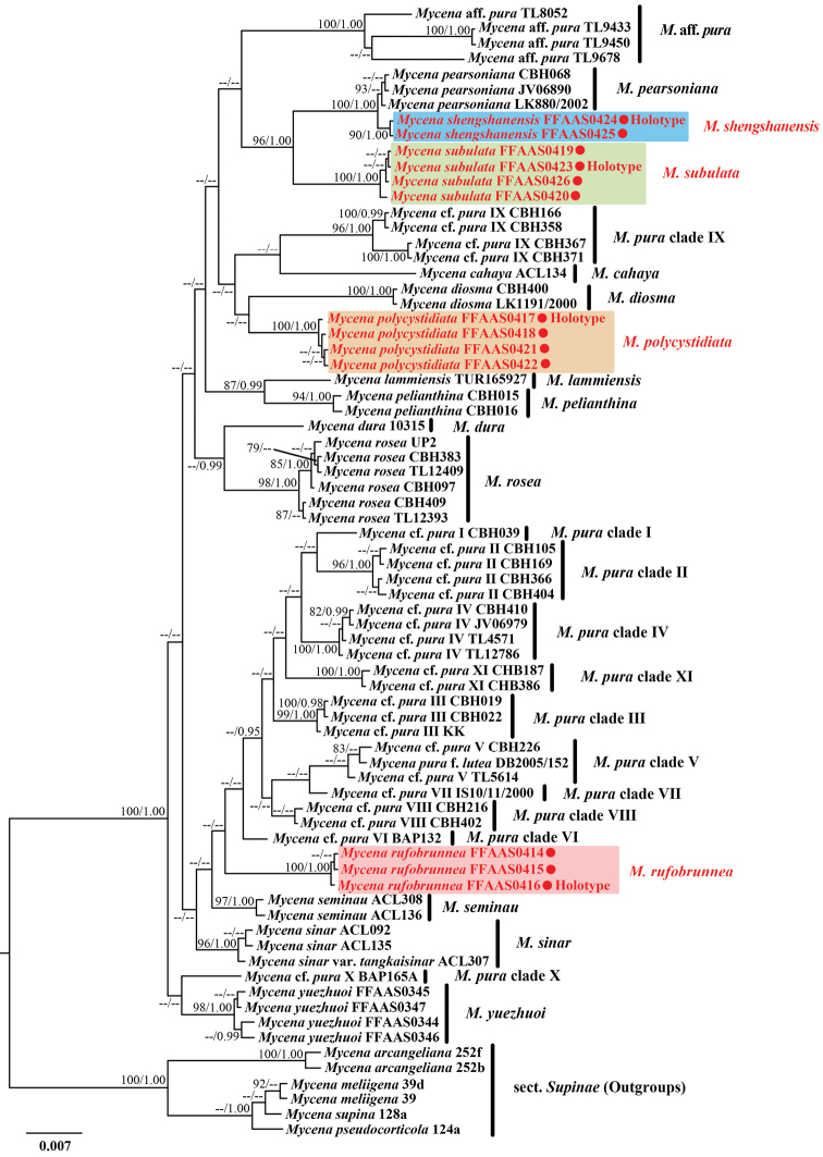
The dataset consisted of 192 sequences, comprising 39 newly-generated sequences (13 ITS, 13 rpb1 and 13 tef1) and 153 sequences (61 ITS, 46 rpb1 and 46 tef1) downloaded from GenBank. In total, 74 accessions of 19 species were included in the dataset. Detailed information for all sequences is presented in Table 1. The aligned dataset contained 1459 nucleotide sites including gaps (229 sites for ITS1, 159 sites for 5.8S, 177 sites for ITS2, 55 sites for rpb1 exons, 295 sites for tef1 exons, 544 sites for intron of rpb1 + introns of tef1), of which 1146 were conserved, 257 were parsimony-informative and 56 were variable, but parsimony-uninformative. For Bayesian Inference (BI), the selected models for each DNA region of the concatenated dataset were as follows: HKY+G for ITS1 and intron of rpb1 + introns of tef1, JC for 5.8S and rpb1 exons, HKY+I+G for ITS2 and SYM+I+G for tef1 exons. The BI and ML analyses resulted in almost identical topologies; thus, the BI topology is presented as a master tree (Fig. 1).
Table 1.
Specimens used in phylogenetic analysis and GenBank accession numbers.
| No. | Species | Specimen voucher | GenBank accession numbers | Locality | Reference | ||
|---|---|---|---|---|---|---|---|
| ITS | rpb1 | tef1 | |||||
| 1. | Mycenaaff.pura | TL8052 | FN394623 | KF723687 | KF723641 | Ecuador | Harder et al. (2010, 2013) |
| 2. | M.aff.pura | TL9433 | FN394622 | KF723688 | KF723642 | Ecuador | Harder et al. (2010, 2013) |
| 3. | M.aff.pura | TL9450 | KJ144653 | KF723689 | KF723643 | Ecuador | Harder et al. (2010, 2013) |
| 4. | M.aff.pura | TL9678 | FN394621 | KF723690 | KF723644 | Ecuador | Harder et al. (2010, 2013) |
| 5. | M.arcangeliana | 252b | JF908401 | – | – | Spain | Osmundson et al. (2013) |
| 6. | M.arcangeliana | 252f | JF908402 | – | – | Spain | Osmundson et al. (2013) |
| 7. | M.cahaya | ACL134 | KF537248 | – | – | Malaysia | Chew et al. (2014) |
| 8. | M.cf.pura I | CBH039 | FN394588 | KF723680 | KF723634 | Denmark | Harder et al. (2010, 2013) |
| 9. | M.cf.pura II | CBH105 | FN394581 | KF723671 | KF723625 | Denmark | Harder et al. (2010, 2013) |
| 10. | M.cf.pura II | CBH169 | FN394579 | KF723672 | KF723626 | Denmark | Harder et al. (2010, 2013) |
| 11. | M.cf.pura II | CBH366 | FN394572 | KF723673 | KF723627 | Denmark | Harder et al. (2010, 2013) |
| 12. | M.cf.pura II | CBH404 | FN394566 | KF723674 | KF723628 | Denmark | Harder et al. (2010, 2013) |
| 13. | M.cf.pura III | CBH019 | FN394605 | KF723675 | KF723629 | Denmark | Harder et al. (2010, 2013) |
| 14. | M.cf.pura III | CBH022 | FN394574 | KF723676 | KF723630 | Denmark | Harder et al. (2010, 2013) |
| 15. | M.cf.pura III | KK | FN394606 | KF723677 | KF723631 | Slovakia | Harder et al. (2010, 2013) |
| 16. | M.cf.pura IV | CBH410 | FN394595 | KF723667 | KF723621 | Denmark | Harder et al. (2010, 2013) |
| 17. | M.cf.pura IV | JV06979 | FN394585 | KF723668 | KF723622 | Denmark | Harder et al. (2010, 2013) |
| 18. | M.cf.pura IV | TL4571 | FN394583 | KF723669 | KF723623 | Denmark | Harder et al. (2010, 2013) |
| 19. | M.cf.pura IV | TL12786 | FN394591 | KF723670 | KF723624 | Sweden | Harder et al. (2010, 2013) |
| 20. | M.cf.pura V | CBH226 | FN394604 | KF723664 | KF723618 | Denmark | Harder et al. (2010, 2013) |
| 21. | M.cf.pura V | TL5614 | FN394602 | KF723666 | KF723620 | Denmark | Harder et al. (2010, 2013) |
| 22. | M.cf.pura VI | BAP132 | FN394561 | KF723660 | KF723614 | USA | Harder et al. (2010, 2013) |
| 23. | M.cf.pura VIII | CBH216 | FN394598 | KF723662 | KF723616 | Denmark | Harder et al. (2010, 2013) |
| 24. | M.cf.pura VIII | CBH402 | FN394599 | KF723663 | KF723617 | Denmark | Harder et al. (2010, 2013) |
| 25. | M.cf.pura IX | CBH166 | FN394607 | KF723701 | KF723655 | Denmark | Harder et al. (2010, 2013) |
| 26. | M.cf.pura IX | CBH358 | FN394608 | KF723702 | KF723656 | Denmark | Harder et al. (2010, 2013) |
| 27. | M.cf.pura IX | CBH367 | KF913022 | KF723703 | KF723657 | Denmark | Harder et al. (2013) |
| 28. | M.cf.pura IX | CBH371 | KF913023 | KF723704 | KF723658 | Denmark | Harder et al. (2013) |
| 29. | M.cf.pura X | BAP165A | FN394563 | KF723698 | KF723652 | USA | Harder et al. (2010, 2013) |
| 30. | M.cf.pura XI | CBH187 | FN394564 | KF723678 | KF723632 | Sweden | Harder et al. (2010, 2013) |
| 31. | M.cf.pura XI | CBH386 | FN394565 | KF723679 | KF723633 | Denmark | Harder et al. (2010, 2013) |
| 32. | M.diosma | CBH400 | FN394617 | KF723699 | KF723653 | Denmark | Harder et al. (2010, 2013) |
| 33. | M.diosma | LK1191/2000 | FN394619 | KF723700 | KF723654 | Germany | Harder et al. (2010, 2013) |
| 34. | M.dura | 10315 | FN394560 | KF723694 | KF723648 | Austria | Harder et al. (2010, 2013) |
| 35. | M.lammiensis | TUR165927 | FN394552 | KF723697 | KF723651 | Finland | Harder et al. (2010, 2013) |
| 36. | M.meliigena | 39 | JF908423 | – | – | Italy | Osmundson et al. (2013) |
| 37. | M.meliigena | 39d | JF908429 | – | – | Italy | Osmundson et al. (2013) |
| 38. | M.pearsoniana | CBH068 | FN394614 | KF723691 | KF723645 | Germany | Harder et al. (2010, 2013) |
| 39. | M.pearsoniana | JV06890 | FN394612 | KF723692 | KF723646 | Denmark | Harder et al. (2010, 2013) |
| 40. | M.pearsoniana | LK880/2002 | FN394613 | KF723693 | KF723647 | Germany | Harder et al. (2010, 2013) |
| 41. | M.pelianthina | CBH015 | FN394549 | KF723695 | KF723649 | Denmark | Harder et al. (2010, 2013) |
| 42. | M.pelianthina | CBH016 | FN394547 | KF723696 | KF723650 | Denmark | Harder et al. (2010, 2013) |
| 43. | M.polycystidiata | FFAAS0417 Holotype | ON427731 | ON468456 | ON468469 | China | This study |
| 44. | M.polycystidiata | FFAAS0418 | ON427732 | ON468457 | ON468470 | China | This study |
| 45. | M.polycystidiata | FFAAS0421 | ON427733 | ON468458 | ON468471 | China | This study |
| 46. | M.polycystidiata | FFAAS0422 | ON427734 | ON468459 | ON468472 | China | This study |
| 47. | M.pseudocorticola | 124a | JF908386 | – | – | Italy | Osmundson et al. (2013) |
| 48. | M.pura | IS10/11/2000 | FN394611 | – | – | USA | Harder et al. (2010) |
| 49. | M.puraf.lutea | DB2005/152 | FN394603 | – | – | Denmark | Harder et al. (2010) |
| 50. | M.rosea | UP2 | FN394550 | – | – | UK | Harder et al. (2010) |
| 51. | M.rosea | CBH097 | FN394556 | KF723681 | KF723635 | Denmark | Harder et al. (2010, 2013) |
| 52. | M.rosea | CBH383 | FN394553 | KF723682 | KF723636 | Denmark | Harder et al. (2010, 2013) |
| 53. | M.rosea | CBH409 | FN394551 | KF723683 | KF723637 | Germany | Harder et al. (2010, 2013) |
| 54. | M.rosea | TL12393 | FN394555 | KF723684 | KF723638 | Denmark | Harder et al. (2010, 2013) |
| 55. | M.rosea | TL12409 | FN394557 | KF723685 | KF723639 | Denmark | Harder et al. (2010, 2013) |
| 56. | M.rufobrunnea | FFAAS0414 | ON427728 | ON468453 | ON468466 | China | This study |
| 57. | M.rufobrunnea | FFAAS0415 | ON427729 | ON468454 | ON468467 | China | This study |
| 58. | M.rufobrunnea | FFAAS0416 Holotype | ON427730 | ON468455 | ON468468 | China | This study |
| 59. | M.seminau | ACL136 | KF537250 | – | – | Malaysia | Chew et al. (2014) |
| 60. | M.seminau | ACL308 | KF537252 | – | – | Malaysia | Chew et al. (2014) |
| 61. | M.shengshanensis | FFAAS0424 Holotype | ON427739 | ON468464 | ON468477 | China | This study |
| 62. | M.shengshanensis | FFAAS0425 | ON427740 | ON468465 | ON468478 | China | This study |
| 63. | M.sinar | ACL092 | KF537247 | – | – | Malaysia | Chew et al. (2014) |
| 64. | M.sinar | ACL135 | KF537249 | – | – | Malaysia | Chew et al. (2014) |
| 65. | M.sinarvar.tangkaisinar | ACL307 | KF537251 | – | – | Malaysia | Chew et al. (2014) |
| 66. | M.subulata | FFAAS0419 | ON427735 | ON468460 | ON468473 | China | This study |
| 67. | M.subulata | FFAAS0420 | ON427736 | ON468461 | ON468474 | China | This study |
| 68. | M.subulata | FFAAS0423 Holotype | ON427737 | ON468462 | ON468475 | China | This study |
| 69. | M.subulata | FFAAS0426 | ON427738 | ON468463 | ON468476 | China | This study |
| 70. | M.supina | 128a | JF908388 | – | – | Italy | Osmundson et al. (2013) |
| 71. | M.yuezhuoi | FFAAS0344 | MW581490 | MW868166 | MW882249 | China | Liu et al. (2021) |
| 72. | M.yuezhuoi | FFAAS0345 | MW581491 | MW868169 | MW882250 | China | Liu et al. (2021) |
| 73. | M.yuezhuoi | FFAAS0346 | MW581492 | MW868168 | MW882251 | China | Liu et al. (2021) |
| 74. | M.yuezhuoi | FFAAS0347 | MW581493 | MW868167 | MW882252 | China | Liu et al. (2021) |
Figure 1.
Bayesian Inference analysis of Mycenasect.Calodontes with ITS, rpb1 and tef1 sequence data. Species in Mycenasect.Supinae served as outgroup. Bootstrap values (BS) from Maximum Likelihood ≥ 75 and Bayesian posterior probabilities (BPP) ≥ 0.95 are shown on each branch (BS/BPP). The new species are marked in red.
The phylogenetic analysis revealed that sect. Calodontes was strong support (BS/Bayesian posterior probability [BPP] = 100/1.00) (Fig. 1). Fifteen species and eleven M.pura complex clades within sect. Calodontes were retrieved. Four new species were resolved as monophyletic, each with strong support: M.polycystidiata (BS/BPP = 100/1.00), M.rufobrunnea (BS/BPP = 100/1.00), M.shengshanensis (BS/BPP = 90/1.00) and M.subulata (BS/BPP = 100/1.00). A sister relationship between M.shengshanensis and M.pearsoniana Dennis ex Singer was well supported. Mycenasubulata was resolved as sister, but genetically distant from M.pearsoniana and M.shengshanensis clade. In addition, the sister relationship of Mycenapolycystidiata and M.rufobrunnea were unresolved.
Taxonomy
. Mycena polycystidiata
Z.W. Liu, Y.P. Ge, L. Zou & Q. Na, sp. nov.
314F9605-EDAA-5568-B959-48406818050D
MycoBank No: 843977
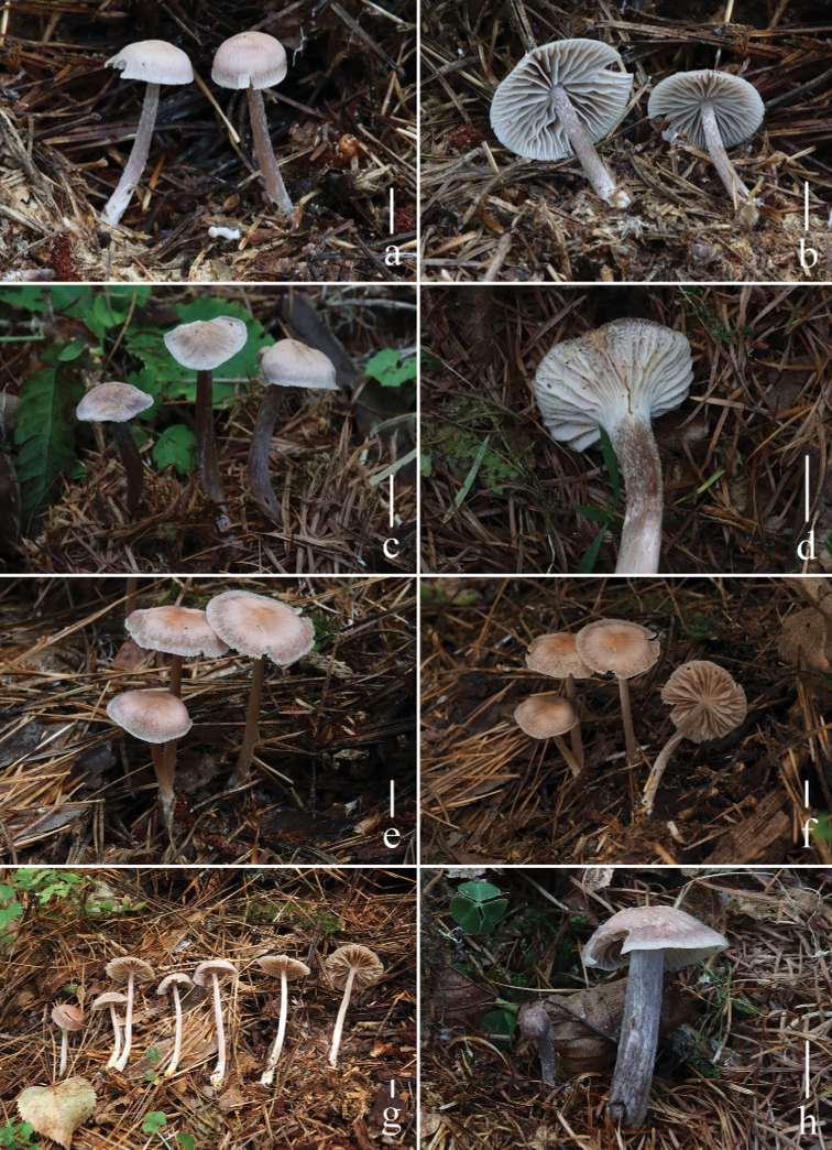
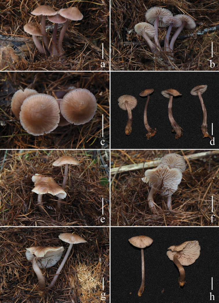
Figure 2.
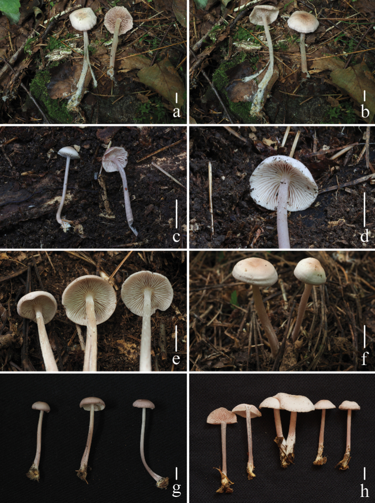
Basidiomata of Mycenapolycystidiata Z.W. Liu, Y.P. Ge, L. Zou & Q. Na a, bFFAAS0422c, dFFAAS0417, holotype e–gFFAAS00421hFFAAS0418 Scale bars: 10 mm (a–h). Photographs a–e, h by Qin Na f, g by Yupeng Ge.
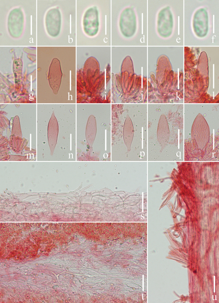
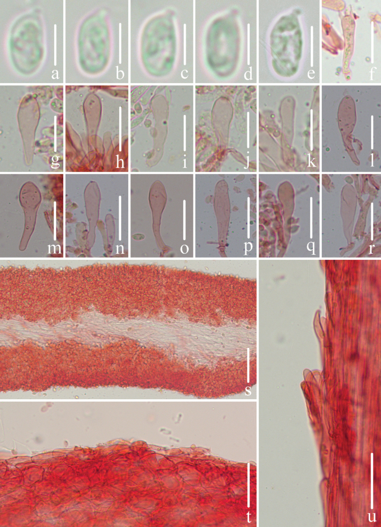
Figure 3.
Microscopic features of Mycenapolycystidiata (FFAAS0417, holotype) a–f basidiospores g basidia h–l cheilocystidia m–r pleurocystidia s pileipellis and hypodermium t lamellar trama u stipitipellis and caulocystidia. Scale bars: 5 μm (a–f); 10 μm (g); 30 μm (h–r); 40 μm (s–u).
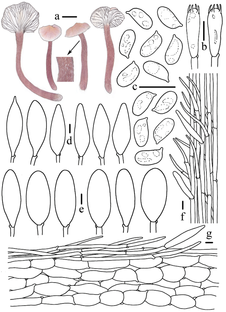
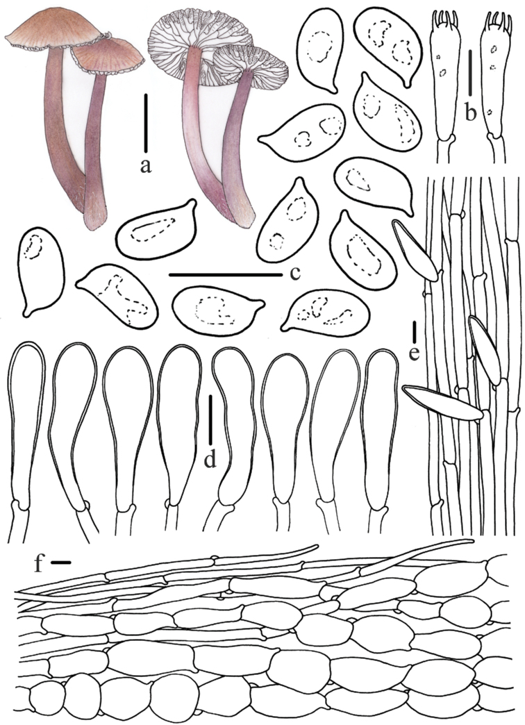
Figure 4.
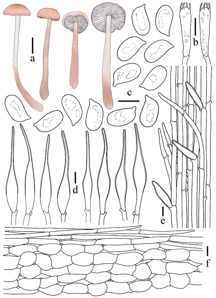
Morphological features of Mycenapolycystidiata (FFAAS0417, holotype) a basidiomata b basidia c basidiospores d pleurocystidia e cheilocystidia f stipitipellis and caulocystidia g pileipellis and hypodermium. Scale bars: 10 mm (a); 10 μm (b–g). Drawings by Zewei Liu.
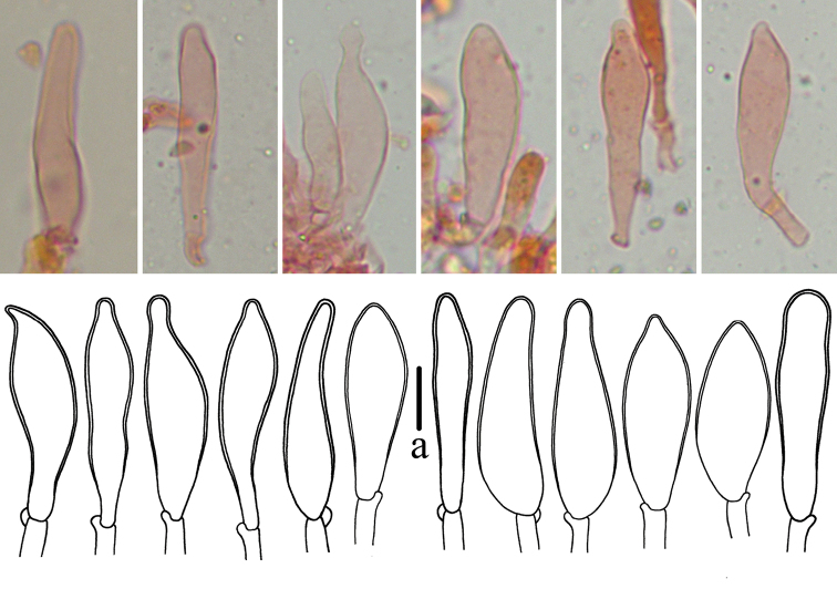
Figure 5.
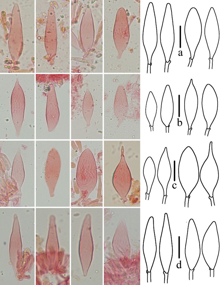
Pleurocystidia of MycenapolycystidiataaFFAAS0422bFFAAS0417, holotype cFFAAS0418dFFAAS0421. Scale bars: 25 μm (a–d).
Diagnosis.
Pileus greyish-rose, umbo brownish-orange, hygrophanous. Stipe pubescent. Pleurocystidia polymorphic in shape. Stipitipellis a cutis, with numerous projecting hyphae.
Holotype.
China. Heilongjiang Province: Liangshui National Nature Reserve, Yichun City, 47°12'74"N, 128°52'86"E, 20 August 2021, Zewei Liu, Yupeng Ge, Qin Na and Shixin Wang, FFAAS0417 (collection number MY0633).
Etymology.
Refers to the variable shape of pleurocystidia.
Description.
Pileus 14–31 mm in diam., campanulate to hemispherical when young, plano-convex with age, with obtuse umbo at centre, margin slightly revolute, at times cracked at mature; umbo brownish-orange (7C3–7C5), disc purplish-grey (13C2, 14B2, 14C2), reddish-grey (12B2, 12C2) to greyish-rose (12B3), near margin reddish-grey (12D2), greyish-ruby (12D3) or purplish-grey (13D3), margin whitish; striate none or indistinct, greyish-ruby (12E3–12E4), towards the centre up to 1/3 diam.; surface dry and rugose, hygrophanous, generally tomentose. Context white, 2 mm thick, fragile. Lamellae emarginate, slightly decurrent when old, 20–28 reaching the stipe, 1–2 tiers of lamellulae, white, irregularly intervenose, edge concolorous, wavy. Stipe 34–73 × 3–7 mm, central, cylindrical, base occasionally compressed with age; apex violet brown (11E3–11E4, 11F4), greyish-ruby (12E4), lower part brownish-grey (11D2, 11E2) to greyish-brown (11D3, 11E3) or purplish-grey (13C2), fragile, hollow; apex to middle densely pubescent, sparser towards base; whitely villose at base. Odour strongly raphanoid, taste indistinct.
Basidiospores (130/5/4) (6.4)6.7–7.4–8.3(8.8) × (3.2)3.5–3.9–4.3(4.6) μm [Q = (1.62)1.72–2.05(2.18), Q = 1.90 ± 0.11] [holotype (70/2/1) (6.7)6.9–7.6–8.5(8.7) × (3.4)3.6–4.0–4.4(4.6) μm, Q = (1.71)1.76–2.05(2.13), Q = 1.90 ± 0.09], elongated ellipsoid to cylindrical, colourless, smooth, thin-walled, amyloid. Basidia 21–31 × 6–8 μm, 4-spored, clavate, hyaline, sterigmata approximately 4 μm in length. Cheilocystidia thin-walled, hyaline, differs in two shapes, mainly utriform, 50–65 × 20–31 μm, some subclavate, 54–78 × 14–19 μm. Pleurocystidia abundant, thin-walled, hyaline, multi-shaped: lanceolate and mostly round to blunt apices, 37–81 × 12–20 μm, lanceolate and acute apices, 51–87 × 14–22 μm, elliptical, 30–86 × 12–31 μm, ovate and acute apices, 49–71 × 15–24 μm, ovate and mostly round to blunt apices, 49–73 × 16–22 μm. Pileipellis a cutis composed of four to five layers cylindrical cells, 51–81 × 4–5 μm, smooth and thin-walled; terminal cells cylindrical or fusiform, 50–69 × 3–22 μm, thin-walled, hyaline. Hypodermium formed by fusiform to subglobose hyphae, 32–69 × 18–54 μm, thin-walled, hyaline. Lamellar trama subregular, dextrinoid. Stipitipellis a cutis composed of cylindrical hyphae 5–8 μm in diam., smooth, thin-walled, with numbers of projecting hyphae 2–6 μm in diam.; caulocystidia 29–74 × 6–19 μm, clavate or fusiform, thin-walled, smooth, hyaline. Clamps present in all tissues.
Habitat.
Scattered on the litter layers in Pinuskoraiensis and Larixgmelinii mixed forests.
Known distribution.
Heilongjiang Province, China.
Additional material examined.
China. Heilongjiang Province: Liangshui National Nature Reserve, Yichun City, 47°12'82"N, 128°52'94"E, 20 August 2021, Zewei Liu, Yupeng Ge, Qin Na and Shixin Wang, FFAAS0418 (collection number MY0634); same location, 21 August 2021, Zewei Liu, Yupeng Ge, Qin Na and Shixin Wang, FFAAS0421 (collection number MY0659); same location, 21 August 2021, Zewei Liu, Yupeng Ge, Qin Na and Shixin Wang, FFAAS0422 (collection number MY0661).
Notes.
Macroscopically, Mycenaluteovariegata Harder & Læssøe and M.pura resemble M.polycystidiata in pileus colour, but the latter possesses more typically utriform cheilocystidia and uncontracted pleuro- and cheilocystidia (Perry 2002; Robich 2003; Harder et al. 2013; Aronsen and Læssøe 2016; Na 2019). Mycenapearsoniana also has a rose to violaceous pileus, but differs from M.polycystidiata in having inamyloid spores and lacking pleurocystidia (Aronsen and Læssøe 2016; Na 2019). Compared with M.polycystidiata, M.sirayuktha Aravind. & Manim. has similar cheilocystidia, but has an obviously greyish-brown striate pileus, inamyloid spores and slightly glutinous pileipellis with finger-like excrescences (Aravindakshan and Manimohan 2015).
The pleurocystidia of M.polycystidiata varied in shape amongst specimens (Fig. 5). In all four specimens, most pleurocystidia were lanceolate and with round to blunt apices, but pleurocystidia with lanceolate and acute apices, elliptical and ovate and acute apices were also observed in FFAAS0417 (Holotype) and FFAAS0418, while elongated lageniform-lanceolate or round apices ovate were detected in FFAAS0421 and FFAAS0422. The multi-shaped pleurocystidia may show a morphological continuum that changes between developmental stages. Nevertheless, the multi-shaped pleurocystidia are unquestionably diagnostic for identification of this species.
. Mycena rufobrunnea
Z.W. Liu, Y.P. Ge & Q. Na, sp. nov.
A5644585-F3C2-536C-B1B0-C0ADB1C6B3C4
MycoBank No: 843978
Figure 6.
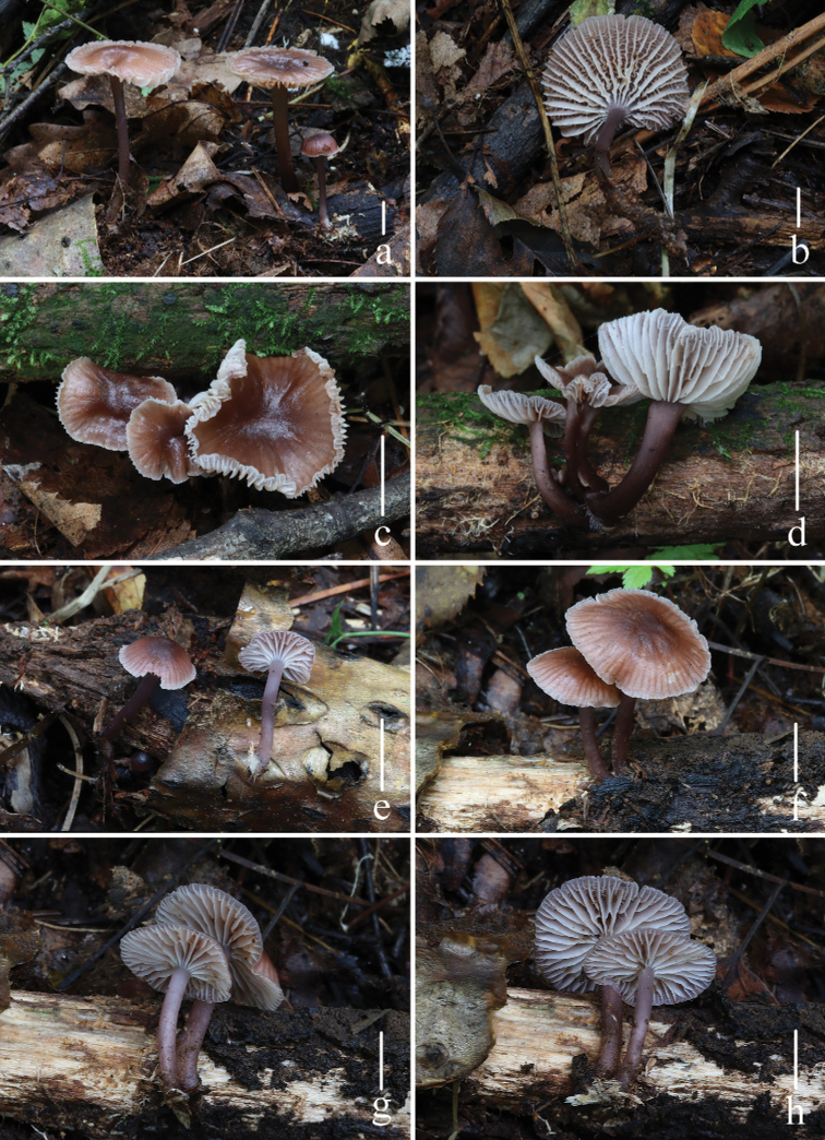
Basidiomata of Mycenarufobrunnea Z.W. Liu, Y.P. Ge & Q. Na a, bFFAAS0414c, dFFAAS0415e–hFFAAS0416, holotype. Scale bars: 10 mm (a–h). Photographs a–h by Qin Na.
Figure 7.
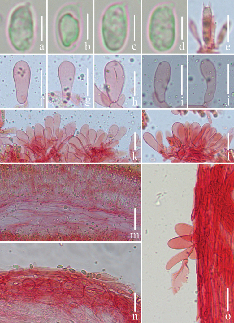
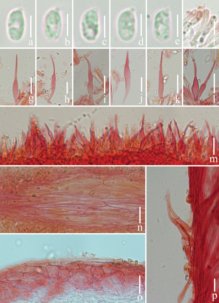
Microscopic features of Mycenarufobrunnea (FFAAS0416, holotype) a–d basidiospores e basidia f–l cheilocystidia m lamellar trama n pileipellis and hypodermiumo stipitipellis and caulocystidia. Scale bars: 5 μm (a–d); 20 μm (e–o).
Figure 8.
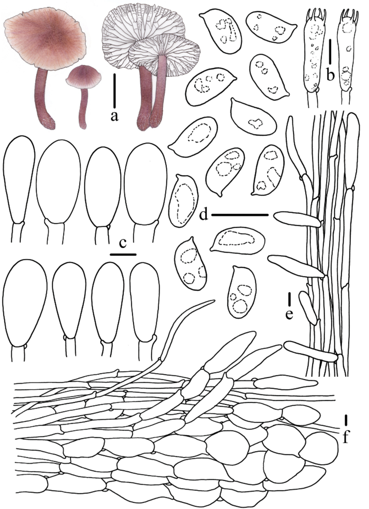
Morphological features of Mycenarufobrunnea (FFAAS0416, holotype) a basidiomata b basidia c cheilocystidia d basidiospores e stipitipellis and caulocystidia f pileipellis and hypodermium. Scale bars: 10 mm (a); 10 μm (b–f). Drawings by Zewei Liu.
Diagnosis.
Pileus dark brown at centre, disc gradually turning paler to reddish-brown to greyish-brown, edge white. Lamellae obviously intervenose. Stipe apex to middle greyish-magenta to dull violet, lower part darker to dark purple or dark magenta. Cheilocystidia utriform, sometimes clavate. Pleurocystidia absent. Caulocystidia clavate or fusiform. Pileipellis with fusiform terminal cells.
Holotype.
China. Jilin Province: Dayangcha, Erdaobaihe Town, Antu County, Yanbian Korean Autonomous Prefecture, 42°20'73"N, 127°56'06"E, 16 August 2021, Zewei Liu, Yupeng Ge, Qin Na and Shixin Wang, FFAAS0416 (collection number MY0581).
Etymology.
Refers to reddish-brown pileus.
Description.
Pileus 12–34 mm in diam., hemispherical to convex when young, then plano-convex, sometimes an unclear umbo at centre, margin slightly revolute, acute to subacute, at times cracked at mature; dark brown (8F6–8F8) at centre, disc gradually turning paler to reddish-brown (8D4–8D5, 8E6–8E8) to greyish-brown (8D3) and turning to whitish at margin; striate reddish-brown (8D4–8D5, 8E6–8E8), towards the centre up to 1/3–1/2 diam.; surface humidus when wet. Context white, 1.5 mm thick, fragile. Lamellae adnexed to emarginate, 20–23 reaching the stipe, 1–3 tiers of lamellulae, white, irregularly intervenose, edge concolorous, slightly serrulate. Stipe 19–62 × 2–6 mm, central, cylindrical, apex to middle greyish-magenta (14E4–14E5) to dull violet (16E3–16E4), lower part darker to dark purple (14F4–14F5) or dark magenta (13F3), fragile, hollow, base slightly swollen with whitish villose. Odour raphanoid, taste indistinct.
Basidiospores (80/4/3) (7.1)7.6–8.4–9.2(9.6) × (3.8)4.0–4.5–5.0 μm [Q = (1.73)1.77–1.98(2.05), Q = 1.88 ± 0.07] [holotype (40/2/1) (7.9)8.1–8.6–9.2(9.4) × 4.2–4.6–5.0 μm, Q = (1.73)1.77–1.96(1.98), Q = 1.87 ± 0.06], elongated ellipsoid to cylindrical, colourless, smooth, thin-walled, amyloid. Basidia 24–34 × 7–10 μm, 4-spored, clavate, hyaline, sterigmata 2–3 μm in length. Cheilocystidia thin-walled, hyaline, utriform, sometimes clavate, 23–44 × 7–17 μm, abundant. Pleurocystidia absent. Pileipellis a cutis composed of four to five slightly interwoven layers of cylindrical cells, 44–70 × 4–7 μm, smooth, thin-walled; terminal cells cylindrical or fusiform, 34–65 × 4–17 μm, thin-walled, hyaline. Hypodermium formed by fusiform, subcylindrical to subglobose hyphae, 15–50 × 12–37 μm, thin-walled, hyaline. Lamellar trama subregular, dextrinoid. Stipitipellis a cutis composed of hyphae 3–9 μm in diam., smooth, thin-walled; caulocystidia common in the apex, sparse in the middle and base, 23–76 × 6–14 μm, clavate and fusiform, thin-walled, hyaline, smooth. Clamps present in all tissues.
Habitat.
Scattered on the decayed logs of Acer, Larix, Pinus, Populus, Quercus and Ulmus mixed forests.
Known distribution.
Jilin Province, China.
Additional material examined.
China. Jilin Province: Dayangcha, Erdaobaihe Town, Antu County, Yanbian Korean Autonomous Prefecture, 42°20'72"N, 127°56'08"E, 16 August 2021, Zewei Liu, Yupeng Ge, Qin Na and Shixin Wang, FFAAS0414 (collection number MY0579); same location, 16 August 2021, Zewei Liu, Yupeng Ge, Qin Na and Shixin Wang, FFAAS0415 (collection number MY0580).
Notes.
Species of sect. Calodontes that are macroscopically similar to Mycenarufobrunnea have been recorded in many regions of the world. Most taxa resemble M.rufobrunnea in pileus colour (Smith 1947; Maas Geesteranus 1992a, 1992b; Grgurinovic 2003; Robich 2003; Chew et al. 2014; Aronsen and Læssøe 2016). Mycenadura Maas Geest. & Hauskn., recorded in Europe, also has a dark brown to greyish-brown pileus, but can be distinguished from M.rufobrunnea in having a white stipe and having pleurocystidia (Robich 2003; Aronsen and Læssøe 2016). Mycenakuehneriana A.H. Sm., which is recorded from the United States and Canada, is distinguished from M.rufobrunnea in that its pileus is pale avellaneous with rose and lilac, almost white when faded and the spores are obviously smaller (5–6 × 2–3 μm) (Smith 1947; Maas Geesteranus 1992a, 1992b). Mycenaclarkeana Grgur. and M.nullawarrensis Grgur., described from Australia, are similar to M.rufobrunnea in having a reddish-brown pileus, but both species have broader spores and possess pleurocystidia (Grgurinovic 2003). Mycenacahaya A.L.C. Chew & Desjardin, M.seminau A.L.C. Chew & Desjardin and M.sinar A.L.C. Chew & Desjardin, known from Malaysia, resemble M.rufobrunnea owing to the brown pileus, but differ in having adnate to subdecurrent lamellae, a yellowish-grey or brownish-orange stem, mucronate cheilocystidia and lack caulocystidia (Chew et al. 2014). Microscopically, utriform or clavate cheilocystidia and absence of pleurocystidia are key characteristics of M.rufobrunnea. Mycenadiosma Krieglst. & Schwöbel has similar cheilocystidia and pleurocystidia are absent or rare, but it has a strongly hygrophanous pileus and a remarkable change in colour (Robich 2003; Aronsen and Læssøe 2016). Mycenapura, M.sirayuktha and M.vinacea Cleland have similar cheilocystidia, but are easily distinguished from M.rufobrunnea by the presence of pleurocystidia (Perry 2002; Grgurinovic 2003; Robich 2003; Aravindakshan and Manimohan 2015; Aronsen and Læssøe 2016; Na 2019).
. Mycena shengshanensis
Z.W. Liu, Y.P. Ge & Q. Na, sp. nov.
53D69B3C-50BD-5DF4-8FBD-BF102F7EE427
MycoBank No: 843979
Figure 9.
Basidiomata of Mycenashengshanensis Z.W. Liu, Y.P. Ge & Q. Na a–dFFAAS0424, holotype e–hFFAAS0425 Scale bars: 10 mm (a–h). Photographs a–c, e–g by Yupeng Ge d, h by Zewei Liu.
Figure 10.
Microscopic features of Mycenashengshanensis (FFAAS0424, holotype) a–e basidiospores f basidia g–r cheilocystidia s lamellar trama t pileipellis and hypodermium u stipitipellis and caulocystidia. Scale bars: 5 μm (a–e); 20 μm (g–r); 40 μm (s–u).
Figure 11.
Morphological features of Mycenashengshanensis (FFAAS0424, holotype) a basidiomata b basidia c basidiospores d cheilocystidia e stipitipellis and caulocystidia f pileipellis and hypodermium. Scale bars: 10 mm (a); 10 μm (b–f). Drawings by Zewei Liu.
Figure 12.
Cheilocystidia of Mycenashengshanensis FFAAS0424, holotype. Scale bars: 10 μm (a).
Diagnosis.
Pileus brown to violet-brown at centre, disc light brown to dull red. Cheilocystidia clavate with slightly inflated apex, thick-walled. Pleurocystidia absent. Caulocystidia clavate with tapered apices, apex to middle thick-walled. Scattered to gregarious under Larixgmelinii.
Holotype.
China. Heilongjiang Province: Shengshan National Nature Reserve, Heihe City, 49°37'45"N, 126°47'39"E, 23 August 2021, Zewei Liu, Yupeng Ge, Qin Na and Shixin Wang, FFAAS0424 (collection number MY0686).
Etymology.
Refers to the type locality.
Description.
Pileus 13–26 mm in diam., when young parabolic to convex, with obtuse umbo at centre, then plano-convex, margin wavy and revolute, at times cracked at mature; centre light brown (7D5–7D6), brown (7E4–7E8), dark brown (8F5–8F6), violet brown (11F4–11F6), disc paler to light brown (7D4–7D5), brown (7E5–7E6), greyish-brown (8D3), reddish-brown (8D4, 8E4), brownish-grey (11C2), dull red (11C3), margin whitish; striate indistinct, brownish-orange (7C3), greyish-brown (7D3), reddish-grey (12D2), greyish-ruby (12E3–12E5), towards the centre up to 1/3–1/2 diam.; surface slightly moist, smooth. Context white, 1–2 mm thick, fragile. Lamellae sinuate to subdecurrent, 19–25 reaching the stipe, 1–3 tiers of lamellulae, white, irregularly intervenose, edge concolorous, wavy and slightly serrulate. Stipe 26–42 × 2–4 mm, central, cylindrical, apex reddish-brown (8E4–8E5), greyish-ruby (12E4–12E5), greyish-brown (11F3), violet brown (11F4–11F5), dark ruby (12F4–12F5), lower part paler to brownish-grey (7C2), brownish-orange (7C3), greyish-brown (11E3), greyish-ruby (12E3), base darker to brown (7E5), reddish-brown (8E4–8E5), greyish-brown (11F3), violet brown (11F4–11F5), greyish-ruby (12E3), dark ruby (12F6–12F8), fragile, hollow, base swollen with white fibrils. Odour raphanoid, taste indistinct.
Basidiospores (60/3/2) (5.9)6.1–6.9–8.1(8.7) × 3.4–4.0–4.4(4.7) μm [Q = 1.62–1.93 (1.97), Q = 1.75 ± 0.09] [holotype (40/2/1) 6.1–7.0–8.2(8.7) × 3.4–4.0–4.4(4.7) μm, Q = (1.62)1.65–1.93(1.97), Q = 1.77 ± 0.08], elongated ellipsoid, colourless, smooth, thin-walled, amyloid. Basidia 22–32 × 6–8 μm, 4-spored, clavate, hyaline, sterigmata 3–4 μm in length. Cheilocystidia moderately thick-walled (0.5–0.6 μm), clavate with slightly inflated apex, 25–63 × 6–12 μm, hyaline. Pleurocystidia absent. Pileipellis a cutis composed of three to four layers of cylindrical cells, 24–57 × 3–5 μm, smooth and thin-walled; terminal cells cylindrical, apically narrow, 28–49 μm in length, apex 1–3 μm and base 2–5 μm in diam., thin-walled, hyaline. Hypodermium formed by fusiform to subglobose hyphae, 19–53 × 13–30 μm, thin-walled, hyaline. Lamellar trama subregular, weakly dextrinoid to dextrinoid. Stipitipellis a cutis composed of hyphae 4–9 μm in diam., smooth, thin-walled; caulocystidia 22–61 × 5–20 μm, clavate with tapered apices, apex to middle thick-walled, smooth, hyaline. Clamps present in all tissues.
Habitat.
Scattered to gregarious on the litter layer in Larixgmelinii.
Known distribution.
Heilongjiang Province, China.
Additional material examined.
China. Heilongjiang Province: Shengshan National Nature Reserve, Heihe City, 23 August 2021, Zewei Liu, Yupeng Ge, Qin Na and Shixin Wang, FFAAS0425 (collection number MY0687).
Notes.
In sect. Calodontes, M.diosma, M.pearsoniana and M.yuezhuoi also have clavate cheilocystidia with a slightly inflated apex and lack pleurocystidia, similar to M.shengshanensis, but M.diosma differs in pileus characters, M.pearsoniana has inamyloid spores and M.yuezhuoi has a more purple pileus and subcellular lamellar trama (Robich 2003; Aronsen and Læssøe 2016; Na 2019; Liu et al. 2021). Clavate cheilocystidia are also present in M.luteovariegata and M.pura, but these species differ in having pleurocystidia (Perry 2002; Robich 2003; Harder et al. 2013; Aronsen and Læssøe 2016; Na 2019). Macroscopically, several species in sect. Calodontes also have a brown with reddish or violet pileus or stipe (Smith 1947; Maas Geesteranus 1992a, 1992b; Grgurinovic 2003; Robich 2003; Aronsen and Læssøe 2016). Mycenadura, M.kuehneriana and M.nullawarrensis are distinguished by basidiospore size (Smith 1947; Maas Geesteranus 1992a, 1992b; Grgurinovic 2003; Robich 2003; Aronsen and Læssøe 2016). Two species, M.seminau and M.sirayuktha, reported from Southeast Asia, are similar to M.shengshanensis owing to the brown pileus, but they differ in having gelatinised or sometimes mucronate cheilocystidia and caulocystidia have not been observed (Chew et al. 2014; Aravindakshan and Manimohan 2015). Fusiform, obclavate, ovate and clavate cheilocystidia with a subcapitate protuberance were observed occasionally, but clavate cheilocystidia with a slightly inflated apex represented the predominant morphological type in M.shengshanensis (Fig. 12).
. Mycena subulata
Z.W. Liu, Y.P. Ge & Q. Na, sp. nov.
E08A7A12-E21E-5CD0-B4D9-2B1C693B32FC
MycoBank No: 843980
Figure 13.
Basidiomata of Mycenasubulata Z.W. Liu, Y.P. Ge & Q. Na a, bFFAAS0419c, dFFAAS0420e–gFFAAS0423, holotype hFFAAS0426 Scale bars: 10 mm (a–h). Photographs a, b, e, f by Yupeng Ge c, d by Qin Na g by Zewei Liu h by Shixin Wang.
Figure 14.
Microscopic features of Mycenasubulata (FFAAS0423, holotype) a–e basidiospores f basidia g–m cheilocystidia n lamellar trama o pileipellis and hypodermium p stipitipellis and caulocystidia. Scale bars: 5 μm (a–e); 10 μm (f); 20 μm (g–p).
Figure 15.
Morphological features of Mycenasubulata (FFAAS0423, holotype) a basidiomata b basidia c basidiospores d cheilocystidia e stipitipellis and caulocystidia f pileipellis and hypodermium. Scale bars: 10 mm (a); 5 μm (c); 10 μm (b, d–f). Drawings by Zewei Liu.
Diagnosis.
Pileus reddish-grey to dull red, slightly hygrophanous. Cheilocystidia thick-walled, slenderly fusiform with distinctly long and narrow protuberance. Stipitipellis a cutis, with projecting hyphae, caulocystidia thick-walled.
Holotype.
China. Heilongjiang Province: Liangshui National Nature Reserve, Yichun City, 47°13'13"N, 128°53'21"E, 21 August 2021, Zewei Liu, Yupeng Ge, Qin Na and Shixin Wang, FFAAS0423 (collection number MY0671).
Etymology.
Refers to cheilocystidia with distinctly long and narrow protuberance.
Description.
Pileus 9–32 mm in diam., convex to campanulate when young, hemispherical to applanate with age, margin sometimes wavy, slightly deflexed; at centre dull red (8C3), brownish-grey (8D2), greyish-brown (8D3), reddish-brown (8D4, 8E4–8E5) and dark brown (8F5), disc paler to reddish-grey (8B2, 9B2), brownish-grey (9C2), dull red (9B3, 9C3), greyish-magenta (13D3), margin light brown (7D4), brown (7E4) or dull red (9C3); striate none or indistinct, reddish-brown (8E4–8E5), towards the centre up to 1/5 diam.; surface dry, unclearly rugose or none, margin slightly hygrophanous. Context white, 1 mm thick, fragile. Lamellae sinuate to subdecurrent, 31–33 reaching the stipe, 1–3 tiers of lamellulae, white, irregularly intervenose, edge concolorous, wavy and slightly serrulate. Stipe 27–75 × 2–5 mm, central, cylindrical, apex to middle brownish-orange (7C3), dull red (8C3), brownish-grey (7D2), greyish-magenta (14D3), lower part brownish-grey (8C2), greyish-brown (8D3), reddish-brown (8D4), fragile, hollow, white granular near apex, base slightly swollen with white fibrils. Odour raphanoid, taste indistinct.
Basidiospores (100/5/4) 6.0–6.7–7.3(7.9) × 3.3–3.8–4.3(4.6) μm [Q = (1.61)1.65–1.87(1.90), Q = 1.76 ± 0.07] [holotype (40/2/1) (6.0)6.2–6.6–7.1 × 3.4–3.7–4.0(4.2) μm, Q = 1.65–1.87(1.90), Q = 1.78 ± 0.07], elongated ellipsoid, colourless, smooth, thin-walled, amyloid. Basidia 23–34 × 5–6 μm, 4-spored, clavate, hyaline, sterigmata 2–3 μm in length. Cheilocystidia moderately thick-walled (0.5–0.6 μm), hyaline, narrowly fusiform with long and narrow protuberance, 43–82 × 4–11 μm, protuberance 14–36 × 1–2 μm. Pleurocystidia absent. Pileipellis a cutis composed of three to four layers of cylindrical cells, 20–89 × 3–7 μm, smooth and thin-walled; terminal cells cylindrical, apically narrow, 24–61 μm in length, the apex 3–4 μm and base 4–7 μm in diam., thin-walled, hyaline. Hypodermium formed by fusiform to subglobose hyphae, 21–41 × 17–25 μm, thin-walled, hyaline. Lamellar trama subregular, dextrinoid. Stipitipellis a cutis composed of hyphae 3–8 μm in diam., smooth, thin-walled, with projecting hyphae 3–8 μm in diam.; caulocystidia 29–47 × 6–12 μm, clavate and apices tapered, thick-walled (0.5–0.6 μm), smooth. Clamps present in all tissues.
Habitat.
Scattered on the litter layer in Pinuskoraiensis, Larixgmelinii and Tilia sp. mixed forests.
Known distribution.
Heilongjiang Province, China.
Additional material examined.
China. Heilongjiang Province: Liangshui National Nature Reserve, Yichun City, 21 August 2021, Zewei Liu, Yupeng Ge, Qin Na and Shixin Wang, FFAAS0419 (collection number MY0654); same location, 21 August 2021, Zewei Liu, Yupeng Ge, Qin Na and Shixin Wang, FFAAS0420 (collection number MY0657); Heilongjiang Province: Taipinggou National Nature Reserve, Hegang City, 3 September 2021, Shixin Wang, FFAAS0426 (collection number MY0795).
Notes.
Cheilocystidia with a long and narrow protuberance is the key microscopic character that distinguishes M.subulata and is uncommon in sect. Calodontes (Smith 1947; Maas Geesteranus 1992a, 1992b; Grgurinovic 2003; Robich 2003; Chew et al. 2014; Aravindakshan and Manimohan 2015; Aronsen and Læssøe 2016; Na 2019; Liu et al. 2021). Mycenalammiensis Harmaja and M.pelianthina (Fr.) Quél. have similar cheilocystidia, but differ from M.subulata by their broader cheilocystidia with purplish-brown contents and having pleurocystidia (Smith 1947; Robich 2003; Aronsen and Læssøe 2016). The cheilocystidia of M.subcorticalis (Cooke & Massee) Sacc. with a protuberance are similar to those of M.subulata. However, M.subcorticalis has larger and inamyloid spores, a gelatinised pileipellis and a stipitipellis with sparse excrescences (Grgurinovic 2003). More rarely, mucronate cheilocystidia and absence of pleurocystidia have been described for M.pearsoniana and its clay pink pileus is similar to that of M.subulata, but M.pearsoniana differs in having a slightly glutinous pileus when wet and inamyloid spores (Aronsen and Læssøe 2016; Na 2019). Other species that are macroscopically similar to M.subulata, namely M.luteovariegata, M.nullawarrensis and M.pura, can be distinguished by cheilocystidia shape and presence of pleurocystidia (Perry 2002; Robich 2003; Grgurinovic 2003; Harder et al. 2013; Aronsen and Læssøe 2016; Na 2019).
Key to species of sect. Calodontes known worldwide
| 1 | Stipe white | 2 |
| – | Stipe coloured | 4 |
| 2 | Pileus white | Mycenasubaquosa |
| – | Pileus coloure | 3 |
| 3 | Pileus pink and lamellae emarginate, pileipellis without inflated terminal cells | Mycenarosea |
| – | Pileus brown and lamellae adnate, pileipellis with fusiform, subcylindrical to lageniform terminal cells | Mycenadura |
| 4 | Lamellae edge with coloured dots | 5 |
| – | Lamellae edge white or without dots | 6 |
| 5 | Caulocystidia present, spores almost broader than 4 μm | Mycenalammiensis |
| – | Caulocystidia absent, spores almost narrower than 4 μm | Mycenapelianthina |
| 6 | Basidiospores inamyloid | 7 |
| – | Basidiospores amyloid | 10 |
| 7 | Pleurocystidia absent | 8 |
| – | Pleurocystidia present | 9 |
| 8 | Stipitipellis and caulocystidia smooth | Mycenapearsoniana |
| – | Stipitipellis and caulocystidia with nodulose excrescences | Mycenasubcorticalis |
| 9 | Pileipellis gelatinised, caulocystidia absent, cheilo- and pleurocytsidia base uncontracted, disc greyish-red or orange white in pileus | Mycenasirayuktha |
| – | Pileipellis not gelatinised, caulocystidia present, cheilo- and pleurocytsidia base contracted, disc wood brown or reddish-brown in pileus | Mycenavinacea |
| 10 | Pleurocystidia present | 11 |
| – | Pleurocystidia absent | 16 |
| 11 | Caulocystidia absent, almost cheilocystidia apically mucronate or subcapitate | Mycenacahaya |
| – | Caulocystidia present, almost cheilocystidia apically broadly rounded | 12 |
| 12 | Caulocystidia with apical excrescences, spores more than 5.6 μm width | Mycenaclarkeana |
| – | Caulocystidia without apical excrescences, spores less than 5.6 μm width | 13 |
| 13 | Cheilocystidia base uncontracted | Mycenapolycystidiata |
| – | Cheilocystidia base contracted | 14 |
| 14 | Stipe brown to dark brown, Qav = 1.5 | Mycenanullawarrensis |
| – | Stipe not brown to dark brown, Qav > 1.5 | 15 |
| 15 | Pileus sulphur yellow to reddish-grey, stipe reddish-grey | Mycenaluteovariegata |
| – | Pileus generally pinkish or purplish, stipe whitish to pinkish-purple | Mycenapura |
| 16 | Caulocystidia absent | 17 |
| – | Caulocystidia present | 18 |
| 17 | Pileus brown to dark brown, spores weakly amyloid | Mycenaseminau |
| – | Pileus brownish-orange to greyish-yellow, spores amyloid | Mycenasinar |
| 18 | Cheilocystidia slender fusiform, with distinctly long and narrow protuberance | Mycenasubulata |
| – | Cheilocystidia clavate, utriform, subfusiform, or subcylindrical, with short mucronate or none | 19 |
| 19 | Spores less than 6 μm length | Mycenakuehneriana |
| – | Spores more than 6 μm length | 20 |
| 20 | Pileus more than 35 mm in diam., lamellae dark brownish-violet to reddish-violet | Mycenadiosma |
| – | Pileus less than 35 mm in diam., lamellae white | 21 |
| 21 | Lamellar trama subcellular, pileus lilac to purple | Mycenayuezhuoi |
| – | Lamellar trama subregular, pileus brownish | 22 |
| 22 | Cheilocystidia utriform, sometimes clavate, thin-walled, lamellae adnexed to emarginate | Mycenarufobrunnea |
| – | Cheilocystidia clavate with slightly inflated apex, thick-walled, lamellae sinuate to subdecurrent | Mycenashengshanensis |
Discussion
Maas Geesteranus (1980) proposed that Mycenasect.Calodontes could be divided into three subsections based on the colour of the lamellar edge and the amyloid reaction of the basidiospores. Subsequently, taxonomists have followed this division, but opinions have differed on the diagnostic characters that support this classification (Grgurinovic 2003; Robich 2003; Harder et al. 2010; Chew et al. 2014). Some taxonomists classified the subsections according to the amyloid reaction of basidiospores, cheilocystidia and pleurocystidia contents and presence or absence of pleurocystidia, but the shapes of cheilocystidia and pleurocystidia were not considered (Grgurinovic 2003; Harder et al. 2010; Chew et al. 2014). Robich (2003) also did not consider the shapes of cheilocystidia and pleurocystidia, but the colour of the lamellar edge and cheilocystidia contents were emphasised to distinguish subsections. According to the historical infrasectional classification of sect. Calodontes, M.polycystidiata could be classified in subsect. Purae, whereas M.rufobrunnea, M.shengshanensis and M.subulata cannot be assigned to a subsection owing to their having amyloid spores and lacking pleurocystidia (Maas Geesteranus 1980; Harder et al. 2010).
Phylogenetic reconstructions do not fully support recognition of three subsections defined by morphological characters; notably, subsect. Violacellae and subsect. Purae are polyphyletic in the phylogenies (Harder et al. 2010, 2012, 2013). Chew et al. (2014) supported the views of Harder et al. (2010, 2012) and the new taxa proposed by the former authors were not assigned to a subsection. Additionally, subsect. Purae was proved to be polyphyletic in our combined analysis of ITS, rpb1 and tef1 dataset, which also supported analysis, based on single gene region (Harder et al. 2013).
The five taxa of Mycenasect.Calodontes recorded from China show obvious differences in pileus colour and in the shapes of cheilocystidia and pleurocystidia (if present) (Liu et al. 2021). The colour of the pileus includes greyish-rose, reddish-grey, purple, reddish-brown and violet-brown and most show a gradual transition with age. Clavate, obclavate, utriform and fusiform cheilocystidia with a long, narrow protuberance are observed, but pleurocystidia are present only in M.polycystidiata. Forms and variations within M.pura complex had a wide range of pileus colour, but the shape of cheilocystidia was highly similar and could be clearly distinguished from the four new taxa (Robich 2003).
In our phylogenetic analysis, four new species all formed separate clades with high support and had obvious genetic distance from other species in sect. Calodontes. Mycenarufobrunnea is more closely related to the phylogenetic species within M.pura complex by Harder et al. (2013). While the other three new species are significantly more distant from M.pura complex genetically, M.shengshanensis and M.subulata, formed a sister relationship with high support from M.pearsoniana; M.polycystidiata clustered with M.diosma, but is poorly supported.
Based on extensive field work in China, most specimens of sect. Calodontes have been observed in coniferous forests or mixed coniferous-broadleaved forests in early autumn (Na 2019; Liu et al. 2021). Specimens of the four new taxa described in the present study were collected from Changbai Mountain and the Lesser Khinggan Mountains in northeast China from mixed broadleaf-Korean pine (Pinuskoraiensis) forests (Zhao et al. 2004; Wang and Guo 2016). In particular, M.polycystidiata and M.subulata were both distributed in the Liangshui National Nature Reserve on the Lesser Khinggan Mountains, where the dominant forest species is P.koraiensis, mixed with fewer Betula, Tilia, Quercus and Picea individuals (She et al. 2022). Moreover, more specimens were located in the northern region of China with an average temperature not more than 20 °C in August. For example, the average temperature is 16.4 °C in Liangshui National Nature Reserve and 16.3 °C in Shengshan National Nature Reserve (Liu 2017). Therefore, we speculate that members of this section in China prefer the climate types Dwa, Dwb and Dwc according to the Köppen climate classification (Kottek et al. 2006; Wang et al. 2020).
Supplementary Material
Acknowledgements
This study was supported by the Natural Science Foundation of Shandong Province (grant No. ZR2020QC001), the National Natural Science Foundation of China (grant No. 3190012), the Natural Science Foundation of Shandong Province (grant No. ZR2019PC028) and the Shandong Agricultural Industry Technology System (grant No. SDAIT-07-03). We sincerely thank Prof. Li Zou and Mr. Shixin Wang (Northeast Forestry University, Harbin, Heilongjiang Province), Mr. Jingyun Song and Mr. Yong Li (Yanhexiang Township Government, Sunwu County, Heilongjiang Province), Ms Yaping Hu (Nanjing Institute of Environmental Sciences, Ministry of Ecology and Environment, Nanjing, Jiangsu Province) and Mr. Bai Wang (Changbai Mountain National Nature Reserve, Jilin Province) for their help during fieldwork.
Citation
Liu Z, Ge Y, Zeng H, Cheng X, Na Q (2022) Four new species of Mycena sect. Calodontes (Agaricales, Mycenaceae) from northeast China. MycoKeys 93: 23–56. https://doi.org/10.3897/mycokeys.93.86580
Funding Statement
Natural Science Foundation of Shandong Province (grant No. ZR2020QC001), National Natural Science Foundation of China (grant No. 3190012), Natural Science Foundation of Shandong Province (grant No. ZR2019PC028), Shandong Agricultural Industry Technology System (grant No. SDAIT-07-03).
References
- Aravindakshan DM, Manimohan P. (2015) Mycenas of Kerala. PhD Thesis, University of Calicut, Kerala, India. 10.13140/RG.2.1.2116.4003 [DOI]
- Aronsen A, Læssøe T. (2016) The Genus Mycena s.l. Fungi of Northern Europe Vol. 5. Narayana Press, Gylling, Denmark, 373 pp. [Google Scholar]
- Berkeley MJ. (1836) Fungi. In: Smith JE (Eds) The English flora. Longman, Hurst, Rees, Orme, Brown, and Green, London, 5(2): 43. 10.5962/bhl.title.6340 [DOI] [Google Scholar]
- Chew AL, Tan YS, Desjardin DE, Musa MY, Sabaratnam V. (2014) Four new bioluminescent taxa of Mycenasect.Calodontes from Peninsular Malaysia. Mycologia 106(5): 976–988. 10.3852/13-274 [DOI] [PubMed] [Google Scholar]
- Clémençon H, Emmett V, Emmett EE. (2004) Cytology and Plectology of the Hymenomycetes. Bibliotheca Mycologica, Vol. 199. J. Cramer, Berlin, Stuttgart, 488 pp. [Google Scholar]
- Edler D, Klein J, Antonelli A, Silvestro D. (2020) RaxmlGUI 2.0: A graphical interface and toolkit for phylogenetic analyses using RAxML. Methods in Ecology and Evolution 12(2): 1–5. 10.1111/2041-210X.13512 [DOI] [Google Scholar]
- Fries EM. (1821) Systema Mycologicum Vol. l. Lundæ, ex officina Berlingiana, Sweden, 520 pp. [Google Scholar]
- Ge YP, Liu ZW, Zeng H, Cheng XH, Na Q. (2021) Updated description of Atheniella (Mycenaceae, Agaricales), including three new species with brightly coloured pilei from Yunnan Province, southwest China. MycoKeys 81: 139–164. 10.3897/mycokeys.81.67773 [DOI] [PMC free article] [PubMed] [Google Scholar]
- Grgurinovic CA. (2003) The Genus Mycena in south-eastern Australia. Fungal Diversity Press, 329 pp.
- Hall TA. (1999) BioEdit: A user-friendly biological sequence alignment editor and analysis program for Windows 95/98/NT. Nucleic Acids Symposium Series 41: 95–98. [Google Scholar]
- Harder CB, Læssøe T, Kjøller R, Frøslev TG. (2010) A comparison between ITS phylogenetic relationships and morphological species recognition within Mycenasect.Calodontes in Northern Europe. Mycological Progress 9(3): 395–405. 10.1007/s11557-009-0648-7 [DOI] [Google Scholar]
- Harder CB, Lodge DJ, Petersen RH, Hughes KW, Blanco JC, Frøslev TG, Læssøe T. (2012) Amyloidity is not diagnostic for species in the Mycenapearsoniana complex (MycenasectionCalodontes). Mycological Progress 11(3): 725–732. 10.1007/s11557-011-0782-x [DOI] [Google Scholar]
- Harder CB, Læssøe T, Frøslev TG, Ekelund F, Rosendahl S, Kjøller R. (2013) A three-gene phylogeny of the Mycenapura complex reveals 11 phylogenetic species and shows ITS to be unreliable for species identification. Fungal Biology 117(11–12): 764–775. 10.1016/j.funbio.2013.09.004 [DOI] [PubMed] [Google Scholar]
- Horak E. (2005) Röhrlinge und Blätterpilze in Europa: Bestimmungsschlüssel für Polyporales, Boletales, Agaricales, Russulales. Elsevier, Spektrum Akad. Verlag, 555 pp. [Google Scholar]
- Katoh K, Rozewicki J, Yamada KD. (2019) MAFFT online service: Multiple sequence alignment, interactive sequence choice and visualization. Briefings in Bioinformatics 20(4): 1160–1166. 10.1093/bib/bbx108 [DOI] [PMC free article] [PubMed] [Google Scholar]
- Kornerup A, Wanscher JH. (1978) Methuen handbook of colour. Eyre Methuen, London, 252 pp. [Google Scholar]
- Kottek M, Grieser J, Beck C, Rudolf B, Rubel F. (2006) World Map of the Koppen-Geiger Climate Classification Updated. Meteorologische Zeitschrift (Berlin) 15(3): 259–263. 10.1127/0941-2948/2006/0130 [DOI] [Google Scholar]
- Kuraku S, Zmasek CM, Nishimura O, Katoh K. (2013) aLeaves facilitates on-demand exploration of metazoan gene family trees on MAFFT sequence alignment server with enhanced interactivity. Nucleic Acids Research 41(W1): W22–W28. 10.1093/nar/gkt389 [DOI] [PMC free article] [PubMed]
- Li Y, Li TH, Yang ZL, Bau T, Dai YC. (2015) Atlas of Chinese Macrofungal Resources. Central Chinese Farmer Press, Zhengzhou, China, 1351 pp. [Google Scholar]
- Liu M. (2017) Research of dynamic changes and responses of Korean pine growth under climate change in northeastern China. PhD Thesis, Northeast Forestry University, Heilongjiang, China.
- Liu ZW, Na Q, Cheng XH, Wu XM, Ge YP. (2021) Mycenayuezhuoi sp. nov. (Mycenaceae, Agaricales), a purple species from the peninsula areas of China. Phytotaxa 511(2): 148–162. 10.11646/phytotaxa.511.2.3 [DOI] [Google Scholar]
- Maas Geesteranus RA. (1980) Studies in Mycenas-15, A tentative subdivision of the genus Mycena in the Northern Hemisphere. Persoonia 11: 93–120. [Google Scholar]
- Maas Geesteranus RA. (1992a) Mycenas of the Northern Hemisphere I. Studies in Mycenas and other papers. Proceedings van de Koninklijke Nederlandse Akademie van Wetenschappen, Amsterdam, North-Holland.
- Maas Geesteranus RA. (1992b) Mycenas of the Northern Hemisphere II. Conspectus of the Mycenas of the Northern Hemisphere. Proceedings van de Koninklijke Nederlandse Akademie van Wetenschappen, Amsterdam, North-Holland.
- Na Q. (2019) Taxonomy and Molecular Phylogeny of Mycena in China. PhD Thesis, Jilin Agriculture University, Jilin, China. 10.27163/d.cnki.gjlnu.2019.000016 [DOI]
- Na Q, Bau T. (2019) Recognition of Mycenasect.Amparoina sect. nov. (Mycenaceae, Agaricales) including four new species and revision of the limits of sect. Sacchariferae. MycoKeys 52: 103–124. 10.3897/mycokeys.52.34647 [DOI] [PMC free article] [PubMed] [Google Scholar]
- Na Q, Hu YP, Liu ZW, Zeng H, Qi LL, Ding H, Cheng XH, Ge YP. (2021) The first reported occurrence of Leucoinocybe (Porotheleaceae, Agaricales) in China: Leucoinocybelishuiensis sp. nov. from Zhejiang Province. Nova Hedwigia 113(3–4): 453–469. 10.1127/nova_hedwigia/2021/0661 [DOI] [Google Scholar]
- Na Q, Hu YP, Zeng H, Song ZZ, Ding H, Cheng XH, Ge YP. (2022) Updated taxonomy on Gerronema (Porotheleaceae, Agaricales) with three new taxa and one new record from China. MycoKeys 89: 87–120. 10.3897/mycokeys.89.79864 [DOI] [PMC free article] [PubMed] [Google Scholar]
- Osmundson TW, Robert VA, Schoch CL, Baker LJ, Smith A, Robich G, Mizzan L, Garbelotto MM. (2013) Filling Gaps in Biodiversity Knowledge for Macrofungi: Contributions and Assessment of an Herbarium Collection DNA Barcode Sequencing Project. PLoS ONE 8(4): e62419. 10.1371/journal.pone.0062419 [DOI] [PMC free article] [PubMed]
- Perry BA. (2002) A taxonomic investigation of Mycena in California. PhD Thesis, San Francisco State University, San Francisco.
- Posada D, Crandall KA. (1998) Modeltest: Testing the model of DNA substitution. Bioinformatics 14(9): 817–818. 10.1093/bioinformatics/14.9.817 [DOI] [PubMed] [Google Scholar]
- Quélet L. (1872) Les champignons de Jura et des Vosges (Vol. 1). Imprimerie et Lithographie de Henri Barbier, 332 pp.
- Robich G. (2003) Mycena D’Europa. Associazione Micologica Bresadola, Fondazione Centro Studi Micologici, Trento, Vicenza, 728 pp. [Google Scholar]
- Ronquist F, Huelsenbeck JP. (2003) MrBayes 3: Bayesian phylogenetic inference under mixed models. Bioinformatics (Oxford, England) 19(12): 1572–1574. 10.1093/bioinformatics/btg180 [DOI] [PubMed] [Google Scholar]
- She DQ, Zhang XT, Xiao L, Zhong ZL, Wang HM, Wang WJ. (2022) Plant beta diversity and its influence factors in the Liangshui National Nature Reserve in the central region of the Xiaoxing’an Mountains. Shengwu Duoyangxing 30(03): 15–26. 10.17520/biods.2021274 [DOI] [Google Scholar]
- Smith AH. (1947) North American species of Mycena. Unversity of Michigan Press, Ann Arbor, Michigan, 687 pp. [Google Scholar]
- Wang RZ, Guo QX. (2016) Woody plants species-area relationships in a broad-leaved Korean pine forest in the Xiaoxing’an Mountains. Acta Ecologica Sinica 36(13): 4091–4098. 10.5846/stxb201410282107 [DOI] [Google Scholar]
- Wang T, Zhou DW, Shen XJ, Fan GH, Zhang H. (2020) Köppen’s climate classification map for China. Journal of the Meteorological Sciences 40(6): 752–760. 10.3969/2019jms.0040 [DOI] [Google Scholar]
- White TJ, Bruns T, Lee S, Taylor J. (1990) Amplification and direct sequencing of fungal ribosomal RNA genes for phylogenetics. In: Innis MA, Gelfand DH, Sninsky JJ, White TJ. (Eds) PCR Protocols: a Guide to Methods and Applications.Academic Press, New York, 315–322. 10.1016/B978-0-12-372180-8.50042-1 [DOI]
- Yu WJ, Chang C, Qin LW, Zeng NK, Wang SX, Fan YG. (2020) Pseudospermacitrinostipes (Inocybaceae), a new species associated with Keteleeria from southwestern China. Phytotaxa 450(1): 8–16. 10.11646/phytotaxa.450.1.2 [DOI] [Google Scholar]
- Zhao SQ, Fang JY, Zong ZJ, Zhu B, Shen MH. (2004) Composition, structure and species diversity of plant communities along an altitudinal gradient on the northern slope of Mt. Changbai, Northeast China. Shengwu Duoyangxing 12(1): 164–173. 10.17520/biods.2004020 [DOI] [Google Scholar]
Associated Data
This section collects any data citations, data availability statements, or supplementary materials included in this article.


