PLOS ONE logoLink to PLOS ONE
. 2023 Jan 19;18(1):e0280412. doi: 10.1371/journal.pone.0280412

Identification and characterization of probiotics isolated from indigenous chicken (Gallus domesticus) of Nepal

Mohan Gupta 1,#, Roji Raut 2,#, Sulochana Manandhar 2, Ashok Chaudhary 2, Ujwal Shrestha 1, Saubhagya Dangol 1, Sudarshan G C 1,3, Keshab Raj Budha 4, Gaurab Karki 5,6, Sandra Díaz-Sánchez 7, Christian Gortazar 7, José de la Fuente 7,8, Pragun Rajbhandari 2, Prajwol Manandhar 2, Rajindra Napit 2,9, Dibesh Karmacharya 2,9,10,*
Editor: Xiaolun Sun11
PMCID: PMC9851537  PMID: 36656809

Abstract

Background

Excessive and irrational use of antibiotics as growth promoters in poultry has been one of key factors contributing to increased emergence of antibiotics resistant bacteria. Several alternatives for antibiotic growth promoters are being sought, and the search for effective probiotics to be used as feed additives is amongst the promising ones. Our study aimed to isolate and test potential probiotics bacteria from cloacal swabs of various indigenous chicken (Gallus domesticus) breeds from rural outskirts of the Kathmandu valley (Nepal).

Methods

Selective isolation of probiotics was conducted by micro-aerophilic enrichment of sample in MRS Broth at 37°C, followed by culturing on MRS agar supplemented with 5 g/L of CaCO3. Isolated bacterial colonies producing transparent halo were selected as potential lactic acid bacteria (LAB), and tested for their antibacterial activity, phenotypic and biochemical characteristics, acidic yield, and tolerance to acid and bile.

Results

A total of 90 potential LAB were isolated from cloacal samples collected from 41 free-ranging chickens of indigenous breeds. Of these, 52 LAB isolates (57%) showed variable antibacterial activity to at least one bacterial pathogen. Of 52 LAB, 46 isolates fulfilled phenotypic and biochemical criteria of Lactobacillus spp. Of these, 37 isolates produced varying percentage yields of lactic acid, 27 isolates showed survival at pH 3.0, and 17 isolates showed survival tolerances in the presence of 0.3% and 0.5% bile salts for 24 hours. Phylogenetic analysis of 16S rDNA sequencing of LAB isolates fulfilling in vitro probiotics properties showed that 3 isolates had genetic identity of 99.38% with Lactobacillus plantarum, while one isolate was genetically similar (99.85%) with the clade of L. reuteri, L. antri and L. panis.

Conclusion

Our study identified four Lactobacillus spp. strains having potential probiotics properties. Further investigations are needed to evaluate these isolates to be used as poultry probiotics feed supplement.

Introduction

An increasing global population and food security needs have imposed a great pressure on poultry and livestock sectors to increase their production utilizing limited resources [1]. Consequently, sub-therapeutic doses of antibiotics are being used widely in poultry as animal growth promoters and for prophylaxis [2]. This is particularly true in low and middle income countries where poultry industries are playing an important role in their national economy [3, 4], and where regulations on the use of antimicrobials are often weak [5]. Such routine and irrational use of antimicrobials has increased the risk of emergence and spread of antimicrobial resistance (AMR) among poultry associated bacterial population, endangering both poultry and human health [6, 7].

Probiotics are both live microorganisms and their metabolites, which when taken orally, provide health benefits to the receiving host by preventing enteric diseases and/or enhancing health performance and productivity [8, 9]. There are numerous ongoing efforts to discover effective probiotics strains for poultry health with similar beneficial effects as antibiotic growth promoters (AGPs). A successful probiotics candidate must be non-pathogenic and produce desirable in vivo benefits while surviving hosts’ gastro-intestinal environment, such as tolerance to acidic pH and high bile salt concentrations. Additionally, such probiotics must have a good adherence to the intestinal epithelium and symbiotic colonization with the natural gut microbiota [10]. Lactic acid bacteria (LAB) including different species and strains of Lactobacillus are one of the most common types of bacterial microorganisms fulfilling these characteristics, and hence are increasingly used as probiotics [11, 12].

Several formulations of probiotics based on different strains of bacterial and non-bacterial agents are widely used in poultry sectors in high income countries [1]. However, the use and particular production of probiotics in poultry sectors in developing countries, like Nepal, is relatively less pronounced. Compared to commercial formulations composed of non-native strains, the use of probiotics bacterial strains isolated from indigenous chicken (Gallus domesticus) breeds may offer unique host-specific advantages of improved gut adherence and survival. In this study, we isolated LAB strains from cloacal swabs of various indigenous chicken breeds from Nepal that survived on natural foraging (without any known exposure to antibiotics), and selected potential probiotics candidates based on in vitro challenge tests for survival in presence of varying bile concentrations and acidic pH, percentage yield of lactic acid and inhibition of selected bacterial pathogens- thereby identifying bacterial isolates with unique and improved probiotics characteristics.

Materials and methods

Sampling was done after obtaining written consents from the farmers. The survey included questions on use of antibiotics and feed source. As this study deals with microorganism (bacteria), and that there is no direct handling of the animals, this study is exempt from any ethical considerations.

Specimen collection and preparation

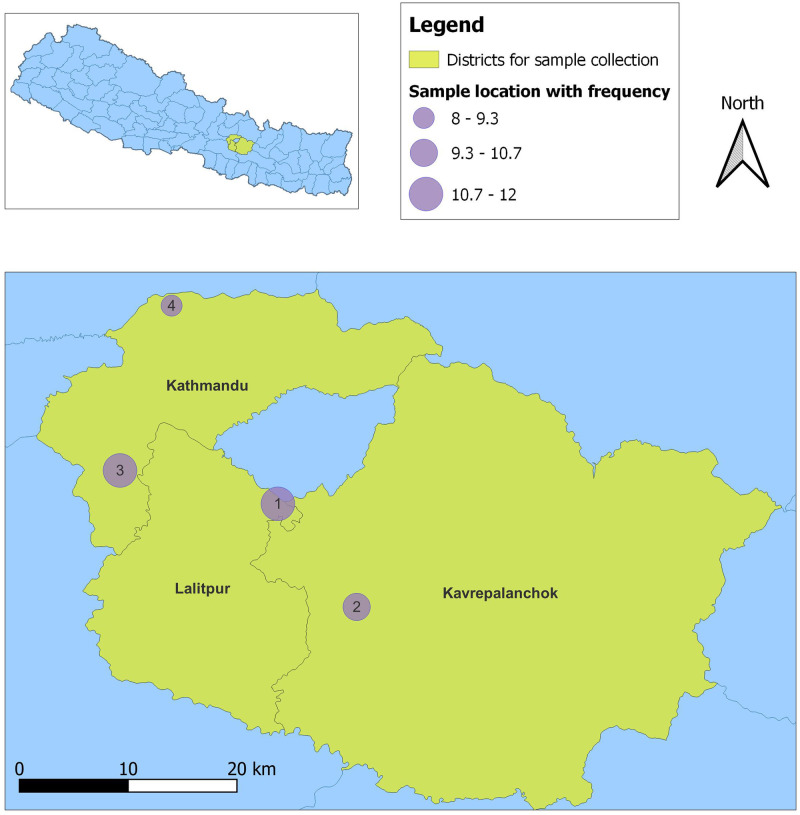
Free-ranging backyard chicken primarily belonging to various indigenous breeds reared in rural outskirt hills of the Kathmandu valley were selected for sampling (Fig 1, S1 Table). The selected birds lacked exposure to commercial and/or antibiotics supplemented feed. Cloacal samples (n = 41) were collected from each chicken using sterile swab sand preserved in cryo-vials containing 50% sterile glycerol. The samples were then transported in cold chain box to the BIOVAC laboratory in Kathmandu and stored at -20°C.

Fig 1. The map of sampling districts (yellow colored) of Nepal showing the sampling locations indicated as numbered circles.

Fig 1

The sample locations are as follows: 1) Lakuribhanjyang, 2) Parthali- Bethankchowk, 3) Chalnakhel-Bosandanda and 4) Jhor- Dhakalchaur. The diameter of sampling locations are shown as varying proportionately with the number of samples collected from that location as indicated in the figure legend.

Selective isolation of LAB isolates

Glycerol stock of cloacal swabs was thawed in room temperature. After vortexing, 50 μl of each sample was inoculated into 5 ml of DeMan, Rogosa and Sharpe (MRS) Broth (HiMedia, India) with 0.5% calcium carbonate (CaCO3). The inoculated broth was incubated at 37°C for 72 hours in carbon-dioxide enriched environment using candle jar incubation method.

After incubation, 50 μL of inoculated broth was streaked on MRS agar (HiMedia, India) with 0.5% calcium carbonate. The agar plates were incubated at 37 °C for 24 hours in carbon-dioxide enriched environment. White to cream colored round colonies (3–4 mm) producing clear hydrolyzing halos were selected as potential LAB isolates. Multiple morphologically distinct colonies producing clear hydrolyzing zones per sample were selected as these could be potentially distinct LAB isolates. The selected colonies were further re-plated on 0.5% CaCO3 supplemented MRS agar for pure culture isolation. The overnight MRS broth culture of each selected LAB isolates were prepared, and subjected to further in vitro screening and challenge tests as below.

Test for anti-bacterial activity

The anti-bacterial activity of selected LAB isolates were tested against a panel of bacterial pathogens using agar well diffusion method [13]. Surface of Muller Hinton Agar (MHA) plates were lawn cultured with 0.5 O.D630 adjusted bacterial suspension of six selected pathogens (Salmonella enterica spp., Escherichia coli, Shigella spp., Klebsiella pneumonia, Citrobacter freundii, and Staphylococcus aureus). After 15–20 minutes, six wells (≥ 30 mm apart) were bored with the broader end of a sterile pipette tip on the inoculated MHA agar. A 100 μl of the overnight MRS broth culture of each selected LAB isolate (adjusted to 0.5 O.D630) was then inoculated into the wells. The broth was allowed to diffuse into the agar for approximately an hour by incubating the covered plates at 4°C in an upright position. After complete diffusion, the plates were incubated in an inverted position at 37°C for 24 hours. Development of clear zone of any size around each well was noted as positive anti-bacterial activity, and sizes of inhibition zone were recorded [14].

Phenotypic bacterial identification

Presumptive phenotypic bacterial identification of the selected LAB isolates demonstrating anti-bacterial activity was performed by using standard microbiological methods, which included Gram staining and biochemical tests for catalase, oxidase, sulfur and indole production [15]. The Gram positive rod shaped bacteria that were negative for catalase, oxidase, sulphur, and indole production were phenotypically identified as Lactobacillus spp.

Test for production of total lactic acid

Lactic acid production by presumptive Lactobacillus spp. isolates was quantified. For this, overnight MRS broth culture of each Lactobacillus spp. was subjected to 10^-1dilution in sterile distilled water. To this, 3 drops of 1% phenolphthalein was added, and was titrated with 0.1 N NaOH solution until a light pink colour was observed [16]. The volume of 0.1 N NaOH required to achieve neutralization was used to calculate the percentage yield of lactic acid using by the following calculation [17, 18]:

Percentageyieldoflacticacid=ActualyieldTheoreticalyieldx100%

The actual yield is the amount of lactic acid formed as a result of bacterial fermentation during overnight incubation, and theoretical yield is the maximum amount of lactic acid presumed to be produced by homo-fermentative LAB strains from available amount of fermentable sugar present in the MRS broth (i.e., 0.02 gm/ml).

Test for acid tolerance

Overnight incubated MRS broth of potential Lactobacillus spp. testing positive for lactic acid production was centrifuged at 1,000 rpm for 10 minutes. Resulting bacterial pellet was re-suspended in 2 ml of sterile saline. One ml of this bacterial suspension was added to 9 ml of sterile artificial gastric juice media (0.2% NaCl, 0.35% pepsin, adjusted to pH 3.0). Another one ml of bacterial suspension was added to 9 ml of sterile MRS broth (adjusted to pH 7.0). The tubes were incubated at 37 °C for 3 hours and O.D600 was measured for both the tubes after incubation [19]. For calculation, bacterial density at O.D600 was empirically equated to the bacterial count in colony forming unit (CFU)/ml as per the growth curve calculation of Lactobacillus plantarum as described by Trabelsi et al. [20]. Acid tolerance ability of bacterial isolates was estimated using the following formula [19]:

Percentacidtolerance=CFU/mlatpH7.0CFU/mlatpH3.0CFU/mlatpH7.0x100%

Test for bile salt tolerance

A set of MRS broth with 0.2% sodium acetate, each supplemented with cattle bile salt (concentrations of 0.0%, 0.3%, 0.5%, and 1%) were prepared. Overnight activated MRS bacterial suspension (20 μl) of presumptive Lactobacillus spp. testing positive for lactic acid production was inoculated in the broth (180 μl) and incubated at 37 °C for 24 hours. After incubation, O.D600 was measured using micro-plate absorbance reader (BioTek, USA). Bacterial absorbance density at O.D600 was empirically equated to the bacterial count in CFU/ml as per the growth curve calculation of Lactobacillus plantarum as described by Trabelsi et al. [20]. The bile tolerance of the test isolates was calculated using the following formula [21]:

Biletolerance=CFU/mlin0.0%bileCFU/mlingiven0.3%,0.5%or1%bileCFU/mlin0.0%bilex100%

Bacterial species identification by 16S rDNA sequencing

Bacterial DNA extraction

Phenotypically identified presumptive Lactobacillus spp. yielding optimal in vitro probiotics characteristics were subjected to bacterial DNA extraction as per kit instructions (Zymo BIOMICS DNA Miniprep kit, USA). The 16S rDNA PCR was performed in a 10 μL reaction containing 1X KAPA HiFi-Hotstart Ready mix (KAPA, USA), 0.2 μM of each forward (5’CCTACGGGNGGCWGCAG3’) and reverse primers (5’GACTACHVGGGTATCTAATCC3’) [22], and 1 μL of template DNA. PCR was conducted at 95 °C/15 minutes for initial denaturation, followed by 40 cycles of each 98 °C/30 s, 65 °C/30 s and 72 °C/30 s, with final extension at 72 °C/5 minutes. The amplicons (550bp) were excised from gel and purified using 1X Agencourt AMPure beads (Beckman Coulter, USA) and sequenced using both the forward and reverse primers on ABI-310 Genetic Analyzer (Applied Biosystems, USA). An M13 sequence was used as a positive control.

After initial quality assessment, the resulting DNA sequences were referenced in the NCBI database using BLAST (Basic Local Alignment Search Tool) [23]. For taxonomic assignment, phylogenetic analysis was performed by comparing sample sequences with a set of Lactobacillus16S rDNA sequences extracted from the NCBI GenBank. The best-fit nucleotide substitution model, K2+G, was selected based on the Bayesian Information Criterion (BIC) score in MEGA X v10.2.5 [24]. Maximum likelihood (ML) phylogeny was inferred using the selected model in MEGA X, with 1000 bootstrap replications. Phylogenetic tree annotation and visualization was performed using FigTree v1.4.4 [25].

Results

A total of 41 cloacal samples were collected from various indigenous breeds of free-ranging backyard chicken from rural outskirt hills of the Kathmandu valley. A total of 90 potential LAB isolates were selected based on CaCO3 hydrolysis in MRS agar.

Of 90 potential LAB isolates, 52 isolates (57%) showed anti-bacterial activity against at least one bacterial pathogen with zone of inhibition ranging from 7 to 18 mm (S2 Table). Majority (69%, 36/52) of LAB isolates inhibited Salmonella spp., followed by inhibition against C. freundi (53%, 28/52), Shigella spp. (44%, 23/52), K. pneumonia (30%, 16/52), E. coli (26%, 14/52) and S. aureus (13%, 7/52). Nine of these LAB isolates (17%) showed inhibition against all tested Enterobacterales bacteria.

Of 52 LAB isolates showing varying anti-bacterial activity, 46 fulfilled basic phenotypic criteria of Lactobacillus spp.; Gram-positive rods giving negative reactions for each sulphur, indole, catalase, and oxidase tests. The selected 46 potential Lactobacillus spp. isolates were further subjected to in vitro screening tests for probiotics properties. Of 46 isolates, 37 produced acid of varying percentage yields ranging from 38 to 81. Among these 37 isolates, 27 isolates showed survival at pH 3.0 with varying survival rates (1.2% to 62.1%). Tolerance to bile salt varied with the bile concentrations. 17 isolates had 0.3% and 0.5% bile salts tolerance, while 14 isolates survived in the presence of 1.0% bile salt. The resulting profile of 52 LAB isolates on in vitro screening tests for probiotics is given S2 Table.

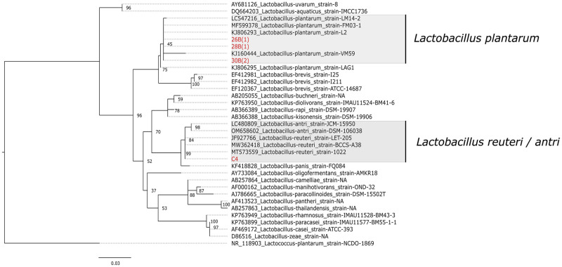
Of 27 phenotypically identified Lactobacillus spp. exhibiting optimal in vitro probiotics properties, 3 isolates (26B, 28B, and 30B) showing anti-bacterial activity against the widest range of tested bacterial pathogens, and one (C4/36(4)) showing high bile tolerance (1.0%) were randomly selected for 16S rDNA sequencing. Based on phylogenetic analyses of 16S rDNA sequences of the isolates, 26B (GenBank accession number ON955508), 28B (GenBank accession number ON955509), and 30B (GenBank accession number ON955510) isolates clustered into the clade of L. plantarum with average p-distance of 0.0061, inferring a genetic similarity of 99.38% with L. plantarum. The isolate C4/36(4) (GenBank accession number ON955511), on the other hand, grouped into the cluster of L. reuteri, L. antri, and L. panis with average p-distance of 0.0014 and a genetic similarity of 99.85% with the given cluster (Fig 2).

Fig 2. Maximum likelihood tree based on 16S rDNA gene sequences of different Lactobacillus spp.

Fig 2

The isolates of the study are indicated in red font. Lactococcus plantarum was chosen as an out-group. The scale bar (0.03) shows the nucleotide substitution rate per site. Bootstrap probabilities as determined for 1000 replicates are shown at nodes. The isolate C4/36(4) has been labeled as C4 in the given phylogenetic tree.

Discussion

In our study, we have isolated and identified potential LAB from 41 non-duplicate cloacal samples collected from a group of diverse backyard chicken breeds reared at a rural outskirt of the Kathmandu valley (Nepal). Of 90 potential LAB isolates, 31 isolates were phenotypically identified to be Lactobacillus spp. fulfilling basic in vitro probiotics characteristics. Of these, at least 4 were genetically confirmed to be Lactobacillus spp. Further in vivo experiments need to be conducted to assess gut adaptation and probiotics performance to develop these isolates as poultry probiotics feed supplement. Industrial production parameters, such as ability to propagate in cheaper growth media, and maintain viability throughout production steps till storage and final application also need to be assessed [10].

With an increasing global threat of antimicrobial resistance (AMR), there is an urgent need to explore alternative solutions to antibiotic use. Routine use of sub-optimal dosages of antimicrobials in poultry sector with an aim to enhance their growth and performance has been one of the drivers of accelerated AMR [26]. The discontinuation of anti-microbial use in poultry, however, as a result of stringent regulation in high income countries, has resulted in poor poultry production [26]. This underscored the importance of prophylactics like antimicrobials for desired poultry yields. There are numerous studies indicating the use of probiotics as alternative solution replacing antimicrobials in poultry industry with similar benefits but without risk of AMR emergence [26]. Additionally, probiotics are also known to inhibit aflatoxigenic molds and degrade aflatoxins [26].

There are various native breeds of chicken found in Nepal, generally known as “local breeds”, but some with specific names like Shakini, Giriraj, Ghantikhuile, Kadaknath and Pwankh Ulte (Dumse) [27]. Based on various factors, gut microbiota of free-ranging indigenous chicken breeds is different from that of commercial breeds [28]. Free-ranging chicken forage on a variety of natural food sources, such as insects and green foliage, which may enrich their microbiome diversity [29]. As a result, compared to commercial breeds, the backyard breeds have higher immunity, and therefore, can be more resilient against various infectious diseases [29].

We have isolated potential probiotics from indigenous chicken breeds. Unlike probiotics that are often isolated from abiotic sources, such as fermented food, the probiotics isolated in our study are host-specific to chicken with potential enhanced intestinal mucosa colonization abilities for greater natural integration into host gut microbiota [30]. However, further in vivo investigation is still needed to fully assess this probiotics feature.

Bacterial infections are one of the major reasons of morbidity and mortality in poultry, especially in commercial broiler and layer breeds- leading to production loss [31]. Colibacillosis and salpingitis caused by pathogenic strains of E. coli, mycoplasmosis caused by Mycoplasma spp., fowl cholera by Pasteurella multocida, necrotic enteritis by Clostridium perfringens, ulcerative enteritis by Clostridium colinum, salmonellosis, fowl typhoid and paratyphoid by diverse serovars of Salmonella spp., omphalitis by coliforms, pododermatitis and staphylococcosis by Staphylococcus aureus, and shigellosis by Shigella spp. are some of the common diseases caused by bacterial infections in poultry [31, 32].

Probiotics are known to protect host from infections through various mechanisms, including inhibiting colonization by pathogens and producing antibacterial metabolites such as acetic acid, lactic acid, alcohols, and bacteriocins [33]. Effective inhibition property against known pathogenic bacteria is one of the important selection criteria for good probiotics candidate. In our study, inhibition of coliform bacteria by LAB isolates was more common compared to inhibition against Gram-positive cocci, where 67% (31/46) phenotypic LAB isolates inhibited Salmonella spp., while 19% (9/46) isolates inhibited all tested enteric bacilli, in contrast to 15% (7/46) isolates that inhibited S. aureus with or without inhibiting enteric bacilli. Taheri et. al also reported higher inhibition of enteric bacilli, where 18% (62/332) of selected LAB strains isolated from gut of broiler chicken inhibited Salmonella enteritidis, Salmonella typhimurium, and E. coli O78:K80 [34]. Kizerwette-Swida et al. reported higher inhibition activity against Gram-positive pathogens including C. perfringes and S. aureus than coliforms including E. coli and Salmonella [35]. In our study, 5 of 46 LAB isolates inhibited all tested Gram-positive cocci and enteric bacilli, 3 (26B, 28B, and 30B) of which were genetically identified to be L. plantarum. Probiotics cultures of L. plantarum has been reported to have efficient antibacterial activity against broad spectrum of bacteria including clinical isolates S. aureus and E. coli via direct cell competitive exclusion as well as production of acids or bacteriocin-like inhibitors [36, 37].

The ability to survive in acidic gut environment, as they pass through stomach and intestine and colonize in the host gut, is an essential property of probiotics [30]. In our study, 27 of 46 isolates fulfilling phenotypic characteristics of LAB isolates showed survival tolerance in simulated gastric juice medium of pH 3.0, with the survival ratio ranging from 1.2% to 62.1%. This infers that the isolates would survive harsh acidic condition of the host gut upon introduction. Feng et al. reported that of 52 potential probiotics strains isolated from gut of piglets, 100% of isolates showed survival at pH 3.0, though 8 isolates failed to survive at an acidic pH of 1.0 [14]. In another study aiming to isolate potential probiotics from poultry fecal samples, of 42 potential isolates, 16% (n = 7) of selected isolates tolerated acidic pH of 2.5 and 4.0 compared to normal pH [38]. Further, lactic acid produced by LAB isolates provides various health benefits including immune-modulatory functions and prevention of diarrheal diseases [1]. In our study, 37 of 46 potential probiotics produced lactic acid with the yield ranging from 38 to 81%.

Bile salts are one of essential constituents of mammalian gut that help solubilize dietary fats. Hydrophobicity and detergent properties of bile salts exert strong antimicrobial effects. Thus, tolerance to bile is considered as one of important qualities of probiotics. Bile concentration varies along duodenum, jejunum and cecum of chicken gut, which were estimated to be 0.17, 0.7, and 0.0085% respectively [39]. In our study, 18 of phenotypic lactobacilli isolates fulfilling probiotics characteristics of acid production and acid tolerance showed 24 hour in vitro survival in the presence of 0.3% and 0.5% bile salts. Shin et al. reported that all 3 sequenced potential probiotics isolates obtained from gut of broiler chicken tolerated the presence of 0.3% bile salts [40]. Oh et al. reported 7 of the potential probiotics isolates tolerated 0.3% bile salts for 24 hours [41].

Conclusion

Our study isolated, characterized and identified 4 Lactobacillus spp. demonstrating optimal in vitro probiotics properties. With further evaluation, these identified Lactobacillus spp. candidates can be developed as effective probiotics which can be used as poultry feed supplement replacing regularly used antimicrobials.

Supporting information

S1 Table. Demographic details of chicken cloacal samples shown by identification number, sampling locations and the chicken breed.

(DOCX)

S2 Table. Laboratory results of 52 LAB isolates on antibacterial inhibition, biochemical test, and in vitro test for potential probiotics strains.

(DOCX)

Acknowledgments

We acknowledge the field and laboratory team of the Center for Molecular Dynamics Nepal (CMDN) for providing logistical, operational and supervisory support throughout the study. We thank the SANN International College for their support. And we express our gratitude to the members of local community where we collected our samples.

Data Availability

All sequence files are available from the https://www.ncbi.nlm.nih.gov/genbank/ database (accession numbers ON955508, ON955509, ON955510, ON955511).

Funding Statement

Partial fund for conducting the laboratory experiments were received as academic thesis support from SANN International College, Purbanchal University (by the authors Mohan Gupta, Ujwal Shrestha, Saubhagya Dangol, and Sudarshan GC). Rest of the fund for conducting this study was obtained from institutions BIOVAC Nepal and Center for Molecular Dynamics Nepal (CMDN). - Roji Raut, Sulochana Manandhar, Ashok Chaudhary, Pragun Rajbhandari, and Prajwol Manandhar received salary from Center for Molecular Dynamics Nepal (CMDN). - Rajindra Napit, and Dibesh Karmacharya received salary from BIOVAC Nepal. The funders had no role in study design, data collection and analysis, decision to publish, or preparation of the manuscript.

References

  • 1.FAO. Probiotics in animal nutrition: production, impact and regulation. Bajagai YS, Klieve AV, Dart PJ, Bryden WL, editors. Rome: FAO Animal Production and Health; 2016.
  • 2.Van TTH, Yidana Z, Smooker PM, Coloe PJ. Antibiotic use in food animals worldwide, with a focus on Africa: Pluses and minuses. Journal of global antimicrobial resistance. 2020;20:170–7. doi: 10.1016/j.jgar.2019.07.031 [DOI] [PubMed] [Google Scholar]
  • 3.Poudel U, Dahal U, Upadhyaya N, Chaudhari S, Dhakal S. Livestock and poultry production in Nepal and current status of vaccine development. Vaccines. 2020;8(2):322. doi: 10.3390/vaccines8020322 [DOI] [PMC free article] [PubMed] [Google Scholar]
  • 4.Jensen HA, editor. Paradigm and visions: network for poultry production and health in developing countries. Proceedings of a Workshop on Poultry as a tool in poverty eradication and promotion of gender equality, Tune Landboskole, Denmark; 1999.
  • 5.Hosain MZ, Kabir SL, Kamal MM. Antimicrobial uses for livestock production in developing countries. Veterinary World. 2021;14(1):210. doi: 10.14202/vetworld.2021.210-221 [DOI] [PMC free article] [PubMed] [Google Scholar]
  • 6.Elliott KA, Kenny C, Madan J. A global treaty to reduce antimicrobial use in livestock. Center for Global Development: Washington, DC, USA. 2017.
  • 7.Lekshmi M, Ammini P, Kumar S, Varela MF. The food production environment and the development of antimicrobial resistance in human pathogens of animal origin. Microorganisms. 2017;5(1):11. doi: 10.3390/microorganisms5010011 [DOI] [PMC free article] [PubMed] [Google Scholar]
  • 8.Fioramonti J, Theodorou V, Bueno L. Probiotics: what are they? What are their effects on gut physiology? Best Practice & Research Clinical Gastroenterology. 2003;17(5):711–24. doi: 10.1016/s1521-6918(03)00075-1 [DOI] [PubMed] [Google Scholar]
  • 9.Roselli M, Finamore A, Britti MS, Konstantinov SR, Smidt H, de Vos WM, et al. The novel porcine Lactobacillus sobrius strain protects intestinal cells from enterotoxigenic Escherichia coli K88 infection and prevents membrane barrier damage. The Journal of nutrition. 2007;137(12):2709–16. doi: 10.1093/jn/137.12.2709 [DOI] [PubMed] [Google Scholar]
  • 10.Collins J, Thornton G, Sullivan G. Selection of probiotic strains for human applications. International dairy journal. 1998;8(5–6):487–90. [Google Scholar]
  • 11.Mountzouris K, Tsitrsikos P, Palamidi I, Arvaniti A, Mohnl M, Schatzmayr G, et al. Effects of probiotic inclusion levels in broiler nutrition on growth performance, nutrient digestibility, plasma immunoglobulins, and cecal microflora composition. Poultry science. 2010;89(1):58–67. doi: 10.3382/ps.2009-00308 [DOI] [PubMed] [Google Scholar]
  • 12.Haddadin M, Abdulrahim S, Hashlamoun E, Robinson R. The effect of Lactobacillus acidophilus on the production and chemical composition of hen’s eggs. Poultry science. 1996;75(4):491–4. doi: 10.3382/ps.0750491 [DOI] [PubMed] [Google Scholar]
  • 13.Tagg J, McGiven A. Assay system for bacteriocins. Applied microbiology. 1971;21(5):943. doi: 10.1128/am.21.5.943-943.1971 [DOI] [PMC free article] [PubMed] [Google Scholar]
  • 14.Feng Y, Qiao L, Liu R, Yao H, Gao C. Potential probiotic properties of lactic acid bacteria isolated from the intestinal mucosa of healthy piglets. Annals of Microbiology. 2017;67(3):239–53. [Google Scholar]
  • 15.Association APH. Compendium for the microbiological examination of foods. American Public Health, Washington, DC. 2015.
  • 16.HENDRATI PM, KUSHARYATI DF, RYANDINI D, OEDJIJONO O. Characterization of Bifidobacteria from infant feces with different mode of birth at Purwokerto, Indonesia. Biodiversitas Journal of Biological Diversity. 2017;18(3):1265–9. [Google Scholar]
  • 17.Fabro M, Milanesio H, Robert L, Speranza J, Murphy M, Rodríguez G, et al. Determination of acidity in whole raw milk: comparison of results obtained by two different analytical methods. Journal of dairy science. 2006;89(3):859–61. [DOI] [PubMed] [Google Scholar]
  • 18.Von Wright A, Axelsson L. Lactic acid bacteria: an introduction. Lactic acid bacteria: CRC Press; 2019. p. 1–16. [Google Scholar]
  • 19.Farid W, Masud T, Sohail A, Naqvi S, Qazalbash MA. Molecular characterization and 16S rRNA sequence analysis of probiotic lactobacillus acidophilus isolated from indigenous Dahi (Yoghurt). International Journal of Bioscience. 2016;9(5):19–27. [Google Scholar]
  • 20.Trabelsi I, Bejar W, Ayadi D, Chouayekh H, Kammoun R, Bejar S, et al. Encapsulation in alginate and alginate coated-chitosan improved the survival of newly probiotic in oxgall and gastric juice. International journal of biological macromolecules. 2013;61:36–42. doi: 10.1016/j.ijbiomac.2013.06.035 [DOI] [PubMed] [Google Scholar]
  • 21.Marhamatizadeh MH, Sayyadi S. Mining of lactic acid bacteria from traditional yogurt (Mast) of Iran for possible industrial probiotic use. Italian Journal of Animal Science. 2019;18(1):663–7. [Google Scholar]
  • 22.Klindworth A, Pruesse E, Schweer T, Peplies J, Quast C, Horn M, et al. Evaluation of general 16S ribosomal RNA gene PCR primers for classical and next-generation sequencing-based diversity studies. Nucleic acids research. 2013;41(1):e1–e. doi: 10.1093/nar/gks808 [DOI] [PMC free article] [PubMed] [Google Scholar]
  • 23.Korf I, Yandell M, Bedell J. Blast: " O’Reilly Media, Inc."; 2003.
  • 24.Kumar S, Stecher G, Li M, Knyaz C, Tamura K. MEGA X: molecular evolutionary genetics analysis across computing platforms. Molecular biology and evolution. 2018;35(6):1547. doi: 10.1093/molbev/msy096 [DOI] [PMC free article] [PubMed] [Google Scholar]
  • 25.Rambaut A. FigTree v1. 3.1 (http://tree.bio.ed.ac.uk/software/figtree/) Institute of Evolutionary Biology. University of Edinburgh, Edinburgh, United Kingdom. 2010.
  • 26.Hernandez-Patlan D, Solis-Cruz B, Hargis BM, Tellez G. Prebiotics and Probiotics—Potential Benefits in Nutrition and Health. Franco-Robles E, Ramírez-Emiliano J, editors: IntechOpen; 2019.
  • 27.Mishra B. Poultry in Nepal: Present Scenario, Indigenous Breeds and Economic Traits 2013. https://bijeshmishra.wordpress.com/2013/01/27/traits-of-economic-importance-in-poultry/.
  • 28.Paul SS, Chatterjee RN, Raju M. Gut Microbial Composition Differs Extensively among Indian Native Chicken Breeds Originated in Different Geographical Locations and a Commercial Broiler Line, but Breed-Specific, as Well as Across-Breed Core Microbiomes, Are Found. 2021;9(2). doi: 10.3390/microorganisms9020391 . [DOI] [PMC free article] [PubMed] [Google Scholar]
  • 29.Kannaki TR, Priyanka E, Haunshi S. Research Note: Disease tolerance/resistance and host immune response to experimental infection with Pasteurella multocida A:1 isolate in Indian native Nicobari chicken breed. Poultry Science. 2021;100(8):101268. doi: 10.1016/j.psj.2021.101268 [DOI] [PMC free article] [PubMed] [Google Scholar]
  • 30.Reuben RC, Roy PC, Sarkar SL, Alam R, Jahid IK. Isolation, characterization, and assessment of lactic acid bacteria toward their selection as poultry probiotics. BMC Microbiology. 2019;19. doi: 10.1186/s12866-019-1626-0 [DOI] [PMC free article] [PubMed] [Google Scholar]
  • 31.Thøfner I, Christensen J-P. Bacterial diseases in poultry. In: Foster N, Kyriazakis I, Barrow P, editors. Advancements and Technologies in Pig and Poultry Bacterial Disease Control: Academic Press; 2021. p. 199–227. [Google Scholar]
  • 32.Swayne DE. Diseases of poultry: John Wiley & Sons; 2013. [Google Scholar]
  • 33.Liu Y, Tran DQ, Rhoads JM. Probiotics in Disease Prevention and Treatment. Journal of clinical pharmacology. 2018;58 Suppl 10(Suppl 10):S164–s79. Epub 2018/09/25. doi: 10.1002/jcph.1121 . [DOI] [PMC free article] [PubMed] [Google Scholar]
  • 34.Taheri HR, Moravej H, Tabandeh F, Zaghari M, Shivazad M. Screening of lactic acid bacteria toward their selection as a source of chicken probiotic. Poultry Science. 2009;88(8):1586–93. doi: 10.3382/ps.2009-00041 [DOI] [PubMed] [Google Scholar]
  • 35.Kizerwetter-Swida M, Binek M. Selection of potentially probiotic Lactobacillus strains towards their inhibitory activity against poultry enteropathogenic bacteria. Polish journal of microbiology. 2005;54(4):287–94. [PubMed] [Google Scholar]
  • 36.Sikorska H, Smoragiewicz W. Role of probiotics in the prevention and treatment of meticillin-resistant Staphylococcus aureus infections. International Journal of Antimicrobial Agents. 2013;42(6):475–81. doi: 10.1016/j.ijantimicag.2013.08.003 [DOI] [PubMed] [Google Scholar]
  • 37.Bindu A, Devi L. In vitro and In Silico Approach For Characterization of Antimicrobial Peptide From Probiotics Against Staphylococcus Aureus and Escherichia Coli. 2021. [DOI] [PubMed] [Google Scholar]
  • 38.Salehizadeh M, Modarressi MH, Mousavi SN, Ebrahimi MT, editors. Evaluation of lactic acid bacteria isolated from poultry feces as potential probiotic and its in vitro competitive activity against Salmonella typhimurium. Veterinary Research Forum; 2020: Faculty of Veterinary Medicine, Urmia University, Urmia, Iran. [DOI] [PMC free article] [PubMed]
  • 39.Lin J, Sahin O, Michel LO, Zhang Q. Critical role of multidrug efflux pump CmeABC in bile resistance and in vivo colonization of Campylobacter jejuni. Infection and immunity. 2003;71(8):4250–9. Epub 2003/07/23. doi: 10.1128/IAI.71.8.4250-4259.2003 . [DOI] [PMC free article] [PubMed] [Google Scholar]
  • 40.Shin M, Han S, Ji A, Kim K, Lee W. Isolation and characterization of bacteriocin-producing bacteria from the gastrointestinal tract of broiler chickens for probiotic use. Journal of applied microbiology. 2008;105(6):2203–12. doi: 10.1111/j.1365-2672.2008.03935.x [DOI] [PubMed] [Google Scholar]
  • 41.Annual report 2018/19. Department of Health Services, Government of Nepal, Ministry of Health and Population, Department of Health Services, 2020.

Decision Letter 0

Xiaolun Sun

8 Sep 2022

PONE-D-22-20489Identification and characterization of probiotics isolated from indigenous chicken (Gallus domesticus) of NepalPLOS ONE

Dear Dr. Karmacharya,

Thank you for submitting your manuscript to PLOS ONE. After careful consideration, we feel that it has merit but does not fully meet PLOS ONE’s publication criteria as it currently stands. Therefore, we invite you to submit a revised version of the manuscript that addresses the points raised during the review process.

I concur with all the comments raised by the reviewers. Please address all of the issues in the revised manuscript.

Please submit your revised manuscript by Oct 23 2022 11:59PM. If you will need more time than this to complete your revisions, please reply to this message or contact the journal office at plosone@plos.org. When you're ready to submit your revision, log on to https://www.editorialmanager.com/pone/ and select the 'Submissions Needing Revision' folder to locate your manuscript file.

Please include the following items when submitting your revised manuscript:

  • A rebuttal letter that responds to each point raised by the academic editor and reviewer(s). You should upload this letter as a separate file labeled 'Response to Reviewers'.

  • A marked-up copy of your manuscript that highlights changes made to the original version. You should upload this as a separate file labeled 'Revised Manuscript with Track Changes'.

  • An unmarked version of your revised paper without tracked changes. You should upload this as a separate file labeled 'Manuscript'.

If you would like to make changes to your financial disclosure, please include your updated statement in your cover letter. Guidelines for resubmitting your figure files are available below the reviewer comments at the end of this letter.

If applicable, we recommend that you deposit your laboratory protocols in protocols.io to enhance the reproducibility of your results. Protocols.io assigns your protocol its own identifier (DOI) so that it can be cited independently in the future. For instructions see: https://journals.plos.org/plosone/s/submission-guidelines#loc-laboratory-protocols. Additionally, PLOS ONE offers an option for publishing peer-reviewed Lab Protocol articles, which describe protocols hosted on protocols.io. Read more information on sharing protocols at https://plos.org/protocols?utm_medium=editorial-email&utm_source=authorletters&utm_campaign=protocols.

We look forward to receiving your revised manuscript.

Kind regards,

Xiaolun Sun

Academic Editor

PLOS ONE

Journal Requirements:

When submitting your revision, we need you to address these additional requirements.

1. Please ensure that your manuscript meets PLOS ONE's style requirements, including those for file naming. The PLOS ONE style templates can be found at 

https://journals.plos.org/plosone/s/file?id=wjVg/PLOSOne_formatting_sample_main_body.pdf and 

https://journals.plos.org/plosone/s/file?id=ba62/PLOSOne_formatting_sample_title_authors_affiliations.pdf

2. Thank you for stating the following financial disclosure: 

"The funders had no role in study design, data collection and analysis, decision to publish, or preparation of the manuscript."

At this time, please address the following queries:

a) Please clarify the sources of funding (financial or material support) for your study. List the grants or organizations that supported your study, including funding received from your institution. 

b) State what role the funders took in the study. If the funders had no role in your study, please state: “The funders had no role in study design, data collection and analysis, decision to publish, or preparation of the manuscript.”

c) If any authors received a salary from any of your funders, please state which authors and which funders.

d) If you did not receive any funding for this study, please state: “The authors received no specific funding for this work.”

Please include your amended statements within your cover letter; we will change the online submission form on your behalf.

3. We note that Figure 1 in your submission contain map images which may be copyrighted. All PLOS content is published under the Creative Commons Attribution License (CC BY 4.0), which means that the manuscript, images, and Supporting Information files will be freely available online, and any third party is permitted to access, download, copy, distribute, and use these materials in any way, even commercially, with proper attribution. For these reasons, we cannot publish previously copyrighted maps or satellite images created using proprietary data, such as Google software (Google Maps, Street View, and Earth). For more information, see our copyright guidelines: http://journals.plos.org/plosone/s/licenses-and-copyright.

We require you to either (1) present written permission from the copyright holder to publish these figures specifically under the CC BY 4.0 license, or (2) remove the figures from your submission:

a. You may seek permission from the original copyright holder of Figure 1 to publish the content specifically under the CC BY 4.0 license.  

We recommend that you contact the original copyright holder with the Content Permission Form (http://journals.plos.org/plosone/s/file?id=7c09/content-permission-form.pdf) and the following text:

“I request permission for the open-access journal PLOS ONE to publish XXX under the Creative Commons Attribution License (CCAL) CC BY 4.0 (http://creativecommons.org/licenses/by/4.0/). Please be aware that this license allows unrestricted use and distribution, even commercially, by third parties. Please reply and provide explicit written permission to publish XXX under a CC BY license and complete the attached form.”

Please upload the completed Content Permission Form or other proof of granted permissions as an ""Other"" file with your submission.

In the figure caption of the copyrighted figure, please include the following text: “Reprinted from [ref] under a CC BY license, with permission from [name of publisher], original copyright [original copyright year].”

b.  If you are unable to obtain permission from the original copyright holder to publish these figures under the CC BY 4.0 license or if the copyright holder’s requirements are incompatible with the CC BY 4.0 license, please either i) remove the figure or ii) supply a replacement figure that complies with the CC BY 4.0 license. Please check copyright information on all replacement figures and update the figure caption with source information. If applicable, please specify in the figure caption text when a figure is similar but not identical to the original image and is therefore for illustrative purposes only.

The following resources for replacing copyrighted map figures may be helpful:

USGS National Map Viewer (public domain): http://viewer.nationalmap.gov/viewer/

The Gateway to Astronaut Photography of Earth (public domain): http://eol.jsc.nasa.gov/sseop/clickmap/

Maps at the CIA (public domain): https://www.cia.gov/library/publications/the-world-factbook/index.html and https://www.cia.gov/library/publications/cia-maps-publications/index.html

NASA Earth Observatory (public domain): http://earthobservatory.nasa.gov/

Landsat: http://landsat.visibleearth.nasa.gov/

USGS EROS (Earth Resources Observatory and Science (EROS) Center) (public domain): http://eros.usgs.gov/#

Natural Earth (public domain): http://www.naturalearthdata.com/

[Note: HTML markup is below. Please do not edit.]

Reviewers' comments:

Reviewer's Responses to Questions

Comments to the Author

1. Is the manuscript technically sound, and do the data support the conclusions?

The manuscript must describe a technically sound piece of scientific research with data that supports the conclusions. Experiments must have been conducted rigorously, with appropriate controls, replication, and sample sizes. The conclusions must be drawn appropriately based on the data presented.

Reviewer #1: Partly

Reviewer #2: Yes

**********

2. Has the statistical analysis been performed appropriately and rigorously?

Reviewer #1: N/A

Reviewer #2: Yes

**********

3. Have the authors made all data underlying the findings in their manuscript fully available?

The PLOS Data policy requires authors to make all data underlying the findings described in their manuscript fully available without restriction, with rare exception (please refer to the Data Availability Statement in the manuscript PDF file). The data should be provided as part of the manuscript or its supporting information, or deposited to a public repository. For example, in addition to summary statistics, the data points behind means, medians and variance measures should be available. If there are restrictions on publicly sharing data—e.g. participant privacy or use of data from a third party—those must be specified.

Reviewer #1: Yes

Reviewer #2: Yes

**********

4. Is the manuscript presented in an intelligible fashion and written in standard English?

PLOS ONE does not copyedit accepted manuscripts, so the language in submitted articles must be clear, correct, and unambiguous. Any typographical or grammatical errors should be corrected at revision, so please note any specific errors here.

Reviewer #1: No

Reviewer #2: Yes

**********

5. Review Comments to the Author

Please use the space provided to explain your answers to the questions above. You may also include additional comments for the author, including concerns about dual publication, research ethics, or publication ethics. (Please upload your review as an attachment if it exceeds 20,000 characters)

Reviewer #1: In this manuscript, Mohan Gupta and his colleagues isolated, characterized, and identified 4 Lactobacillus spp. from indigenous chicken of Nepal demonstrating optimal probiotic activities in vitro, which may be used as antibiotic additive alternatives. However, lack of in vivo experiment and novel findings limit the integrality of this paper. Language modification is recommended. The other comments can be found below:

1. At line 4, please make sure your short title is “Isolation and identification of probiotics from native chicken breeds of Nepal” or “Isolation and identification of probiotics from chicken of Nepal”? Please keep consistent.

2. Please give references for sentences at line 76-77 and 77-78.

3. At line 107, 110, 120, 124, 190, 205, 206, 207, there should be spaces between words “with0.5%”, “India)with0.5%”, “O.D630adjustedbacterial”, “0.5O.D630”, “fromvarious”, “37produced”, “37isolates, 27isolates”, “17isolates”. I will not list all of those kind of error, please correct throughout the manuscript.

4. At line 88, there shouldn’t be a space before “Sampling”.

5. At lines 126-128, what’s the criterion you used to determine whether the LAB isolates have anti-bacterial activity or not? Although you mentioned in the results part at line 195 that “pathogen with zone of inhibition ranging from 7 to 16mm”, please give a reference, and is the same criterion (zone range) applied for every pathogen?

6. How these 90 potential LAB isolates distributed among the 41 cloacal samples? How do you distinguish the LAB isolates from same cloacal/plate sample?

7. At lines 199-200, please delete “of which 5 isolates also inhibited S. aureus.”, it’s repetitive.

8. According to your description from lines 136-146, please make clear at line 205, the 38%-81% is acid yield or percentage of acid yield.

9. At lines 151-153, to calculate the acid tolerance, why not use two pH for same medium, here you use gastric juice media and MRS.

10. From line 202-209, looks like not all the LAB isolates applied to all the screening methods, please clarify how many isolates were tested in each method. In an other words, are these tests applied sequentially?

11. At line 223, please change “a” to “as”.

12. According to the formula at line 167, how you get value above 100% for the bile tolerance data at Table S2? Also, is that reasonable that the tolerance increases as the bile concentration increase, for example, data for 22(3)?

13. Conclusion for C4/36(4), although it has high tolerance to bile, but one of the important characteristics of probiotics is the ability to inhibit pathogen growth, looks it doesn’t fit.

14. It’s not necessary to put sentences at lines 301-302 in conclusion part.

Reviewer #2: Gupta et al., have presented important findings on characterising on probiotics for countering the effect of gut pathogens and rising AMR related global issues. I found this article very well designed and experiments are well executed. i

**********

6. PLOS authors have the option to publish the peer review history of their article (what does this mean?). If published, this will include your full peer review and any attached files.

If you choose “no”, your identity will remain anonymous but your review may still be made public.

Do you want your identity to be public for this peer review? For information about this choice, including consent withdrawal, please see our Privacy Policy.

Reviewer #1: No

Reviewer #2: No

**********

[NOTE: If reviewer comments were submitted as an attachment file, they will be attached to this email and accessible via the submission site. Please log into your account, locate the manuscript record, and check for the action link "View Attachments". If this link does not appear, there are no attachment files.]

While revising your submission, please upload your figure files to the Preflight Analysis and Conversion Engine (PACE) digital diagnostic tool, https://pacev2.apexcovantage.com/. PACE helps ensure that figures meet PLOS requirements. To use PACE, you must first register as a user. Registration is free. Then, login and navigate to the UPLOAD tab, where you will find detailed instructions on how to use the tool. If you encounter any issues or have any questions when using PACE, please email PLOS at figures@plos.org. Please note that Supporting Information files do not need this step.

PLoS One. 2023 Jan 19;18(1):e0280412. doi: 10.1371/journal.pone.0280412.r002

Author response to Decision Letter 0


21 Sep 2022

Point by point response to editor’s comments:

1. Please ensure that your manuscript meets PLOS ONE's style requirements, including those for file naming.

- The file names have been corrected.

2. a) Please clarify the sources of funding (financial or material support) for your study. List the grants or organizations that supported your study, including funding received from your institution.

- Partial fund for conducting the laboratory experiments were received as academic thesis support from SANN International College, Purbanchal University (by the authors Mohan Gupta, Ujwal Shrestha, Saubhagya Dangol, and Sudarshan GC). Rest of the fund for conducting this study was obtained from institutions BIOVAC Nepal and Center for Molecular Dynamics Nepal (CMDN).

b) State what role the funders took in the study.

- The funders had role in data analysis and preparation of the manuscript study design.

c) If any authors received a salary from any of your funders, please state which authors and which funders.

- Roji Raut, Sulochana Manandhar, Ashok Chaudhary, Pragun Rajbhandari, and Prajwol Manandhar received salary from Center for Molecular Dynamics Nepal (CMDN).

- Rajindra Napit, and Dibesh Karmacharya received salary from BIOVAC Nepal.

3. Question on Figure 1:

- Figure 1 is not a copy right material. Instead, this has been created by the authors using the program Q-GIS (version 3.22.7) based on the recorded GPS locations. We permit the figure to be used freely by anyone for download, copy, distribute and any other way.

Response to the comments of Reviewer 1

1. At line 4, please make sure your short title is “Isolation and identification of probiotics from native chicken breeds of Nepal” or “Isolation and identification of probiotics from chicken of Nepal”? Please keep consistent.

- This has been corrected in the revised manuscript now by replacing with the former phrase.

2. Please give references for sentences at line 76-77 and 77-78.

- The reference to the first sentence was provided. For the second sentence, the use, and particularly production of probiotics in poultry and other livestock sectors in Nepal is relatively much less practiced. This sentence has been accordingly modified in the revised manuscript now.

3. At line 107, 110, 120, 124, 190, 205, 206, 207, there should be spaces between words “with0.5%”, “India) with 0.5%”, “O.D630adjustedbacterial”, “0.5O.D630”, “fromvarious”, “37produced”, “37isolates, 27isolates”, “17isolates”. I will not list all of those kind of error, please correct throughout the manuscript.

- We apologies for the typo. It seems like when the Microsoft word document is shared between authors who have different versions of operating system, the problem of word spacing automatically appears. We have checked all the text word by word in the revised manuscript now. Thank you for pointing this out.

4. At line 88, there shouldn’t be a space before “Sampling”.

- This has been corrected in the revised manuscript now.

5. At lines 126-128, what’s the criterion you used to determine whether the LAB isolates have anti-bacterial activity or not? Although you mentioned in the results part at line 195 that “pathogen with zone of inhibition ranging from 7 to 16mm”, please give a reference, and is the same criterion (zone range) applied for every pathogen?

- The production of clear zone of any size around the inoculated wells was taken as an indication of antibacterial activity imposed by that LAB isolate against the given pathogen inoculated as a lawn culture. This criterion has been applied for every tested pathogen. In this study, the observed zones of inhibition ranged between 7 mm to 16 mm. This has been clarified in the revised manuscript, and referenced too.

6. How these 90 potential LAB isolates distributed among the 41 cloacal samples? How do you distinguish the LAB isolates from same cloacal/plate sample?

- Multiple morphologically distinct colonies producing clear hydrolyzing zones and ranging from white to cream in colour and measuring 3-4 mm in size were selected as these could be potentially distinct LAB isolates. Because we used cloacal swab as source sample, it is expected to harbor diverse culturable microbiome, many of which could be potential LAB strains. This has been clarified in the revised manuscript now.

7. At lines 199-200, please delete “of which 5 isolates also inhibited S. aureus.”, it’s repetitive.

- This has been responded accordingly in the revised manuscript now.

8. According to your description from lines 136-146, please make clear at line 205, the 38%-81% is acid yield or percentage of acid yield.

- This has been clarified in the revised manuscript now.

9. At lines 151-153, to calculate the acid tolerance, why not use two pH for same medium, here you use gastric juice media and MRS.

- We wanted to test if the bacterial growth invitro in its favorable MRS medium was comparable to the actual gastric juice present invivo. The use of two pH for same medium would have been easier by simply adjusting the pH to 3 and 7, however with our long term motive to establish it as a feed supplement, we used gastric juice media for more realistic comparision.

10. From line 202-209, looks like not all the LAB isolates applied to all the screening methods, please clarify how many isolates were tested in each method. In an other words, are these tests applied sequentially.

- Yes, not all LAB isolates were subjected to all screening methods. Only those isolates testing positive in each screening step were tested further in the next following test. The order of the tests was as mentioned sequentially in the Methods section. That is, the exact sequence were the selective isolation in CaCO3 media (N=90), followed by antimicrobial susceptibility test (N=52), phenotypic test (Gram staining and biochemical tests)(N=46), and test for lactic acid production (N=37). Finally, these 37 isolates fulfilling all above screening parameters were subjected to tolerance tests to acid and bile salt.

The sequence of screening has been mentioned in methods. For further clarification, explanations have been added in the methods section under ‘Test for acid tolerance’ and ‘Test for bile tolerance’.

11. At line 223, please change “a” to “as”.

-This has been responded accordingly in the revised manuscript now.

12. According to the formula at line 167, how you get value above 100% for the bile tolerance data at Table S2? Also, is that reasonable that the tolerance increases as the bile concentration increase, for example, data for 22(3)?

- The formula for bile tolerance is based on absorbance defined by CFU. The value above 100% signifies, the bacterial number with bile has doubled faster than without bile. The increase in bile concentration doesn’t necessarily increase the bile tolerance, infact, less than 50% of the isolates showed no tolerance to bile with even lesser isolates able to tolerate 1% bile. A similar pattern was shown in the works by Anukam & Koyama, 2007 whereby the CFU increased upto 0.5% bile concentration and decreased on further increased bile cocncentration. Here, we selected the samples with highest tolerance at 0.3% and reduced bile tolerance at 0.5% and 1% bile concentration. The reason we did not select 22(3).

13. Conclusion for C4/36(4), although it has high tolerance to bile, but one of the important characteristics of probiotics is the ability to inhibit pathogen growth, looks it doesn’t fit.

- Although C4/36(4) doesn’t fit the most essential characteristic of pathogen growth, we have selected it for molecular screening. The isolate doesn’t seem to have any significant feature to be an isolate of choice for molecular screening as it showed no pathogen inhibition invitro. Still we have chosen it because, we are unaware of the behavior of the bacteria invivo , the actual mechanism by which the bacteria inhibits the pathogen. So, if the bacteria has a high enough tendency to tolerate bile, it may show a different behavior invivo with pathogen interaction; which is again still our hypothesis. So, having 3 isolates with all the desired characteristics, we have selected one entirely different isolate. The molecular screening indeed showed the isolate to be homologous to L.reuteri, one very potential probiotic candidate.

14. It’s not necessary to put sentences at lines 301-302 in conclusion part.

-This has been responded accordingly in the revised manuscript now.

Attachment

Submitted filename: Rebuttal Letter_21Sep22_edit.docx

Decision Letter 1

Xiaolun Sun

10 Nov 2022

PONE-D-22-20489R1Identification and characterization of probiotics isolated from indigenous chicken (Gallus domesticus) of NepalPLOS ONE

Dear Dr. Karmacharya,

Thank you for submitting your manuscript to PLOS ONE. After careful consideration, we feel that it has merit but does not fully meet PLOS ONE’s publication criteria as it currently stands. Therefore, we invite you to submit a revised version of the manuscript that addresses the points raised during the review process.

==============================I concur with all the comments raised by the reviewers. please address these issues in the revised manuscript.

==============================

Please submit your revised manuscript by Dec 25 2022 11:59PM. If you will need more time than this to complete your revisions, please reply to this message or contact the journal office at plosone@plos.org. When you're ready to submit your revision, log on to https://www.editorialmanager.com/pone/ and select the 'Submissions Needing Revision' folder to locate your manuscript file.

Please include the following items when submitting your revised manuscript:

  • A rebuttal letter that responds to each point raised by the academic editor and reviewer(s). You should upload this letter as a separate file labeled 'Response to Reviewers'.

  • A marked-up copy of your manuscript that highlights changes made to the original version. You should upload this as a separate file labeled 'Revised Manuscript with Track Changes'.

  • An unmarked version of your revised paper without tracked changes. You should upload this as a separate file labeled 'Manuscript'.

If you would like to make changes to your financial disclosure, please include your updated statement in your cover letter. Guidelines for resubmitting your figure files are available below the reviewer comments at the end of this letter.

If applicable, we recommend that you deposit your laboratory protocols in protocols.io to enhance the reproducibility of your results. Protocols.io assigns your protocol its own identifier (DOI) so that it can be cited independently in the future. For instructions see: https://journals.plos.org/plosone/s/submission-guidelines#loc-laboratory-protocols. Additionally, PLOS ONE offers an option for publishing peer-reviewed Lab Protocol articles, which describe protocols hosted on protocols.io. Read more information on sharing protocols at https://plos.org/protocols?utm_medium=editorial-email&utm_source=authorletters&utm_campaign=protocols.

We look forward to receiving your revised manuscript.

Kind regards,

Xiaolun Sun

Academic Editor

PLOS ONE

Journal Requirements:

Please review your reference list to ensure that it is complete and correct. If you have cited papers that have been retracted, please include the rationale for doing so in the manuscript text, or remove these references and replace them with relevant current references. Any changes to the reference list should be mentioned in the rebuttal letter that accompanies your revised manuscript. If you need to cite a retracted article, indicate the article’s retracted status in the References list and also include a citation and full reference for the retraction notice.

[Note: HTML markup is below. Please do not edit.]

Reviewers' comments:

Reviewer's Responses to Questions

Comments to the Author

1. If the authors have adequately addressed your comments raised in a previous round of review and you feel that this manuscript is now acceptable for publication, you may indicate that here to bypass the “Comments to the Author” section, enter your conflict of interest statement in the “Confidential to Editor” section, and submit your "Accept" recommendation.

Reviewer #1: (No Response)

**********

2. Is the manuscript technically sound, and do the data support the conclusions?

The manuscript must describe a technically sound piece of scientific research with data that supports the conclusions. Experiments must have been conducted rigorously, with appropriate controls, replication, and sample sizes. The conclusions must be drawn appropriately based on the data presented.

Reviewer #1: Partly

**********

3. Has the statistical analysis been performed appropriately and rigorously?

Reviewer #1: N/A

**********

4. Have the authors made all data underlying the findings in their manuscript fully available?

The PLOS Data policy requires authors to make all data underlying the findings described in their manuscript fully available without restriction, with rare exception (please refer to the Data Availability Statement in the manuscript PDF file). The data should be provided as part of the manuscript or its supporting information, or deposited to a public repository. For example, in addition to summary statistics, the data points behind means, medians and variance measures should be available. If there are restrictions on publicly sharing data—e.g. participant privacy or use of data from a third party—those must be specified.

Reviewer #1: Yes

**********

5. Is the manuscript presented in an intelligible fashion and written in standard English?

PLOS ONE does not copyedit accepted manuscripts, so the language in submitted articles must be clear, correct, and unambiguous. Any typographical or grammatical errors should be corrected at revision, so please note any specific errors here.

Reviewer #1: (No Response)

**********

6. Review Comments to the Author

Please use the space provided to explain your answers to the questions above. You may also include additional comments for the author, including concerns about dual publication, research ethics, or publication ethics. (Please upload your review as an attachment if it exceeds 20,000 characters)

Reviewers: In this revised manuscript, two main questions should be clarified. Firstly, please clarify the fundings, institutions, authors, and their roles in this paper, secondly, please make a clear explanation of the equations you used in this study, it’s better to give any references if someone used that before. I checked the references you cited, they only had word description, without results/equation explanation.

1. Multiple institutions were listed, but only three of them provided fundings. Please list the roles and responsibilities of institutions from France, USA, Spain, Australia played in this paper. Also, please list the contribution of each author.

2. At “Financial Disclosure” region, you claimed that “The funders had no role in study design, data collection and analysis, decision to publish, or preparation of the manuscript.” But when you answered the question 1b) of the editor, you claimed that “The funders had role in data analysis and preparation of the manuscript study design”. Which is the correct one.

3. The short title is essentially same as the title, please change it to a concise one.

4. At lines 78-80, the sentences should be rephrased as “However, the use and particular production of probiotics in poultry sectors in developing countries, like Nepal, is relatively less pronounced.”

5. At lines 140, the title was “Test for production of total lactic acid”, but the formula and results you demonstrated were percentage yield of lactic acid. Please make an explanation and provide the method you used to calculate the actual yield of lactic acid.

6. At lines 158-160, 168-170, different bacteria have different behavior, you need to make your own curve of the relationship of CFU/ml and OD value. And the reference you cited, looks like the calculation they did was wrong, according to their equation, OD1 was equal to 7.968*10^8 CFU/ml.

7. At line 162, there should be a space between “mlat”. And the equation should be Percent acid tolerance=(CFU/ml at pH3)/(CFU/ml at pH7), if you want to mean the percent of bacteria survive in acids. Same problem for the equation at line 171. If my understanding of these calculations is correct, you should adjust your results’ data.

8. What’s the reason not identify LAB first (Phenotypic bacterial identification), then do the analysis of antibacterial activity?

9. At line 199, from Table S2, looks zone of inhibition ranging was from 7 to 18 mm.

10. At lines 199-201, of the six pathogenic bacteria you tested, only Staphylococcus aureus is G positive, you can describe the results you had, but it’s unrepresentative.

11. At line 214, where did the number “31” come from? Since only 27 isolates from the 37 acid-producing isolates can survive in pH3.

12. Table S2, table note, please change CaCO3 to CaCO3.

**********

7. PLOS authors have the option to publish the peer review history of their article (what does this mean?). If published, this will include your full peer review and any attached files.

If you choose “no”, your identity will remain anonymous but your review may still be made public.

Do you want your identity to be public for this peer review? For information about this choice, including consent withdrawal, please see our Privacy Policy.

Reviewer #1: No

**********

[NOTE: If reviewer comments were submitted as an attachment file, they will be attached to this email and accessible via the submission site. Please log into your account, locate the manuscript record, and check for the action link "View Attachments". If this link does not appear, there are no attachment files.]

While revising your submission, please upload your figure files to the Preflight Analysis and Conversion Engine (PACE) digital diagnostic tool, https://pacev2.apexcovantage.com/. PACE helps ensure that figures meet PLOS requirements. To use PACE, you must first register as a user. Registration is free. Then, login and navigate to the UPLOAD tab, where you will find detailed instructions on how to use the tool. If you encounter any issues or have any questions when using PACE, please email PLOS at figures@plos.org. Please note that Supporting Information files do not need this step.

PLoS One. 2023 Jan 19;18(1):e0280412. doi: 10.1371/journal.pone.0280412.r004

Author response to Decision Letter 1


21 Dec 2022

In this revised manuscript, two main questions should be clarified. Firstly, please clarify the fundings, institutions, authors, and their roles in this paper.

Response: Funding: Partial fund for conducting the laboratory experiments were received as academic thesis support from SANN International College, Purbanchal University (by the authors Mohan Gupta, Ujwal Shrestha, Saubhagya Dangol, and Sudarshan GC). Rest of the logistical support for conducting this study was obtained from institutions BIOVAC Nepal and Center for Molecular Dynamics Nepal (CMDN).

The authors, their affiliated institutions, and contribution in this study are already clearly mentioned in the PLOSone manuscript submission portal during paper submission.

Secondly, please make a clear explanation of the equations you used in this study, it’s better to give any references if someone used that before. I checked the references you cited, they only had word description, without results/equation explanation.

Response:

The equations used in the study have been explained in the manuscript and cited accordingly.

Multiple institutions were listed, but only three of them provided fundings. Please list the roles and responsibilities of institutions from France, USA, Spain, Australia played in this paper. Also, please list the contribution of each author.

Response: All the other institutions provided intellectual and technical support in study design and manuscript preparation.

2. At “Financial Disclosure” region, you claimed that “The funders had no role in study design, data collection and analysis, decision to publish, or preparation of the manuscript.” But when you answered the question 1b) of the editor, you claimed that “The funders had role in data analysis and preparation of the manuscript study design”. Which is the correct one?

Response: The statement “The funders had role in data analysis and preparation of the manuscript and study design” is correct and amended accordingly in the paper.

3. The short title is essentially same as the title, please change it to a concise one.

Response: The short title has been replaced as “Characterization of probiotics from indigenous chicken”

4. At lines 78-80, the sentences should be rephrased as “However, the use and particular production of probiotics in poultry sectors in developing countries, like Nepal, is relatively less pronounced.”

Response: This has been corrected accordingly.

5. At lines 140, the title was “Test for production of total lactic acid”, but the formula and results you demonstrated were percentage yield of lactic acid. Please make an explanation and provide the method you used to calculate the actual yield of lactic acid.

Response:

Actual yield of lactic acid was calculated using the formula,

Actual lactic acid present in 100 ml of bacterial broth = percent yield of lactic acid = (Vg × N × 90 × 100)/Vm,

Where, Vg= volume of NaOH solution added for titration

N= concentration of sodium hydroxide standardized solution expressed in Eq/L

90= equivalent weight of lactic acid

Vm= volume of LAB broth culture used for titration

As gram equivalent weight of NaOH is 40, and we used 0.1 N of NaOH,

And, balanced chemical equation for reaction between NaOH and Lactic acid is as:

C3H6O3 + NaOH → NaC3H5O3 + H2O

ie. 1 mole of NaOH requires 1 mole of Lactic acid for neutralization.

Since, 1 mole of NaOH = 40gm and 1 mole of Lactic acid = 90.08g

40gm NaOH requires 90.08g Lactic acid

Ie. 1 gm of NaOH requires 2.25 gm Lactic acid

Thus, actual lactic acid yield = (Vg X 4 X 2.25)/1000 x Vm

Here, Vm= 2 as we used 2 ml of LAB culture for initial titration.

Thus, actual lactic acid yield = (Vg X 4 X 2.25)/1000 x 2

= Vg X 0.0045

For example, for our sample 1 (1), volume of NaOH required for titration was 2.5 ml.

Thus, % lactic acid yield for this sample 1 (1) = actual yield/theoretical yield X 100

% lactic acid yield for sample 1 (1) = (2.5 X 0.0045) X 100 = 56.25

0.02

This formula has been detailed in the article “Fabro M, Milanesio H, Robert L, Speranza J, Murphy M, Rodríguez G, et al. Determination of acidity in whole raw milk: comparison of results obtained by two different analytical methods. Journal of dairy science. 2006;89(3):859-61.” which we have cited in the paper.

6. At lines 158-160, 168-170, different bacteria have different behavior, you need to make your own curve of the relationship of CFU/ml and OD value. And the reference you cited, looks like the calculation they did was wrong, according to their equation, OD1 was equal to 7.968*10^8 CFU/ml.

Response:

As per the suggestion, we did perform the growth curve for one of our bacterial isolate 28B (1) that is Lactobacillus plantarum, and obtained the growth curve as shown in Figure 1.

Figure 1. Growth curve analysis of isolate 28B(1)

Further, we established the OD and CFU relationship as shown in Figure 2. We obtained the equation for relationship as y=7.3249+4.3782.

Figure 2. Correlation between CFU and OD

Using this equation, we could obtain similar percentage increase in bile (table 1) and acid tolerance (table 2) as we obtained using the equation from the paper (Trabelsi et.al. 2013).

Table 1. Comparison of bile tolerance using two equations

Since no larger variation was observed in our final data, the OD and CFU correlation seems to be consistent irrespective of the reading.

7. At line 162, there should be a space between “mlat”. And the equation should be Percent acid tolerance=(CFU/ml at pH3)/(CFU/ml at pH7), if you want to mean the percent of bacteria survive in acids.

Same problem for the equation at line 171. If my understanding of these calculations is correct, you should adjust your results’ data.

Response:

We calculated percent acid tolerance as (CFU/ml at pH 7.0-CFU/ml at pH 3.0)/(CFU/ml at pH 7.0) x100% as calculated in a cited paper “Farid W, Masud T, Sohail A, Naqvi S, Qazalbash MA. Molecular characterization and 16S rRNA sequence analysis of probiotic lactobacillus acidophilus isolated from indigenous Dahi (Yoghurt). International Journal of Bioscience. 2016;9(5):19-27”

8. What’s the reason not identify LAB first (Phenotypic bacterial identification), then do the analysis of antibacterial activity?

Response: We thank the reviewer for the suggestion, which is in fact a standard approach. We, on the other hand, adopted a reverse approach where we first assessed the antibacterial activity followed by phenotypic bacterial identification. It was because we wanted to isolate potential LAB having antibacterial activity. In earlier pilot study, we found that large proportion of phenotypically identified LAB isolates lacked antibacterial activity. Thus, to reduce our work load and resources, we first performed antibacterial assessment of potential LAB isolates, and only those producing inhibitory activity were subjected to phenotypic bacterial identification.

9. At line 199, from Table S2, looks zone of inhibition ranging was from 7 to 18 mm.

Response: This has been corrected now.

10. At lines 199-201, of the six pathogenic bacteria you tested, only Staphylococcus aureus is G positive, you can describe the results you had, but it’s unrepresentative.

Response: Yes, we agree on this. This sentence has been removed now.

11. At line 214, where did the number “31” come from? Since only 27 isolates from the 37 acid-producing isolates can survive in pH3.

Response: This has been corrected now.

12. Table S2, table note, please change CaCO3 to CaCO3.

Response: This has been corrected now.

Attachment

Submitted filename: Rebuttal_21Dec22.docx

Decision Letter 2

Xiaolun Sun

29 Dec 2022

Identification and characterization of probiotics isolated from indigenous chicken (Gallus domesticus) of Nepal

PONE-D-22-20489R2

Dear Dr. Karmacharya,

We’re pleased to inform you that your manuscript has been judged scientifically suitable for publication and will be formally accepted for publication once it meets all outstanding technical requirements.

Within one week, you’ll receive an e-mail detailing the required amendments. When these have been addressed, you’ll receive a formal acceptance letter and your manuscript will be scheduled for publication.

An invoice for payment will follow shortly after the formal acceptance. To ensure an efficient process, please log into Editorial Manager at http://www.editorialmanager.com/pone/, click the 'Update My Information' link at the top of the page, and double check that your user information is up-to-date. If you have any billing related questions, please contact our Author Billing department directly at authorbilling@plos.org.

If your institution or institutions have a press office, please notify them about your upcoming paper to help maximize its impact. If they’ll be preparing press materials, please inform our press team as soon as possible -- no later than 48 hours after receiving the formal acceptance. Your manuscript will remain under strict press embargo until 2 pm Eastern Time on the date of publication. For more information, please contact onepress@plos.org.

Kind regards,

Xiaolun Sun, PhD

Academic Editor

PLOS ONE

Acceptance letter

Xiaolun Sun

5 Jan 2023

PONE-D-22-20489R2

Identification and characterization of probiotics isolated from indigenous chicken (Gallus domesticus) of Nepal

Dear Dr. Karmacharya:

I'm pleased to inform you that your manuscript has been deemed suitable for publication in PLOS ONE. Congratulations! Your manuscript is now with our production department.

If your institution or institutions have a press office, please let them know about your upcoming paper now to help maximize its impact. If they'll be preparing press materials, please inform our press team within the next 48 hours. Your manuscript will remain under strict press embargo until 2 pm Eastern Time on the date of publication. For more information please contact onepress@plos.org.

If we can help with anything else, please email us at plosone@plos.org.

Thank you for submitting your work to PLOS ONE and supporting open access.

Kind regards,

PLOS ONE Editorial Office Staff

on behalf of

Dr. Xiaolun Sun

Academic Editor

PLOS ONE

Associated Data

    This section collects any data citations, data availability statements, or supplementary materials included in this article.

    Supplementary Materials

    S1 Table. Demographic details of chicken cloacal samples shown by identification number, sampling locations and the chicken breed.

    (DOCX)

    S2 Table. Laboratory results of 52 LAB isolates on antibacterial inhibition, biochemical test, and in vitro test for potential probiotics strains.

    (DOCX)

    Attachment

    Submitted filename: Rebuttal Letter_21Sep22_edit.docx

    Attachment

    Submitted filename: Rebuttal_21Dec22.docx

    Data Availability Statement

    All sequence files are available from the https://www.ncbi.nlm.nih.gov/genbank/ database (accession numbers ON955508, ON955509, ON955510, ON955511).


    Articles from PLOS ONE are provided here courtesy of PLOS

    RESOURCES