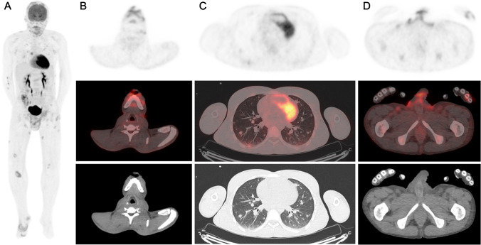
A 30-year-old man presented painful disseminated skin and genital lesions with pyrexia. The lesions appeared 1 month prior to the admission as vesicles, progressively evolving into confluent and crusted lesions. Polymerase chain reaction (PCR) assay of the lesions swab was positive for monkeypox and negative for herpes simplex virus. Laboratory investigations showed elevated C-reactive protein and lymphopenia. Human immunodeficiency virus 1/2 antibody/antigen test was reactive. CD4 count was 23.9 cells/mm3. Chest radiograph revealed multiple lung opacities. Serology was negative for syphilis, cryptococcus, toxoplasmosis, and positive for cytomegalovirus (CMV) but CMV PCR was negative. Sputum and bronchoalveolar lavage (BAL) were negative for tuberculosis and pneumocystis. [18F]-FDG PET/CT was performed for the detection of a clinically occult AIDS-related disease. Maximal intensity projection images (A) and fused axial images (B, C, D) revealed multiple hypermetabolic cutaneous lesions on the scalp, face, and limbs with corresponding skin thickening on CT; a large hypermetabolic pubic lesion extending in the subcutaneous tissues and scrotum; and numerous lung nodules with mild [18F]-FDG uptake. Mildly hypermetabolic peripheral lymph nodes were also observed. Lung nodules were compatible with viral lung disease occurring in immunocompromised patients [1, 2]. Serology, sputum, and BAL results did not suggest another etiology for this nodular pattern. Lung nodules were hence assumed as Monkeypox-related. Patterns of inflammation in lungs and of immune activation in lymph nodes on PET/CT have been previously described in non-human primates [3, 4]. To our knowledge, this is the first case of disseminated monkeypox disease with skin and lung lesions shown on [18F]-FDG PET/CT.
Author contribution
All authors made individual contributions to authorship. RM: original writing-draft, review of the literature. BH: Management of the patient. All authors reviewed, commented, and approved the final draft.
Declarations
Consent for publication
Written informed consent was obtained from the patient.
Conflict of interest
The authors declare no competing interests.
Footnotes
This article is part of the Topical Collection on Infection and inflammation.
Publisher's note
Springer Nature remains neutral with regard to jurisdictional claims in published maps and institutional affiliations.
References
- 1.Tanaka N, Kunihiro Y, Yanagawa N. Infection in immunocompromised hosts: imaging. J Thorac Imaging. 2018;33(5):306–321. doi: 10.1097/RTI.0000000000000342. [DOI] [PubMed] [Google Scholar]
- 2.Franquet T. Imaging of pulmonary viral pneumonia. Radiology. 2011;260(1):18–39. doi: 10.1148/radiol.11092149. [DOI] [PubMed] [Google Scholar]
- 3.Dyall J, Johnson RF, Chen DY, Huzella L, Ragland DR, Mollura DJ, et al. Evaluation of monkeypox disease progression by molecular imaging. J Infect Dis. 2011;204(12):1902–1911. doi: 10.1093/infdis/jir663. [DOI] [PMC free article] [PubMed] [Google Scholar]
- 4.Thomasson D, Seidel J, Ragland DR, Byrum R, Reba RC, Hammoud D, et al. crossm lymphoid tissue serves as a predictor of disease outcome in the nonhuman primate model of monkeypox virus infection. 2017;91(21):1–13. [DOI] [PMC free article] [PubMed]


