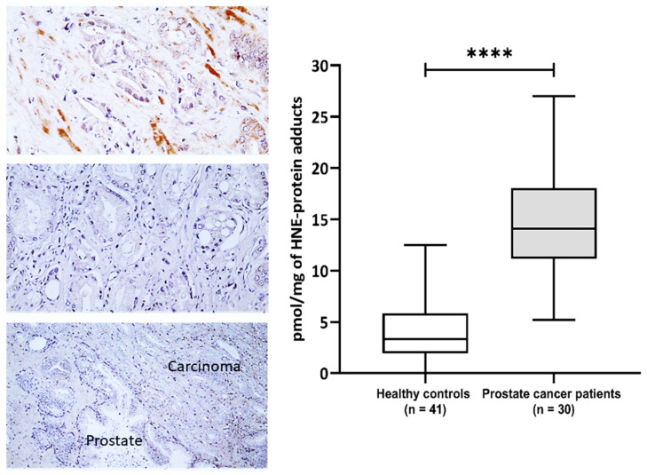
Figure 1.
The 4-hydroxynonenal (4-HNE) modification of plasma and prostate cancer tissue proteins. Left—Immunohistochemistry of prostate carcinoma specimens obtained by monoclonal antibody specific for the 4-HNE-protein adducts visualized the 4-HNE presence by brown color (DAB staining), while negative cells were contrast-stained blue by hematoxylin. Photo on the top shows rare immunopositivity of the stromal cells observed only for one patient/cancer (200×), the middle photo shows the most usually observed absence of 4-HNE in cancer and in stromal cells (200×), while the lower photo shows the absence of 4-HNE both in cancer and in the adjacent prostate tissue (100×). Right—Plasma concentration of 4-HNE–protein adducts (pmol/mg of protein) in samples of healthy controls (n = 41) and prostate cancer patients (n = 30). Results are presented as a box and whisker plot. The line in the box represents the median, while the interquartile range (IQR) box represents the middle quartiles (the 75th minus the 25th percentile). The whiskers on either side of the IQR box represent the lowest and highest quartiles of the data. The ends of the whiskers represent the maximum and minimum of the data. **** p < 0.0001.

