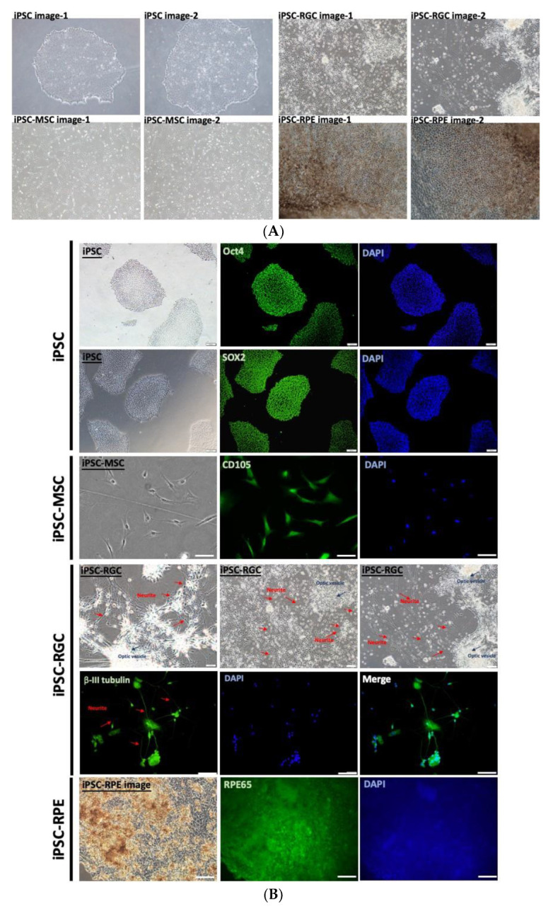
Figure 1.

Characterization of iPSCs, iPSC-MSCs, iPSC-RGCs, and iPSC-RPEs with optimal differentiation. (A) Representative images of iPSCs, iPSC-MSCs, iPSC-RGCs, and iPSC-RPEs obtained by phase-contrast microscopy. (B) Validation of the indicated cell types by immunofluorescent staining of the indicated lineage-specific markers. Nuclei stained with DAPI. Scale bars = 100 μm.
