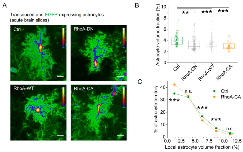
Figure 4.
Increasing astrocytic RhoA activity in vivo reduces the amount of fine astrocytic processes. For testing the effect on astrocyte morphology, the four rAAVs were injected into dorsal hippocampi of mice in which astrocytes sparsely express EGFP. Astrocyte morphology was investigated in acute slices obtained from these mice by visualizing transduced EGFP-expressing astrocytes using 2PE fluorescence microscopy. (A) Sample images (tdTomato signal not illustrated, scale bar 10 µm). (B) The volume fraction (VF) of the astrocyte excluding the soma and major branches was used to quantify the effect of RhoA variants on astrocyte morphology. Expression of all RhoA variants significantly reduced the astrocyte VF (Kruskal–Wallis test, p < 0.001; Dunn post hoc tests, p < 0.01, <0.001 and <0.001 for RhoA-DN, RhoA-WT, and RhoA-CA, respectively, ** for p < 0.01 and *** for p < 0.001). (C) For analysing the subcellular effects of RhoA expression, individual astrocytes were divided into evenly spaced regions of interest (see Materials and Methods), and the distribution of subcellular volume fractions was determined for each cell. Average distributions are calculated and displayed for RhoA-CA and control rAAVs. They reveal a shift towards lower local volume fractions when astrocytes express RhoA-CA (two-way repeated measures ANOVA, p < 0.001 for Ctrl vs. RhoA-CA; post hoc Tukey test *** for p < 0.001 and n.s. for p > 0.90; p > 0.90 for local volume fractions > 12.5%). For (B,C), slices were analysed from at least 3 animals per condition (n = 51, 57, 51, and 33 cells for Ctrl, RhoA-DN, RhoA-WT, and RhoA-CA, respectively). SO, SP, SR represent stratum oriens, stratum pyramidale, stratum radiatum, respectively. Box and Whisker plots: box indicates the 25th and 75th percentiles, whiskers the 5th and 95th percentiles, the horizontal line in the box the median, the filled circle the mean, and hollow circles the individual data points.

