Abstract
Fragile X syndrome (FXS) is a global neurodevelopmental disorder caused by the expansion of CGG trinucleotide repeats (≥200) in the Fragile X Messenger Ribonucleoprotein 1 (FMR1) gene. FXS is the hallmark of Fragile X-associated disorders (FXD) and the most common monogenic cause of inherited intellectual disability and autism spectrum disorder. There are several animal models used to study FXS. In the FXS model of Drosophila, the only ortholog of FMR1, dfmr1, is mutated so that its protein is missing. This model has several relevant phenotypes, including defects in the circadian output pathway, sleep problems, memory deficits in the conditioned courtship and olfactory conditioning paradigms, deficits in social interaction, and deficits in neuronal development. In addition to FXS, a model of another FXD, Fragile X-associated tremor/ataxia syndrome (FXTAS), has also been established in Drosophila. This review summarizes many years of research on FXD in Drosophila models.
Keywords: Fragile X syndrome, FXTAS, FMR1 gene, FMRP, Drosophila melanogaster
1. Introduction
When Morgan (1910) began his pioneering experiments in genetics in the early 1900s while working with fruit flies, he probably had no idea how important they would become over time, including studying the molecular pathogenesis of human diseases. As a model system, many advantages of the fruit fly, D. melanogaster, have made it the most important eukaryotic organism for understanding basic genetic principles, inheritance mechanisms, chromosomes and genes, and mutagenesis. These advantages include a small body size (2–3 mm), easy and inexpensive cultivation and maintenance in the laboratory, a large number of offspring per mating (~100 eggs per day), and a rapid life cycle (about ten days at 25 °C) [1]. In addition, the fruit fly, D. melanogaster, is an excellent example of the 3R principle (Replacement, Refinement, Reduction), which replaces the use of higher laboratory animals in research studies [2]. The 3R principle is based on the belief that animal species have a certain degree of intrinsic value that must be considered in order to adequately consider animal welfare [3].
The genome of D. melanogaster was sequenced and published in 2000 [4,5]. It has a size of about 180 Mb, of which about 120 Mb is euchromatin [4,6]. The predicted number of protein-coding genes is more than 14,000, together with about 3000 non-coding genes [7,8]. About 60% of all human genes and 75% of genes related to human diseases have their homologues in the fruit fly [9,10]. Although fruit flies and humans possess completely different anatomical features, they share similar and important cellular and molecular processes and biological pathways [11,12]. These highly conserved pathways are membrane excitability, synaptic plasticity, and neuronal signaling. In addition, classes of neurotransmitters, such as adrenergic, dopaminergic, serotonergic, and histaminergic, are highly conserved, too [13,14]. Some structures in the Drosophila brain also have their counterparts in mammals, such as the mushroom bodies involved in learning and memory, which correspond to the mammalian hippocampus [15]. Furthermore, because the fruit fly brain is compact and consists of more than 100,000 neurons involved in various behaviors, it is used as a model for “screening therapeutic drugs for various human neuropathies” [16].
Evolutionary conservation of gene functions has shown that some mechanisms in fruit flies apply to more complex versions of human behavioral processes [17]. In addition, the conservation of these genetic processes has also provided important insights into the underlying mechanisms of human diseases due to the alteration of typical neuronal functions [18,19]. Recent studies are also directed toward mechanisms involved in rare diseases [20]. Finally, Drosophila is an indispensable model organism for the study of the Fragile X Messenger Ribonucleoprotein 1 (FMR1) gene triplet repeats expansion in humans, clinically known as Fragile X syndrome (FXS) [21]. The Drosophila model has proven useful for early-stage pharmacological screening of drug candidates identified in the fly that need to be tested in a mammalian model of FXS [22].
2. Fragile X Syndrome
The FMR1 gene, which encodes the FMR1 protein (FMRP), is located on chromosome Xq27.3, and there are between 5 and 44 CGG trinucleotide repeats in the normal promoter region of the FMR1 gene [23,24,25]. The expansion of the CGG triplet repeats (≥200) in the 5′ untranslated region (UTR) of the FMR1 gene is a full mutation (FM). It causes FXS, the most common form of inherited intellectual disability (ID), and is a monogenic cause of autism spectrum disorder (ASD) [26]. Hypermethylation triggered by the FM of the FMR1 gene results in transcriptional silencing and a reduction or absence of FMRP [27,28]. FXS affects approximately 1 in 5000 males and 1 in 8000 females [29].
The product of the FMR1 gene, FMRP, is produced mainly in neurons (particularly in the cortex, Purkinje cells, and hippocampus) and testes and plays an important role in normal brain development [30]. FMRP is an RNA-binding protein that is mainly localized in the cytoplasm and carries a Nuclear Localization Signal (NLS) and a Nuclear Exportation Signal (NES) [31]. Fragile X Related 1 (FXR1) and Fragile X Related 2 (FXR2) are two paralogues that are highly homologous to FMRP [32]. FMRP has a specific mechanism of action: it enters the nucleus, interacts with pre-messenger ribonucleoprotein complexes (pre-mRNP), and brings them into the cytoplasm. The FMRP-mRNP complexes are associated with polyribosomes and play an important role in the translation of proteins in the neuronal soma and dendritic spines [33]. This protein also plays a role in post-transcriptional regulation and is a component of stress granules and P-bodies (reviewed in [34]). The FMRP-mRNPs complexes are also a component of RNA granules, where they mediate binding between mRNAs and kinesins and play a role in cell transport [35]. In the last decade, other FMRP targets have also been identified, and studies have shown that FMRP also plays a role in the microRNA (miRNA) and Piwi-interacting RNA (piRNA) pathways [21,36,37]. Some of these targets are involved in neurodevelopmental disorders such as idiopathic ASD and other neuronal pathologies [21].
As mentioned above, FMRP deficiency is associated with a wide range of neurobehavioral clinical features of FXS, which include physical, cognitive, and behavioral abnormalities (i.e., 50–60% are diagnosed with ASD) [38]. Characteristic physical features such as an elongated face, large or protruding ears, a high-arched palate, joint hypermobility, and macroorchidism at and after puberty are present in most individuals with FXS [39]. Behavioral features include shyness, social anxiety, attention deficits, hyperactivity, disturbed eye gaze, sensory overexcitation, aggressive behavior, sleep problems, hand flapping, repetitive behaviors, and obsessive-compulsive disorder [26,40,41,42,43]. Seizures occur in about 15–20% of people with FXS [44], while obesity and gastrointestinal dysfunction are diagnosed in over 30% of patients [45]. Men with FXS have an average Full Scale IQ (FSIQ) between 40 and 50 and are more severely affected than women with FXS, who have an average FSIQ between 70 and 80, although 1/3 have an IQ below 70 [46].
3. Drosophila Model for the Study of Fragile X Syndrome
Numerous animal models of FXS include Fmr1 knock-out (KO) mice (discovered more than 20 years ago), rats, the D. melanogaster model of FXS, and Fmr1 KO zebrafish [21,47]. All these models are based on the disruption or KO of the FMR1 gene homologue. However, there are no naturally occurring animal models for FXS [22]. This review focuses on using the Drosophila FXS model to study FXS.
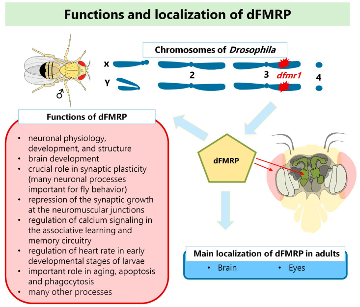
The FMR1 homolog in Drosophila was identified in 2000 and designated dfmr1 [48]. In the meantime, there have been a few different names for this homolog (for more details, please visit flybase.org, accessed on 1. December 2022), and the current official name is Fmr1. Nevertheless, in this review, we mark it as dfmr1 to avoid confusion and to distinguish it from the human FMR1 gene. dfmr1 is 8.7 kb long, and all of its functional domains are highly conserved, with two KH domains being 35% identical and 60% similar between dfmr1 and human FMR1 [48]. The encoded protein, dFMRP, is found in adults’ brains, eyes, and mushroom bodies [21,49,50,51,52]. Some studies have also described dFMRP in Drosophila embryos and larvae [21]. The function of dFMRP in neuronal physiology, development, and structure is extensively studied in fruit fly larvae and adults. According to FlyBase data, dFMRP is involved in more than 50 biological processes with neuronal and non-neuronal functions in Drosophila [21]. The most important of these is its critical role in synaptic plasticity [21]. dFMRP plays an important role in aging, apoptosis, phagocytosis, and many other processes, too [21]. Figure 1 shows a schematic representation of the main localizations and the functions of dFMRP.
Figure 1.
Functions and localization of dFMRP in D. melanogaster (adapted from reference [21]) Aberrations: dfmr1-Fragile X Messenger Ribonucleoprotein 1 gene in Drosophila; dFMRP-dfmr1 protein in D. melanogaster.
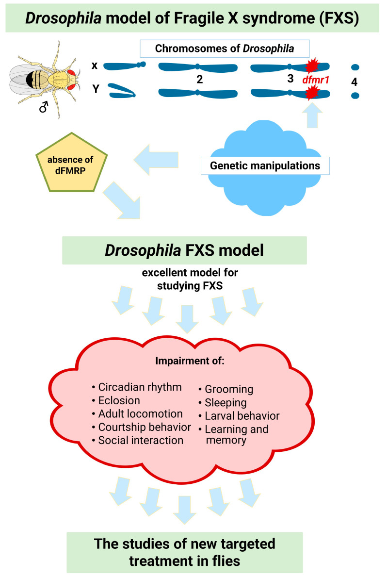
The range of genetic tools available when using Drosophila is greater than for any other multicellular organism, and there is a wide repertoire of available genetic manipulations [12]. Point mutations in dfmr1 or the absence of all or most of the coding region of the dfmr1 gene form the basis of the Drosophila FXS model [49,50,53,54,55]. Therefore, the first dfmr1 mutant, i.e., the Drosophila FXS model, was generated by inaccurately excising a P element upstream of the dfmr1 locus [49].
dfmr1 mutants exhibit defective neuronal architecture and synaptic function. Furthermore, these mutants exhibit abnormally organized synapses in the peripheral and central nervous systems. The absence of dFMRP causes marked synaptic overgrowth at the neuromuscular junctions of Drosophila larvae [49,52,56,57]. The larval crawling pattern is also altered in Drosophila FXS models [58]. In another study, dfmr1 larvae showed reduced motility during reorientation but normal motility during active crawling [59]. In addition, dfmr1 mutants show altered behaviors such as irregular circadian rhythms, decreased male courtship activity, increased locomotion, learning and memory deficits, autism-like behaviors such as abnormal grooming, and social deficits [50,60,61,62,63]. Since the symptoms of FXS may be linked to the phenotypes of dfmr1 mutants [21], the fruit fly model of FXS (Figure 2) is a robust model for studying FXS [22].
Figure 2.
Drosophila model of fragile X syndrome (adapted from reference [21]). Aberrations: dfmr1-Fragile X Messenger Ribonucleoprotein 1 gene in Drosophila; dFMRP-dfmr1 protein in Drosophila).
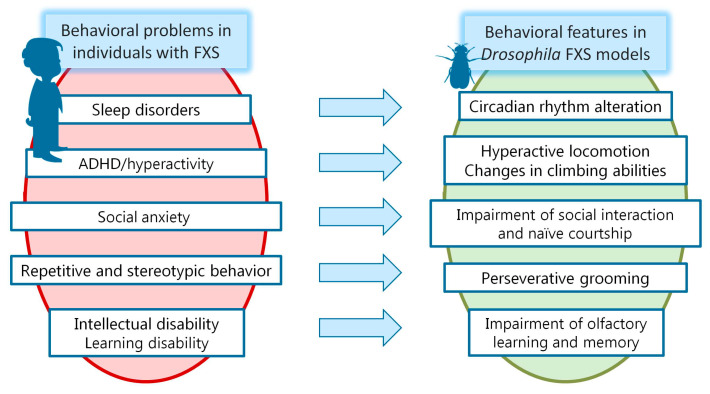
Here we have discussed the pathological phenotype observed in humans with FXS. Figure 3 shows the main behavioral phenotypes in humans with FXS and the overlapping loss-of-function phenotypes in dfmr1 mutants.
Figure 3.
Major phenotypes in humans with FXS and overlapping loss-of-function phenotypes in dfmr1 mutants.
3.1. Impaired Circadian Rhythms and Sleep Problems in FXS
Sleep problems are common in individuals with FXS. To illustrate, between 23% and 46% of individuals (predominantly younger males) with FXS have relatively mild sleep difficulties [43]. Another study found that the prevalence of sleep problems in individuals with FXS and ASD is about 80% higher than in the general population [64]. These problems may lead to circadian fluctuations and altered glucose homeostasis in individuals with FXS [65]. A consistent behavioral abnormality in the FXS model of Drosophila is altered circadian rhythm behavior (reviewed in [21]). The altered circadian rhythm in this animal model potentially mimics the sleep abnormalities observed in patients with FXS [50]. Circadian rhythms in Drosophila models manifest in locomotor activity, sleep/wake patterns, and various physiological and metabolic processes. Xu and colleagues (2012) described a role for dfmr1 in miRNA pathways, pointing to the altered expression of selected miRNAs in the circadian abnormalities associated with the loss of dfmr1 in flies [66]. Furthermore, Bushey and colleagues described that dfmr1 regulates sleep needs in Drosophila FXS models [67].
A study by Dockendorff et al. (2002) showed that pupae from Drosophila larval control groups retained their ability to eclose. They were kept under 12 h of light/12 h of darkness conditions (for five days and then in complete darkness for six days), mostly locked into their circadian gap early in the morning with a duration of 23.5 h. In contrast, Drosophila FXS models under these conditions generally locked in later in the day and had a longer eclosion time with lower amplitude [54]. In another study, eclosion time in the dfmr1 mutant remained dependent on the circadian rhythm, which was delayed by 6–8 h [50]. In contrast, Inoue et al. (2002) showed that the emergence of dfmr1B55 pupae was dependent on the circadian rhythm, with a similar phase and amplitude to wild type (wt) [53]. This inconsistency could be explained by the discovery made by Sekine et al. (2008) that the variations in eclosion time in FXS model flies are a consequence of the genetic background and not the deletion itself. When dfmr1B55 mutants were backcrossed to Canton S (CS) and yellow white (YW) flies, mutants with the same deletion from different genetic backgrounds were produced. These mutants did not exhibit eclosion abnormalities, suggesting that the eclosion deficit was due to the genetic background [68]. In addition, another study showed that several different types of Drosophila FXS models often do not survive because they fail to eclose. This phenomenon was particularly dominant in the dfxrΔ50 and dfxrΔ113 alleles, where 99% of flies failed to hatch [50].
To investigate circadian rhythms in more detail, the investigators conducted similar studies to see if the FXS flies exhibited circadian defects in locomotor activity. Interestingly, the locomotor activity of the Drosophila FXS models under 12 h light/12 h dark was similar to that of the control groups. However, once the flies in the control groups became accustomed to the regular alternation of day and night, they could maintain their rhythmic locomotor behavior for a certain period, even when walking freely in complete darkness. In contrast, the FXS models lacked this ability [4,50,53,69]. Notably, 20–30% of FXS models remained rhythmic despite constant darkness [68]. When dfmr1B55 mutants were backcrossed to CS and YW Drosophila strains for seven generations, the mutants maintained arrhythmic behavior in both genetic backgrounds, demonstrating that in this case, the genetic background does not affect circadian defects [68]. Nevertheless, a recent study challenged previous findings on circadian rhythms, suggesting that they may be influenced by excessive grooming in FXS Drosophila [70].
Drosophila FXS models have prolonged sleep compared to wt primarily due to an increased number of sleep episodes. On average, females slept 3 h longer and males 4 h longer than flies from control groups. After the end of the dark period, they woke up later than the control groups and slept longer during the day. In addition, their sleep was deeper, and they were less likely to wake up briefly [67]. Even their naps during the day were unusually deep and resembled night sleep [71]. In addition, null mutants showed defects in recovery from sleep deprivation by having shorter sleep episodes and a lower amount of recovered sleep. In contrast, overexpression of dfmr1 results in shorter sleep [67].
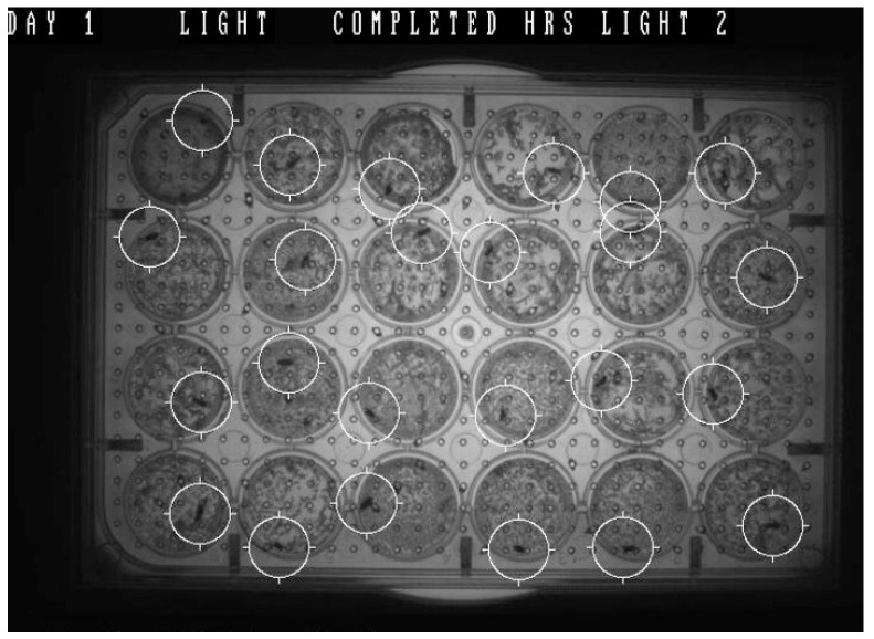
Nowadays, there are a variety of software and systems for tracking locomotor activity and circadian rhythms in small model organisms such as Drosophila. The circadian rhythms of flies may be measured by recording their locomotor activity over time, and they are normally entrained by light intensity. Such advanced systems can be used to measure the activity of experimental models and also control (and change) the lighting regime. Figure 4 shows an example of tracking Drosophila in one of these types of software.
Figure 4.
An example of the study of circadian rhythms in Drosophila using the software. (The image is from Protic’s lab: www.polyfrax.com, accessed on 1. December 2022.). A multi-well plate was prepared. Depending on experimental needs, the plates can be 24-, 48-, or 96- well. The flies were placed individually in each well, and an air-permeable cover was put over the top of the well plate. The flies were then individually housed in a humid environment with some available nutrition. The well plate was loaded with flies and inserted into the system’s chamber. The software was set up to control the environment (temperature and lighting) and the measure of the distance traveled by each fly. The white cycles show the flies as software targets in 24 wells. The activity (measured as distance traveled) is measured and recorded as data during the experiment.
3.2. Hyperactivity and Attention Deficit/Hyperactivity Disorder in FXS
Attention deficit/hyperactivity disorder (ADHD) is one of the most common behavioral problems in individuals with FXS [39,72]. Thus, hyperactivity of locomotion has been used to assess drug efficacy in preclinical studies with animal models of FXS. In addition, hyperactive locomotion/climbing in a Drosophila model can mimic hyperactivity or ADHD in individuals [73,74,75].
The measurement of overall activity in the Drosophila FXS model has been inconsistent across studies. According to Dockendorff et al. (2002), total activity in Drosophila FXS models was not significantly different from control groups [54], but in another study, Morales et al. (2002) found that it decreased [50]. Another study showed lower locomotor activity in the dfmr1B55 mutant than in the wt. dfmrB55 by covering the space less well overall and making more stops (mostly at specific points) than the wt. In the same study, dfmr13 covered the space more evenly but had more stops than the wt [62]. In addition, Drosophila FXS models had defects in flight ability, which were measured in the flight experiment [49]. An interesting finding linking Drosophila FXS models to FXS in humans and its model in Fmr1 KO mice was relatively high bursts of activity. This behavior in Drosophila may correspond to the hyperactivity in the Fmr1 KO mouse and humans with FXS [54].
One study examined changes in climbing ability in Drosophila FXS models with aging. Flies with a deletion in dfmr1 were compared with two strains commonly used for laboratory purposes and one strain specific for its longevity. The climbing performance of the Drosophila FXS models decreased dramatically with age. Interestingly, the fastest 5-day-old Drosophila FXS models showed the same performance as other genotypes studied, but their climbing ability dramatically reduced at 25 days of age after eclosion. In contrast, the other genotypes gradually declined with age. Drosophila FXS models were found to be poor climbers at all ages in population studies and had the highest failure rate in completing the task among the flies studied [75].
3.3. Other Autistic-Like Behaviors in FXS
It is known that 50–60% of boys and 20% of girls with FXS are diagnosed with ASD [72,76]. Here we have discussed additional behavioral phenotypes that occur in the Drosophila FXS model that overlap with autistic phenotypes in individuals with FXS.
Social interaction in the Drosophila FXS model. Impaired social interactions are a characteristic feature of ASD [77]. The study’s results examining social interaction between two female Drosophila FXS models and their interaction with the wt model indicated normal receptive but altered expressive social behavior in the Drosophila FXS models. One possible explanation is that the models do not exhibit appropriate motor behavior or chemical signals necessary for social interaction [62]. Interestingly, dfmr13 did not spend much time at the boundary, regardless of whether the other chamber contained a mutant fly of the same type or the wt fly. Measuring the distance between the flies and the possibility that two flies were less than 5 mm apart confirmed these results [62].
Grooming in the FXS model of Drosophila. Excessive grooming in dfmr1 mutant flies appears to reflect the hyperactive and ASD-like features of FXS observed in mice and humans [78].
According to the study data, the frequency and duration of grooming were significantly higher in 1-day-old mutants than in wt. The grooming index, i.e., the time spent grooming during a given time interval, was also significantly increased. The grooming pattern was altered such that the mutants groomed the posterior parts excessively, while the grooming of the anterior parts was similar to wt [70]. In another study, grooming increased progressively with age [78]. Moreover, excessive grooming was found to be persistent and structured. Namely, in the mutants, the switching between grooming pairs was excessively repeated during a run. Moreover, these FXS flies tended to start another grooming session with the same body part they had finished in the last session. Perseverative grooming corresponds to repetitive and stereotypical behaviors characteristic of ASD [70].
Courtship behavior in the FXS model of Drosophila. Inappropriate courtship behavior is described in individuals with ASD [79]. Naive courtship behavior is also altered in Drosophila FXS models [54,80,81,82,83]. Indeed, courtship in Drosophila consists of several phases leading to copulation. The male orients himself toward the female and follows her, then taps her with his legs, vibrates with his wing, licks her genital region, and finally attempts copulation [84]. Naive courtship behavior is estimated by the courtship index (CI), the percentage of time the male spends courting the female. In the Drosophila FXS models, the CI index was reduced, and they could not sustain courtship sufficiently to achieve more advanced phases of courtship, such as wing vibration, genital licking, and copulation attempts [54]. In the normal Drosophila population, other males sometimes court immature males. Still, FXS males courted immature males for less time than the control groups and persisted for less time in advanced phases [54]. The fact that immature male pheromones differ from female pheromones led to the conclusion that the courtship defect is not the result of specific sensory defects that may be present in Drosophila FXS models [54]. Furthermore, in one study, naïve courtship was impaired in both young and aged Drosophila FXS models, but the difference from the control groups was less marked in aged Drosophila. The explanation for this finding is naïve courtship also decreases with age in wt flies [80].
3.4. ID in FXS and Learning and Memory Impairment in the FXS Model of Drosophila
It is known that FXS is the leading cause of the inherited form of ID, and the deficit/absence of FMRP has been described as the core cause of ID in FXS. Although the relationship between FMR1 expansion, gene methylation, and FMRP deficit is well known, the relationship between FMRP and ID needs more studies [85,86]. Only some assays exist to study learning and memory in Drosophila FXS models.
Almost 20 years ago, McBride and colleagues (2005) were the first to study learning and memory in Drosophila dfmr1 mutants. They used the courtship paradigm to study associative memory. The complicated procedure of the experiment involved one hour of training a virgin male with an unresponsive female. Continuous rejection should teach memory-intact males to stop courting. Time spent courting was reduced in dfmr1 mutants and control groups, implying that FXS flies can learn during training. This form of memory is a mixture of associative and non-associative memory [82]. Although learning is preserved in young FXS flies, FXS models lose this function at 20 days of age when tested in the same assay [80]. To examine only associative memory, these researchers placed flies that had learned to be rejected in the chamber with receptive females. Tests were performed immediately after training. The results showed that the males in the Drosophila FXS models forgot what they had learned and attempted to copulate as often as the control groups. This study was the first to identify defects in immediate recall memory, memory lasting 0–2 min after training, and short-term memory that lasts up to one hour after training in FXS Drosophila [82]. Long-term memory can also be examined using tests based on the courtship paradigm. Using this test, Banerjee et al. (2010) demonstrated that long-term training memory is impaired in FXS models of Drosophila [81].
Another popular method for assessing memory in Drosophila is based on classical conditioning. In this test, flies were exposed to two odors. The first odor was followed by an electric shock, while the second was not. The trained flies were then placed in a T-maze to choose between the previously exposed odors. Learning and memory were assessed in the next step by measuring the percentage of flies in the chamber with an odor that was not followed by the electric shock. Learning was assessed immediately after training, and memory after a while, depending on the type of memory [69]. Caffe et al. (2012) found that learning ability decreased significantly in FXS models of Drosophila compared to wt [87]. This test could estimate long-term memory when performed repeatedly. When the training was performed ten times without interruption, a decremental cycloheximide-insensitive memory (ARM) was formed. The same procedure, with a 15-min break between training sessions, would also develop ARM, but non-decremental cycloheximide-sensitive long-term memory (LTM) was also developed. The latter form of memory was dependent on protein synthesis. When Drosophila FXS models were placed in the T-maze one day after repeated training, they showed defects in LTM but not ARM. Finally, silencing of dfmr1 only in mushroom bodies showed the same results [61].
4. D. melanogaster as a Model Organism to Study Fragile X-Associated Tremor/Ataxia Syndrome (FXTAS)
As described above, the normal range of CGG repeats in the human FMR1 gene is between 5 and 44. The premutation range (PM) of the FMR1 gene is characterized by 55–200 CGG repeats, and those carriers typically do not present with FXS symptoms. However, they are associated with three other disorders: Fragile X-associated primary ovarian insufficiency (FXPOI) [88], Fragile X-associated tremor/ataxia syndrome (FXTAS) [89,90], and Fragile X-associated neuropsychiatric disorders (FXAND) [91].
FXTAS is a progressive neurodegenerative disorder that occurs in approximately 40% of males and 16% of female carriers. Individuals diagnosed with FXTAS present with a progressive intention tremor, difficulty with ambulation, ataxia, deficits in executive function, and brain atrophy associated with elevated FMR1 mRNA levels [90,92]. The prevalence of FXTAS increases with age. A study of PM men showed that 17% were affected at age 50, 38% at age 60, 47% at age 70, and 75% at age 80 [93]. In contrast to FM of FMR1, which results in transcriptional silencing of FMR1 mRNA and a concomitant loss of FMRP, in FXTAS, there are normal FMRP levels or a modest reduction in the high PM repeat range. However, in PM carriers, there is a dramatic increase in FMR1 mRNA levels, leading to mRNA toxicity and the pathogenesis of FXTAS [72,94]. Elevated mRNA levels lead to increased Ca2+ levels in the neuron and subsequent mitochondrial dysfunction. Proteins and RNAs are sequestered in inclusion bodies. The formation of R-loops leads to DNA damage. RAN translation leads to the production of toxic polyglycine-containing (FMRpolyG) proteins [72,94].
Jin and colleagues first described the Drosophila model of FXTAS in 2003. The PM range in dfmr1 alone is sufficient to cause neurodegeneration [95]. Therefore, FXTAS has been modeled in Drosophila by overexpressing 90 CGG repeats fused with a green fluorescent protein (GFP), resulting in neuron-specific degeneration and inclusion formation [95,96]. Notably, FMRpolyG is toxic and directly influences the toxicity of CGG repeat constructs in Drosophila [97]. In addition, Jin and colleagues described that the CGG-induced neurodegenerative phenotype in the Drosophila FXTAS model could be rescued by overexpression of purα [98]. Other studies identified some tropomyosin and RNA-binding proteins as genetic modifiers of neurodegeneration in the Drosophila FXTAS model [98,99].
Flies with modest PM in dfmr1 (rCGG90 repeats) exclusively in neurons do not reach adulthood. Lethality occurs mainly during embryonic development prior to larval formation [95]. This model shows deficits in locomotion and retinal degeneration [95].
5. Conclusions and Future Perspectives
Currently, there is no cure for FXS in patients, but the studies of medication use in Drosophila have been helpful in initiating medication core modifier clinical trials that work well in the fly and transitioning such treatments to humans with FXS. Examples of this include the minocycline studies in flies that were translated to humans, demonstrating behavioral benefits [100]. Another example is the benefit of metformin in flies [101], which led to clinical trials in humans with FXS [102,103,104,105]. This review has emphasized the simplicity and cost-effectiveness of preclinical trials in flies to encourage young researchers to further the studies of new targeted treatments in flies that can be translated to patients with FXS. Since the lack of FMRP has profound effects on many systems paralleled in flies and humans, this animal model will be utilized many times in the future to guide new treatments for FXS and possibly FXTAS.
Acknowledgments
This publication is supported by the Science Fund of the Republic of Serbia, Program IDEA, GRANT No. 7673781, “Polyphenols as potential targeted treatments in D. melanogaster model of fragile X syndrome”, POLYFRAX_Drosophila and by The Serbian Ministry of Education, Science and Technological Development (Contract Number: 451-03-68/2022-14/200178). The authors are solely responsible for the content of this publication, and this content does not express the attitudes of the Science Fund of the Republic of Serbia.
Author Contributions
Conceptualization, D.P., J.T. and D.B.B.; investigation, J.T., V.M., M.P., S.P.-L., S.M. and S.C.; writing—original draft preparation, D.P., J.T., V.M., M.P., S.P.-L., S.M. and S.C.; writing—review and editing, D.P., D.B.B. and R.H. All authors have read and agreed to the published version of the manuscript.
Institutional Review Board Statement
Not applicable.
Informed Consent Statement
Not applicable.
Data Availability Statement
Not applicable.
Conflicts of Interest
Dejan B. Budimirovic has received funding from Zynerba Pharmaceuticals (ZYN2-CL-017, ZYN2-CL-033) as a principal investigator on clinical trials in FXS. He also consulted on clinical trial outcome measures in FXS (Seaside, Ovid). All the above funding has been directed to the Kennedy Krieger Institute/the Johns Hopkins Medical Institutions; Dejan B. Budimirovic receives no personal funds, and the Institute has no relevant financial interest in any of the commercial entities listed. Other authors declare no conflict of interest.
Funding Statement
This research was funded by the Science Fund of the Republic of Serbia, Program IDEA, GRANT No. 7673781 and by The Serbian Ministry of Education, Science and Technological Development (Contract Number: 451-03-68/2022-14/200178).
Footnotes
Disclaimer/Publisher’s Note: The statements, opinions and data contained in all publications are solely those of the individual author(s) and contributor(s) and not of MDPI and/or the editor(s). MDPI and/or the editor(s) disclaim responsibility for any injury to people or property resulting from any ideas, methods, instructions or products referred to in the content.
References
- 1.King R.C. Genetics. 2nd ed. Oxford University Press; New York, NY, USA: 1965. pp. 1–462. [Google Scholar]
- 2.Fuentes E., Sivakumar N., Selvik L.-K., Arch M., Cardona P.J., Ioerger T.R., Dragset M.S. Drosophila melanogaster is a powerful host model to study mycobacterial virulence. bioRxiv. 2022 doi: 10.1101/2022.05.12.491628. [DOI] [Google Scholar]
- 3.Russell W.M.S., Burch R.L. The Principles of Humane Experimental Technique. Methuen & Co, Ltd.; North Yorkshire, UK: 1959. p. 238. [Google Scholar]
- 4.Adams M.D., Celniker S.E., Holt R.A., Evans C.A., Gocayne J.D., Amanatides P.G., Scherer S.E., Li P.W., Hoskins R.A., Galle R.F., et al. The genome sequence of Drosophila melanogaster. Science. 2000;287:2185–2195. doi: 10.1126/science.287.5461.2185. [DOI] [PubMed] [Google Scholar]
- 5.Myers E.W., Sutton G.G., Delcher A.L., Dew I.M., Fasulo D.P., Flanigan M.J., Kravitz S.A., Mobarry C.M., Reinert K.H., Remington K.A., et al. A whole-genome assembly of Drosophila. Science. 2000;287:2196–2204. doi: 10.1126/science.287.5461.2196. [DOI] [PubMed] [Google Scholar]
- 6.Celniker S.E., Wheeler D.A., Kronmiller B., Carlson J.W., Halpern A., Patel S., Adams M., Champe M., Dugan S.P., Frise E., et al. Finishing a whole-genome shotgun: Release 3 of the Drosophila melanogaster euchromatic genome sequence. Genome Biol. 2002;3:research0079.1. doi: 10.1186/gb-2002-3-12-research0079. [DOI] [PMC free article] [PubMed] [Google Scholar]
- 7.Brown J.B., Boley N., Eisman R., May G.E., Stoiber M.H., Duff M.O., Booth B.W., Wen J., Park S., Suzuki A.M., et al. Diversity and dynamics of the Drosophila transcriptome. Nature. 2014;512:393–399. doi: 10.1038/nature12962. [DOI] [PMC free article] [PubMed] [Google Scholar]
- 8.Kaufman T.C. A Short History and Description of Drosophila melanogaster Classical Genetics: Chromosome Aberrations, Forward Genetic Screens, and the Nature of Mutations. Genetics. 2017;206:665–689. doi: 10.1534/genetics.117.199950. [DOI] [PMC free article] [PubMed] [Google Scholar]
- 9.Yamamoto S., Jaiswal M., Charng W.-L., Gambin T., Karaca E., Mirzaa G., Wiszniewski W., Sandoval H., Haelterman N.A., Xiong B., et al. A Drosophila Genetic Resource of Mutants to Study Mechanisms Underlying Human Genetic Diseases. Cell. 2014;159:200–214. doi: 10.1016/j.cell.2014.09.002. [DOI] [PMC free article] [PubMed] [Google Scholar]
- 10.Ugur B., Chen K., Bellen H.J. Drosophila tools and assays for the study of human diseases. Dis. Model. Mech. 2016;9:235–244. doi: 10.1242/dmm.023762. [DOI] [PMC free article] [PubMed] [Google Scholar]
- 11.Gehring W.J., Kloter U., Suga H. Current Topics in Developmental Biology. Volume 88. Elsevier; Amsterdam, The Netherlands: 2009. Chapter 2 Evolution of the Hox Gene Complex from an Evolutionary Ground State; pp. 35–61. [DOI] [PubMed] [Google Scholar]
- 12.Jennings B.H. Drosophila–a versatile model in biology & medicine. Mater. Today. 2011;14:190–195. [Google Scholar]
- 13.O’Kane C.J. Drosophila as a model organism for the study of neuropsychiatric disorders. Curr. Top Behav. Neurosci. 2011;7:37–60. doi: 10.1007/7854_2010_110. [DOI] [PubMed] [Google Scholar]
- 14.Nitta Y., Sugie A. Studies of neurodegenerative diseases using Drosophila and the development of novel approaches for their analysis. Fly. 2022;16:275–298. doi: 10.1080/19336934.2022.2087484. [DOI] [PMC free article] [PubMed] [Google Scholar]
- 15.Campbell R.A.A., Turner G.C. The mushroom body. Curr. Biol. 2010;20:R11–R12. doi: 10.1016/j.cub.2009.10.031. [DOI] [PubMed] [Google Scholar]
- 16.Yamaguchi M., Yoshida H. Drosophila as a Model Organism. In: Yamaguchi M., editor. Drosophila Models for Human Diseases. Volume 1076. Springer; Singapore: 2018. pp. 1–10. Advances in Experimental Medicine and, Biology. [DOI] [PubMed] [Google Scholar]
- 17.Bellen H.J., Tong C., Tsuda H. 100 years of Drosophila research and its impact on vertebrate neuroscience: A history lesson for the future. Nat. Rev. Neurosci. 2010;11:514–522. doi: 10.1038/nrn2839. [DOI] [PMC free article] [PubMed] [Google Scholar]
- 18.Fortini M.E., Bonini N.M. Modeling human neurodegenerative diseases in Drosophila: On a wing and a prayer. Trends Genet. 2000;16:161–167. doi: 10.1016/S0168-9525(99)01939-3. [DOI] [PubMed] [Google Scholar]
- 19.Muqit M.M.K., Feany M.B. Modelling neurodegenerative diseases in Drosophila: A fruitful approach? Nat. Rev. Neurosci. 2002;3:237–243. doi: 10.1038/nrn751. [DOI] [PubMed] [Google Scholar]
- 20.Verheyen E.M. The power of Drosophila in modeling human disease mechanisms. Dis. Model. Mech. 2022;15:dmm049549. doi: 10.1242/dmm.049549. [DOI] [PMC free article] [PubMed] [Google Scholar]
- 21.Drozd M., Bardoni B., Capovilla M. Modeling Fragile X Syndrome in Drosophila. Front. Mol. Neurosci. 2018;11:124. doi: 10.3389/fnmol.2018.00124. [DOI] [PMC free article] [PubMed] [Google Scholar]
- 22.Curnow E., Wang Y. New Animal Models for Understanding FMRP Functions and FXS Pathology. Cells. 2022;11:1628. doi: 10.3390/cells11101628. [DOI] [PMC free article] [PubMed] [Google Scholar]
- 23.Verkerk A.J., Pieretti M., Sutcliffe J.S., Fu Y.H., Kuhl D.P., Pizzuti A., Reiner O., Richards S., Victoria M.F., Zhang F.P., et al. Identification of a gene (FMR-1) containing a CGG repeat coincident with a breakpoint cluster region exhibiting length variation in fragile X syndrome. Cell. 1991;65:905–914. doi: 10.1016/0092-8674(91)90397-H. [DOI] [PubMed] [Google Scholar]
- 24.Weiler I.J., Irwin S.A., Klintsova A.Y., Spencer C.M., Brazelton A.D., Miyashiro K., Comery T.A., Patel B., Eberwine J., Greenough W.T. Fragile X mental retardation protein is translated near synapses in response to neurotransmitter activation. Proc. Natl. Acad. Sci. USA. 1997;94:5395–5400. doi: 10.1073/pnas.94.10.5395. [DOI] [PMC free article] [PubMed] [Google Scholar]
- 25.Feng Y., Gutekunst C.A., Eberhart D.E., Yi H., Warren S.T., Hersch S.M. Fragile X mental retardation protein: Nucleocytoplasmic shuttling and association with somatodendritic ribosomes. J. Neurosci. 1997;17:1539–1547. doi: 10.1523/JNEUROSCI.17-05-01539.1997. [DOI] [PMC free article] [PubMed] [Google Scholar]
- 26.Hagerman R.J., Hagerman P.J. Testing for fragile X gene mutations throughout the life span. JAMA. 2008;300:2419–2421. doi: 10.1001/jama.2008.684. [DOI] [PMC free article] [PubMed] [Google Scholar]
- 27.Sutcliffe J.S., Nelson D.L., Zhang F., Pieretti M., Caskey C.T., Saxe D., Warren S.T. DNA methylation represses FMR-1 transcription in fragile X syndrome. Hum. Mol. Genet. 1992;1:397–400. doi: 10.1093/hmg/1.6.397. [DOI] [PubMed] [Google Scholar]
- 28.Saldarriaga W., Tassone F., González-Teshima L.Y., Forero-Forero J.V., Ayala-Zapata S., Hagerman R. Fragile X Syndrome. Colomb. Med. 2014;45:190–198. doi: 10.25100/cm.v45i4.1810. [DOI] [PMC free article] [PubMed] [Google Scholar]
- 29.Tassone F., Iong K.P., Tong T.H., Lo J., Gane L.W., Berry-Kravis E., Nguyen D., Mu L.Y., Laffin J., Bailey D.B., et al. FMR1 CGG allele size and prevalence ascertained through newborn screening in the United States. Genome Med. 2012;4:100. doi: 10.1186/gm401. [DOI] [PMC free article] [PubMed] [Google Scholar]
- 30.Devys D., Lutz Y., Rouyer N., Bellocq J.P., Mandel J.L. The FMR-1 protein is cytoplasmic, most abundant in neurons and appears normal in carriers of a fragile X premutation. Nat. Genet. 1993;4:335–340. doi: 10.1038/ng0893-335. [DOI] [PubMed] [Google Scholar]
- 31.Bardoni B., Mandel J.L., Fisch G.S. FMR1 gene and fragile X syndrome. Am. J. Med. Genet. 2000;97:153–163. doi: 10.1002/1096-8628(200022)97:2<153::AID-AJMG7>3.0.CO;2-M. [DOI] [PubMed] [Google Scholar]
- 32.Hoogeveen A.T., Willemsen R., Oostra B.A. Fragile X syndrome, the Fragile X related proteins, and animal models. Microsc. Res. Tech. 2002;57:148–155. doi: 10.1002/jemt.10064. [DOI] [PubMed] [Google Scholar]
- 33.Bardoni B., Davidovic L., Bensaid M., Khandjian E.W. The fragile X syndrome: Exploring its molecular basis and seeking a treatment. Expert Rev. Mol. Med. 2006;8:1–16. doi: 10.1017/S1462399406010751. [DOI] [PubMed] [Google Scholar]
- 34.Maurin T., Zongaro S., Bardoni B. Fragile X Syndrome: From molecular pathology to therapy. Pt 2Neurosci. Biobehav. Rev. 2014;46:242–255. doi: 10.1016/j.neubiorev.2014.01.006. [DOI] [PubMed] [Google Scholar]
- 35.Davidovic L., Jaglin X.H., Lepagnol-Bestel A.M., Tremblay S., Simonneau M., Bardoni B., Khandjian E.W. The fragile X mental retardation protein is a molecular adaptor between the neurospecific KIF3C kinesin and dendritic RNA granules. Hum Mol. Genet. 2007;16:3047–3058. doi: 10.1093/hmg/ddm263. [DOI] [PubMed] [Google Scholar]
- 36.Kelley K., Chang S.J., Lin S.L. Mechanism of repeat-associated microRNAs in fragile X syndrome. Neural Plast. 2012;2012:104796. doi: 10.1155/2012/104796. [DOI] [PMC free article] [PubMed] [Google Scholar]
- 37.Specchia V., D’Attis S., Puricella A., Bozzetti M.P. dFmr1 Plays Roles in Small RNA Pathways of Drosophila melanogaster. Int. J. Mol. Sci. 2017;18:1066. doi: 10.3390/ijms18051066. [DOI] [PMC free article] [PubMed] [Google Scholar]
- 38.Budimirovic D.B., Subramanian M. Neurobiology of Autism and Intellectual Disability: Fragile X Syndrome. In: Johnston M., Michael A.M. Jr., Fatemi M.H., Ali M.B.A., editors. Neurobiology of Disease. 2nd ed. Oxford University Press; New York, NY, USA: 2016. pp. 375–384. [Google Scholar]
- 39.Hagerman R.J., Berry-Kravis E., Hazlett H.C., Bailey D.B., Jr., Moine H., Kooy R.F., Tassone F., Gantois I., Sonenberg N., Mandel J.L., et al. Fragile X syndrome. Nat. Rev. Dis. Primers. 2017;3:17065. doi: 10.1038/nrdp.2017.65. [DOI] [PubMed] [Google Scholar]
- 40.Bailey D.B., Jr., Raspa M., Olmsted M., Holiday D.B. Co-occurring conditions associated with FMR1 gene variations: Findings from a national parent survey. Am. J. Med. Genet. A. 2008;146A:2060–2069. doi: 10.1002/ajmg.a.32439. [DOI] [PubMed] [Google Scholar]
- 41.Kaufmann W.E., Kidd S.A., Andrews H.F., Budimirovic D.B., Esler A., Haas-Givler B., Stackhouse T., Riley C., Peacock G., Sherman S.L., et al. Autism Spectrum Disorder in Fragile X Syndrome: Cooccurring Conditions and Current Treatment. Pediatrics. 2017;139((Suppl. S3)):S194–S206. doi: 10.1542/peds.2016-1159F. [DOI] [PMC free article] [PubMed] [Google Scholar]
- 42.Budimirovic D., Haas-Givler B., Blitz R., Esler A., Kaufmann W., Sudhalter V., Stackhouse T.M., Scharfenaker S.K., Berry-Kravis E. Autism Spectrum Disorder in Fragile X Syndrome. [(accessed on 5 October 2022)]. Available online: https://fragilex.org/wp-content/uploads/2012/08/Autism-Spectrum-Disorder-in-Fragile-X-Syndrome-2014-Nov.pdf.
- 43.Budimirovic D.B., Protic D.D., Delahunty C.M., Andrews H.F., Choo T.H., Xu Q., Berry-Kravis E., Kaufmann W.E., Consortium F. Sleep problems in fragile X syndrome: Cross-sectional analysis of a large clinic-based cohort. Am. J. Med. Genet. A. 2022;188:1029–1039. doi: 10.1002/ajmg.a.62601. [DOI] [PMC free article] [PubMed] [Google Scholar]
- 44.Berry-Kravis E., Filipink R.A., Frye R.E., Golla S., Morris S.M., Andrews H., Choo T.H., Kaufmann W.E., Consortium F. Seizures in Fragile X Syndrome: Associations and Longitudinal Analysis of a Large Clinic-Based Cohort. Front. Pediatr. 2021;9:736255. doi: 10.3389/fped.2021.736255. [DOI] [PMC free article] [PubMed] [Google Scholar]
- 45.Choo T.H., Xu Q., Budimirovic D., Lozano R., Esler A.N., Frye R.E., Andrews H., Velinov M. Height and BMI in fragile X syndrome: A longitudinal assessment. Obesity. 2022;30:743–750. doi: 10.1002/oby.23368. [DOI] [PMC free article] [PubMed] [Google Scholar]
- 46.Hagerman R.J., Hagerman P.J. Fragile X Syndrome and Premutation Disorders. Mac Keith Press; London, UK: 2020. [Google Scholar]
- 47.Dahlhaus R. Of Men and Mice: Modeling the Fragile X Syndrome. Front. Mol. Neurosci. 2018;11:41. doi: 10.3389/fnmol.2018.00041. [DOI] [PMC free article] [PubMed] [Google Scholar]
- 48.Wan L., Dockendorff T.C., Jongens T.A., Dreyfuss G. Characterization of dFMR1, a Drosophila melanogaster homolog of the fragile X mental retardation protein. Mol. Cell Biol. 2000;20:8536–8547. doi: 10.1128/MCB.20.22.8536-8547.2000. [DOI] [PMC free article] [PubMed] [Google Scholar]
- 49.Zhang Y.Q., Bailey A.M., Matthies H.J., Renden R.B., Smith M.A., Speese S.D., Rubin G.M., Broadie K. Drosophila fragile X-related gene regulates the MAP1B homolog Futsch to control synaptic structure and function. Cell. 2001;107:591–603. doi: 10.1016/S0092-8674(01)00589-X. [DOI] [PubMed] [Google Scholar]
- 50.Morales J., Hiesinger P.R., Schroeder A.J., Kume K., Verstreken P., Jackson F.R., Nelson D.L., Hassan B.A. Drosophila Fragile X Protein, DFXR, Regulates Neuronal Morphology and Function in the Brain. Neuron. 2002;34:961–972. doi: 10.1016/S0896-6273(02)00731-6. [DOI] [PubMed] [Google Scholar]
- 51.Sudhakaran I.P., Hillebrand J., Dervan A., Das S., Holohan E.E., Hulsmeier J., Sarov M., Parker R., VijayRaghavan K., Ramaswami M. FMRP and Ataxin-2 function together in long-term olfactory habituation and neuronal translational control. Proc. Natl. Acad. Sci. USA. 2014;111:E99–E108. doi: 10.1073/pnas.1309543111. [DOI] [PMC free article] [PubMed] [Google Scholar]
- 52.Pan L., Zhang Y.Q., Woodruff E., Broadie K. The Drosophila fragile X gene negatively regulates neuronal elaboration and synaptic differentiation. Curr. Biol. 2004;14:1863–1870. doi: 10.1016/j.cub.2004.09.085. [DOI] [PubMed] [Google Scholar]
- 53.Inoue S., Shimoda M., Nishinokubi I., Siomi M.C., Okamura M., Nakamura A., Kobayashi S., Ishida N., Siomi H. A role for the Drosophila fragile X-related gene in circadian output. Curr. Biol. 2002;12:1331–1335. doi: 10.1016/S0960-9822(02)01036-9. [DOI] [PubMed] [Google Scholar]
- 54.Dockendorff T.C., Su H.S., McBride S.M., Yang Z., Choi C.H., Siwicki K.K., Sehgal A., Jongens T.A. Drosophila lacking dfmr1 activity show defects in circadian output and fail to maintain courtship interest. Neuron. 2002;34:973–984. doi: 10.1016/S0896-6273(02)00724-9. [DOI] [PubMed] [Google Scholar]
- 55.McBride S.M., Holloway S.L., Jongens T.A. Using Drosophila as a tool to identify Pharmacological Therapies for Fragile X Syndrome. Drug Discov. Today Technol. 2012;10:e129–e136. doi: 10.1016/j.ddtec.2012.09.005. [DOI] [PMC free article] [PubMed] [Google Scholar]
- 56.Schenck A., Bardoni B., Langmann C., Harden N., Mandel J.L., Giangrande A. CYFIP/Sra-1 controls neuronal connectivity in Drosophila and links the Rac1 GTPase pathway to the fragile X protein. Neuron. 2003;38:887–898. doi: 10.1016/S0896-6273(03)00354-4. [DOI] [PubMed] [Google Scholar]
- 57.Specchia V., Puricella A., D’Attis S., Massari S., Giangrande A., Bozzetti M.P. Drosophila melanogaster as a Model to Study the Multiple Phenotypes, Related to Genome Stability of the Fragile-X Syndrome. Front. Genet. 2019;10:10. doi: 10.3389/fgene.2019.00010. [DOI] [PMC free article] [PubMed] [Google Scholar]
- 58.Xu K., Bogert B.A., Li W., Su K., Lee A., Gao F.B. The fragile X-related gene affects the crawling behavior of Drosophila larvae by regulating the mRNA level of the DEG/ENaC protein pickpocket1. Curr. Biol. 2004;14:1025–1034. doi: 10.1016/j.cub.2004.05.055. [DOI] [PubMed] [Google Scholar]
- 59.Günther M.N., Nettesheim G., Shubeita G.T. Quantifying and predicting Drosophila larvae crawling phenotypes. Sci. Rep. 2016;6:27972. doi: 10.1038/srep27972. [DOI] [PMC free article] [PubMed] [Google Scholar]
- 60.Sehgal A., Price J.L., Man B., Young M.W. Loss of circadian behavioral rhythms and per RNA oscillations in the Drosophila mutant timeless. Science. 1994;263:1603–1606. doi: 10.1126/science.8128246. [DOI] [PubMed] [Google Scholar]
- 61.Bolduc F.V., Bell K., Cox H., Broadie K.S., Tully T. Excess protein synthesis in Drosophila Fragile X mutants impairs long-term memory. Nat. Neurosci. 2008;11:1143–1145. doi: 10.1038/nn.2175. [DOI] [PMC free article] [PubMed] [Google Scholar]
- 62.Bolduc F.V., Valente D., Nguyen A.T., Mitra P.P., Tully T. An assay for social interaction in Drosophila fragile X mutants. Fly. 2010;4:216–225. doi: 10.4161/fly.4.3.12280. [DOI] [PMC free article] [PubMed] [Google Scholar]
- 63.Gantois I., Popic J., Khoutorsky A., Sonenberg N. Metformin for Treatment of Fragile X Syndrome and Other Neurological Disorders. Annu. Rev. Med. 2019;70:167–181. doi: 10.1146/annurev-med-081117-041238. [DOI] [PubMed] [Google Scholar]
- 64.Wirojanan J., Jacquemont S., Diaz R., Bacalman S., Anders T.F., Hagerman R.J., Goodlin-Jones B.L. The efficacy of melatonin for sleep problems in children with autism, fragile X syndrome, or autism and fragile X syndrome. J. Clin. Sleep Med. 2009;5:145–150. doi: 10.5664/jcsm.27443. [DOI] [PMC free article] [PubMed] [Google Scholar]
- 65.Lumaban J.G., Nelson D.L. The Fragile X proteins Fmrp and Fxr2p cooperate to regulate glucose metabolism in mice. Hum. Mol. Genet. 2015;24:2175–2184. doi: 10.1093/hmg/ddu737. [DOI] [PMC free article] [PubMed] [Google Scholar]
- 66.Xu S., Poidevin M., Han E., Bi J., Jin P. Circadian rhythm-dependent alterations of gene expression in Drosophila brain lacking fragile X mental retardation protein. PLoS ONE. 2012;7:e37937. doi: 10.1371/journal.pone.0037937. [DOI] [PMC free article] [PubMed] [Google Scholar]
- 67.Bushey D., Tononi G., Cirelli C. The Drosophila Fragile X Mental Retardation Gene Regulates Sleep Need. J. Neurosci. 2009;29:1948–1961. doi: 10.1523/JNEUROSCI.4830-08.2009. [DOI] [PMC free article] [PubMed] [Google Scholar]
- 68.Sekine T., Yamaguchi T., Hamano K., Siomi H., Saez L., Ishida N., Shimoda M. Circadian phenotypes of Drosophila fragile x mutants in alternative genetic backgrounds. Zoolog. Sci. 2008;25:561–571. doi: 10.2108/zsj.25.561. [DOI] [PubMed] [Google Scholar]
- 69.McBride S.M., Bell A.J., Jongens T.A. Behavior in a Drosophila Model of Fragile X. In: Denman R.B., editor. Modeling Fragile X Syndrome. Volume 54. Springer; Berlin/Heidelberg, Germany: 2012. pp. 83–117. [DOI] [PubMed] [Google Scholar]
- 70.Andrew D.R., Moe M.E., Chen D., Tello J.A., Doser R.L., Conner W.E., Ghuman J.K., Restifo L.L. Spontaneous motor-behavior abnormalities in two Drosophila models of neurodevelopmental disorders. J. Neurogenet. 2021;35:1–22. doi: 10.1080/01677063.2020.1833005. [DOI] [PubMed] [Google Scholar]
- 71.Van Alphen B., Yap M.H., Kirszenblat L., Kottler B., van Swinderen B. A dynamic deep sleep stage in Drosophila. J. Neurosci. 2013;33:6917–6927. doi: 10.1523/JNEUROSCI.0061-13.2013. [DOI] [PMC free article] [PubMed] [Google Scholar]
- 72.Salcedo-Arellano M.J., Dufour B., McLennan Y., Martinez-Cerdeno V., Hagerman R. Fragile X syndrome and associated disorders: Clinical aspects and pathology. Neurobiol. Dis. 2020;136:104740. doi: 10.1016/j.nbd.2020.104740. [DOI] [PMC free article] [PubMed] [Google Scholar]
- 73.Kashima R., Redmond P.L., Ghatpande P., Roy S., Kornberg T.B., Hanke T., Knapp S., Lagna G., Hata A. Hyperactive locomotion in a Drosophila model is a functional readout for the synaptic abnormalities underlying fragile X syndrome. Sci. Signal. 2017;10:eaai8133. doi: 10.1126/scisignal.aai8133. [DOI] [PMC free article] [PubMed] [Google Scholar]
- 74.Santos A.R., Kanellopoulos A.K., Bagni C. Learning and behavioral deficits associated with the absence of the fragile X mental retardation protein: What a fly and mouse model can teach us. Learn. Mem. 2014;21:543–555. doi: 10.1101/lm.035956.114. [DOI] [PMC free article] [PubMed] [Google Scholar]
- 75.Martinez V.G., Javadi C.S., Ngo E., Ngo L., Lagow R.D., Zhang B. Age-related changes in climbing behavior and neural circuit physiology in Drosophila. Dev. Neurobiol. 2007;67:778–791. doi: 10.1002/dneu.20388. [DOI] [PubMed] [Google Scholar]
- 76.Hagerman P.J. The fragile X prevalence paradox. J. Med. Genet. 2008;45:498–499. doi: 10.1136/jmg.2008.059055. [DOI] [PMC free article] [PubMed] [Google Scholar]
- 77.Volkmar F.R., Pauls D. Autism. Lancet. 2003;362:1133–1141. doi: 10.1016/S0140-6736(03)14471-6. [DOI] [PubMed] [Google Scholar]
- 78.Tauber J.M., Vanlandingham P.A., Zhang B. Elevated Levels of the Vesicular Monoamine Transporter and a Novel Repetitive Behavior in the Drosophila Model of Fragile X Syndrome. PLoS ONE. 2011;6:e27100. doi: 10.1371/journal.pone.0027100. [DOI] [PMC free article] [PubMed] [Google Scholar]
- 79.Lough E., Hanley M., Rodgers J., South M., Kirk H., Kennedy D.P., Riby D.M. Violations of Personal Space in Young People with Autism Spectrum Disorders and Williams Syndrome: Insights from the Social Responsiveness Scale. J. Autism. Dev. Disord. 2015;45:4101–4108. doi: 10.1007/s10803-015-2536-0. [DOI] [PubMed] [Google Scholar]
- 80.Choi C.H., McBride S.M., Schoenfeld B.P., Liebelt D.A., Ferreiro D., Ferrick N.J., Hinchey P., Kollaros M., Rudominer R.L., Terlizzi A.M., et al. Age-dependent cognitive impairment in a Drosophila fragile X model and its pharmacological rescue. Biogerontology. 2010;11:347–362. doi: 10.1007/s10522-009-9259-6. [DOI] [PMC free article] [PubMed] [Google Scholar]
- 81.Banerjee P., Schoenfeld B.P., Bell A.J., Choi C.H., Bradley M.P., Hinchey P., Kollaros M., Park J.H., McBride S.M., Dockendorff T.C. Short- and long-term memory are modulated by multiple isoforms of the fragile X mental retardation protein. J. Neurosci. 2010;30:6782–6792. doi: 10.1523/JNEUROSCI.6369-09.2010. [DOI] [PMC free article] [PubMed] [Google Scholar]
- 82.McBride S.M., Choi C.H., Wang Y., Liebelt D., Braunstein E., Ferreiro D., Sehgal A., Siwicki K.K., Dockendorff T.C., Nguyen H.T., et al. Pharmacological rescue of synaptic plasticity, courtship behavior, and mushroom body defects in a Drosophila model of fragile X syndrome. Neuron. 2005;45:753–764. doi: 10.1016/j.neuron.2005.01.038. [DOI] [PubMed] [Google Scholar]
- 83.Chang S., Bray S.M., Li Z., Zarnescu D.C., He C., Jin P., Warren S.T. Identification of small molecules rescuing fragile X syndrome phenotypes in Drosophila. Nat. Chem. Biol. 2008;4:256–263. doi: 10.1038/nchembio.78. [DOI] [PubMed] [Google Scholar]
- 84.Hall J.C. The mating of a fly. Science. 1994;264:1702–1714. doi: 10.1126/science.8209251. [DOI] [PubMed] [Google Scholar]
- 85.Kim K., Hessl D., Randol J.L., Espinal G.M., Schneider A., Protic D., Aydin E.Y., Hagerman R.J., Hagerman P.J. Association between IQ and FMR1 protein (FMRP) across the spectrum of CGG repeat expansions. PLoS ONE. 2019;14:e0226811. doi: 10.1371/journal.pone.0226811. [DOI] [PMC free article] [PubMed] [Google Scholar]
- 86.Budimirovic D.B., Schlageter A., Filipovic-Sadic S., Protic D.D., Bram E., Mahone E.M., Nicholson K., Culp K., Javanmardi K., Kemppainen J., et al. A Genotype-Phenotype Study of High-Resolution FMR1 Nucleic Acid and Protein Analyses in Fragile X Patients with Neurobehavioral Assessments. Brain Sci. 2020;10:694. doi: 10.3390/brainsci10100694. [DOI] [PMC free article] [PubMed] [Google Scholar]
- 87.Coffee R.L., Jr., Williamson A.J., Adkins C.M., Gray M.C., Page T.L., Broadie K. In vivo neuronal function of the fragile X mental retardation protein is regulated by phosphorylation. Hum. Mol. Genet. 2012;21:900–915. doi: 10.1093/hmg/ddr527. [DOI] [PMC free article] [PubMed] [Google Scholar]
- 88.Sullivan S.D., Welt C., Sherman S. FMR1 and the continuum of primary ovarian insufficiency. Semin. Reprod. Med. 2011;29:299–307. doi: 10.1055/s-0031-1280915. [DOI] [PubMed] [Google Scholar]
- 89.Hagerman P. Fragile X-associated tremor/ataxia syndrome (FXTAS): Pathology and mechanisms. Acta Neuropathol. 2013;126:1–19. doi: 10.1007/s00401-013-1138-1. [DOI] [PMC free article] [PubMed] [Google Scholar]
- 90.Hagerman R.J., Leehey M., Heinrichs W., Tassone F., Wilson R., Hills J., Grigsby J., Gage B., Hagerman P.J. Intention tremor, parkinsonism, and generalized brain atrophy in male carriers of fragile X. Neurology. 2001;57:127–130. doi: 10.1212/WNL.57.1.127. [DOI] [PubMed] [Google Scholar]
- 91.Hagerman R.J., Protic D., Rajaratnam A., Salcedo-Arellano M.J., Aydin E.Y., Schneider A. Fragile X-Associated Neuropsychiatric Disorders (FXAND) Front. Psychiatry. 2018;9:564. doi: 10.3389/fpsyt.2018.00564. [DOI] [PMC free article] [PubMed] [Google Scholar]
- 92.Cabal-Herrera A.M., Tassanakijpanich N., Salcedo-Arellano M.J., Hagerman R.J. Fragile X-Associated Tremor/Ataxia Syndrome (FXTAS): Pathophysiology and Clinical Implications. Int. J. Mol. Sci. 2020;21:4391. doi: 10.3390/ijms21124391. [DOI] [PMC free article] [PubMed] [Google Scholar]
- 93.Jacquemont S., Hagerman R.J., Leehey M.A., Hall D.A., Levine R.A., Brunberg J.A., Zhang L., Jardini T., Gane L.W., Harris S.W., et al. Penetrance of the fragile X-associated tremor/ataxia syndrome in a premutation carrier population. JAMA. 2004;291:460–469. doi: 10.1001/jama.291.4.460. [DOI] [PubMed] [Google Scholar]
- 94.Giulivi C., Napoli E., Tassone F., Halmai J., Hagerman R. Plasma metabolic profile delineates roles for neurodegeneration, pro-inflammatory damage and mitochondrial dysfunction in the FMR1 premutation. Biochem. J. 2016;473:3871–3888. doi: 10.1042/BCJ20160585. [DOI] [PMC free article] [PubMed] [Google Scholar]
- 95.Jin P., Zarnescu D.C., Zhang F., Pearson C.E., Lucchesi J.C., Moses K., Warren S.T. RNA-mediated neurodegeneration caused by the fragile X premutation rCGG repeats in Drosophila. Neuron. 2003;39:739–747. doi: 10.1016/S0896-6273(03)00533-6. [DOI] [PubMed] [Google Scholar]
- 96.Qurashi A., Liu H., Ray L., Nelson D.L., Duan R., Jin P. Chemical screen reveals small molecules suppressing fragile X premutation rCGG repeat-mediated neurodegeneration in Drosophila. Hum. Mol. Genet. 2012;21:2068–2075. doi: 10.1093/hmg/dds024. [DOI] [PMC free article] [PubMed] [Google Scholar]
- 97.Todd P.K., Oh S.Y., Krans A., He F., Sellier C., Frazer M., Renoux A.J., Chen K.C., Scaglione K.M., Basrur V., et al. CGG repeat-associated translation mediates neurodegeneration in fragile X tremor ataxia syndrome. Neuron. 2013;78:440–455. doi: 10.1016/j.neuron.2013.03.026. [DOI] [PMC free article] [PubMed] [Google Scholar]
- 98.Jin P., Duan R., Qurashi A., Qin Y., Tian D., Rosser T.C., Liu H., Feng Y., Warren S.T. Pur alpha binds to rCGG repeats and modulates repeat-mediated neurodegeneration in a Drosophila model of fragile X tremor/ataxia syndrome. Neuron. 2007;55:556–564. doi: 10.1016/j.neuron.2007.07.020. [DOI] [PMC free article] [PubMed] [Google Scholar]
- 99.Sofola O.A., Jin P., Qin Y., Duan R., Liu H., de Haro M., Nelson D.L., Botas J. RNA-binding proteins hnRNP A2/B1 and CUGBP1 suppress fragile X CGG premutation repeat-induced neurodegeneration in a Drosophila model of FXTAS. Neuron. 2007;55:565–571. doi: 10.1016/j.neuron.2007.07.021. [DOI] [PMC free article] [PubMed] [Google Scholar]
- 100.Leigh M.J., Nguyen D.V., Mu Y., Winarni T.I., Schneider A., Chechi T., Polussa J., Doucet P., Tassone F., Rivera S.M., et al. A randomized double-blind, placebo-controlled trial of minocycline in children and adolescents with fragile x syndrome. J. Dev. Behav. Pediatr. 2013;34:147–155. doi: 10.1097/DBP.0b013e318287cd17. [DOI] [PMC free article] [PubMed] [Google Scholar]
- 101.Monyak R.E., Emerson D., Schoenfeld B.P., Zheng X., Chambers D.B., Rosenfelt C., Langer S., Hinchey P., Choi C.H., McDonald T.V., et al. Insulin signaling misregulation underlies circadian and cognitive deficits in a Drosophila fragile X model. Mol. Psychiatry. 2017;22:1140–1148. doi: 10.1038/mp.2016.51. [DOI] [PMC free article] [PubMed] [Google Scholar]
- 102.Biag H.M.B., Potter L.A., Wilkins V., Afzal S., Rosvall A., Salcedo-Arellano M.J., Rajaratnam A., Manzano-Nunez R., Schneider A., Tassone F., et al. Metformin treatment in young children with fragile X syndrome. Mol. Genet. Genomic Med. 2019;7:e956. doi: 10.1002/mgg3.956. [DOI] [PMC free article] [PubMed] [Google Scholar]
- 103.Dy A.B.C., Tassone F., Eldeeb M., Salcedo-Arellano M.J., Tartaglia N., Hagerman R. Metformin as targeted treatment in fragile X syndrome. Clin. Genet. 2018;93:216–222. doi: 10.1111/cge.13039. [DOI] [PMC free article] [PubMed] [Google Scholar]
- 104.Protic D., Aydin E.Y., Tassone F., Tan M.M., Hagerman R.J., Schneider A. Cognitive and behavioral improvement in adults with fragile X syndrome treated with metformin-two cases. Mol. Genet. Genomic Med. 2019;7:e00745. doi: 10.1002/mgg3.745. [DOI] [PMC free article] [PubMed] [Google Scholar]
- 105.Protic D., Kaluzhny P., Tassone F., Hagerman R. Prepubertal Metformin Treatment in Fragile X Syndrome Alleviated Macroorchidism: A Case Study. Adv. Clin. Transl. Res. 2019;3:100021. [Google Scholar]
Associated Data
This section collects any data citations, data availability statements, or supplementary materials included in this article.
Data Availability Statement
Not applicable.


