Abstract
Meliaceae plants are found worldwide in tropical or subtropical climates. They are important ethnobotanically as sources of traditional medicine, with 575 species and 51 genera. Previous research found that microorganisms are plant pioneers to produce secondary metabolites with diverse compound structures and bioactivities. Several plants of the Meliaceae family contain secondary metabolites isolated from endophytic fungi. Furthermore, related articles from 2002 to 2022 were collected from SciFinder, Google Scholar, and PubMed. About 276 compounds were isolated from endophytic fungi such as terpenoids, polyketides, lactones, pyrones, quinone, anthraquinones, xanthones, coumarines, isocoumarines, resorcylic acid lactones, cytochalasins, aromatics, ester, quinols, alkaloids, nitro compound, fatty acids, and sugars with bioactivities such as antioxidant, antibacterial, antifungal, anti-influenza, neuroprotective activities, anti-HIV, cytotoxic, allelopathic, anti-inflammatory, antifeedant effects, and BSLT toxicity. Meanwhile, secondary metabolites isolated from endophytic fungi were reported as one of the sources of active compounds for medicinal chemistry. This comprehensive review summarizes the ethnobotanical uses and secondary metabolites derived from Meliaceae endophytic fungi.
Keywords: endophytic fungi, Meliaceae, secondary metabolites, phytochemistry, bioactivities
1. Introduction
Meliaceae is the mahogany family in tropical and subtropical regions such as the Himalayas, South America, Central America, Africa, South Asia, and Southeast Asia [1]. In this genus, the major secondary metabolites are sesquiterpenoid, triterpenoid, and limonoid, with minor compounds including flavonoid, lignans, chromone, and phenolic [2,3]. Many secondary metabolites that are valuable in pharmaceuticals have been isolated from the endophytic microorganisms of Meliaceae. Endophytes live in plant tissues without causing obvious disease at any time in the host. They produce bioactive substances that enhance the growth of the host plant. Endophytic fungi play important ecological roles in protecting plants from various biotic (pathogen damage) and abiotic stresses (such high salinity, temperature, and drought) [4,5,6].
Due to their extensive biological activities, fungal secondary metabolites possess unique chemical structures and are considered one of the best repositories for drug discovery from natural sources. Many endophytes produce bioactive products that block the growth of other organisms. In some cases, they can synthesize products similar to those produced by plants [7,8,9,10]. Plant growth promotion may be due to the ability to produce more growth-promoting metabolites [11,12]. Endophytic fungi are polyphyletic microorganisms that live in plant tissues, for examples Fusarium fujikuroi, Colletotrichum sp., and Talaromyces verruculosus. Generally, these microbial entities have been ignored as ecosystem components and are currently considered a new biodiversity richness. Endophyte biosynthetic study gained traction due to the discovery of capable strains synthesizing plant compounds [13,14,15]. Therefore, this study aims to provide an overview of the structural diversity of secondary metabolites in endophytic fungi. It focuses on the compounds found in Meliaceae endophytic fungi and their bioactivity. It also covers all compounds representing metabolic products isolated from endophytic fungi.
2. Methodology and Botany
This study started with a search for literature on the endophytic fungi isolated from the Meliaceae family and confirmed using a plant database (www.theplantlist.org (accessed on 29 November 2022)). Scifinder, PubMed, Google Scholar, Mendeley, and Scopus were used to collect articles on endophytic fungi isolated from Meliaceae based on biological and phytochemical properties. Meliaceae grows under lowland primary forests, particularly in Malesia, accounting for up to 17% of all trees with trunk diameters more significant than 10 cm found in Sumatran forests [16]. Some people actively seek out the bitterness of the bark, which has been known and used medicinally for centuries. One of these families includes the Munronia pinnata (Sapindales, Meliaceae) plant, a key ingredient in South Asian Materia Medica. The bark and leaves of Azadirachta indica (Rutales, Meliaceae), also known as neem, are effective insecticides, and the tree has a variety of applications, including the reclamation of abandoned land. The young shoots are used as vegetables (Sadao) and sold in markets worldwide, including Australia. Another example is Lansium domesticum (Sa-pindales, Meliaceae), which produces highly commercialized fruit used in traditional medicine as antidiarrheal, antimalarial, and anti-feeding drugs [17,18,19,20].
3. Phytochemistry
3.1. Overview of Isolated Compounds Derived from Endophytic Fungi Meliaceae
The Meliaceae family produces major secondary metabolites of the terpenoid group and minor secondary metabolites aromatic group. This can be seen in several plants belonging to the Meliaceae family, namely Chisoseton macrophyllus, which contains limonoids and triterpenoids; Xylocarpus granatum produces limonoids, protolimonoids, and flavonols; Melia azedarach shows flavonoids, terpenoids, steroids, and anthraquinones, and Toona sinensis contains terpenoids, phenylpropanoids, and flavonoids. Azadirachta indica produces alkaloids, steroids, flavonoids, and terpenoids; Trichilia monedalpha gives phenolic acids, terpenes, steroids, and limonoids. At the same time, Dysoxylum and Lansium domesticum produce triterpenoids, and limonoid, sesquiterpenoid, and steroid compounds. On the other hand, Swietenia contains terpenoid compounds, the most dominant of which are lignans and limonoids.
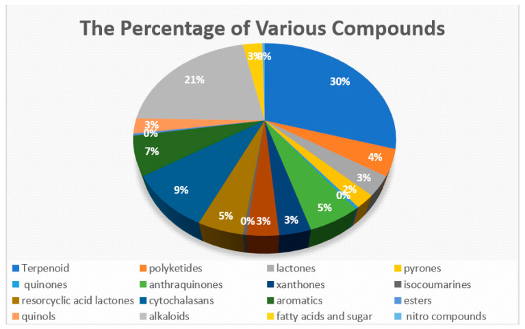
The group of compounds successfully isolated from the Meliaceae family showed a relationship with the group of compounds successfully isolated from endophytic fungi. This is shown by the compounds that were successfully isolated, and all of them entered into a group that had been successfully isolated from their host. The 276 compounds have been isolated from the endophytic fungi from the stembark, leaves, fruits, barks, seeds, and roots of Meliaceae, based on the literature from 2002 to 2022 (Table S1). C. macrophyllus [21], X. granatum [22], M. azedarach [23], T. sinensis [24], C. tagal [25], A. indica [26], T. monadelpha [27], T. longipes [28], D. binectariferum [29], and L. domesticum [30] are members of the Meliaceae family with endophytic fungi that create secondary metabolites. The 276 secondary metabolites isolated are composed of 82 terpenoids, 12 polyketides, nine lactones, six pyrones, one quinone, 15 anthraquinones, nine xanthones, 10 isocoumarines, 13 resorcylic acid lactones, 25 cytochalasins, 18 aromatics, one ester, seven quinols, 59 alkaloids, one nitro compound, and eight fatty acids and sugars (Figure 1) [31,32,33,34,35,36,37,38,39,40,41,42,43,44,45,46,47,48,49,50,51,52,53,54,55,56,57,58,59,60,61,62,63,64].
Figure 1.
The percentage of various compounds which have been isolated from endophytic fungi in Meliaceae [31,32,33,34,35,36,37,38,39,40,41,42,43,44,45,46,47,48,49,50,51,52,53,54,55,56,57,58,59,60,61,62,63].
3.2. Triterpenoid and Sesquiterpenoid Compounds
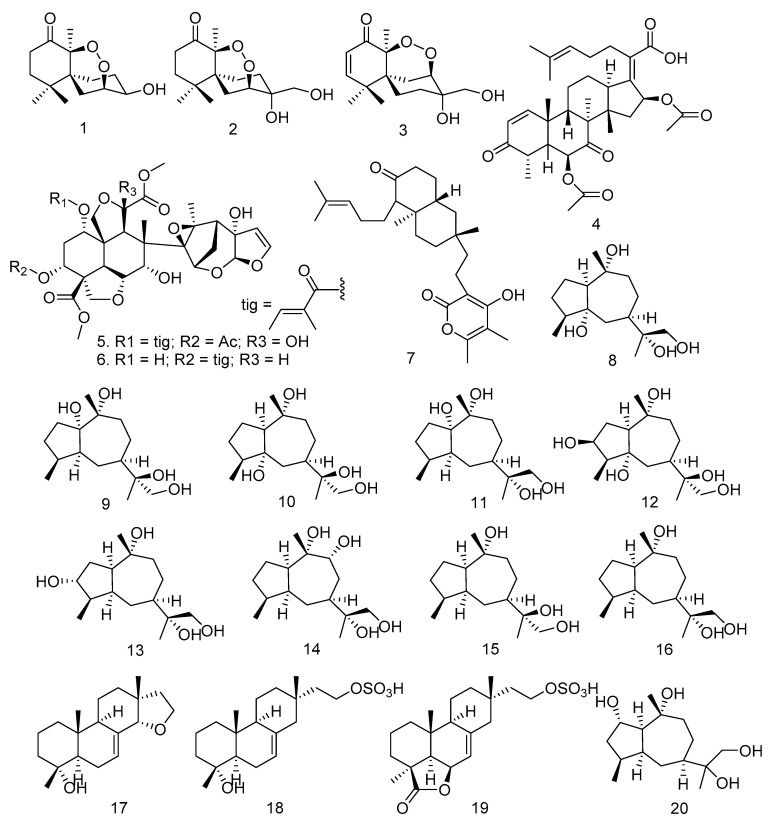
Based on previous study, 82 terpenoids have been isolated from endophytic fungi isolated from the Meliaceae family. The isolated terpenoids are classified into sesquiterpenoids, triterpenoids, steroids, and meroterpenoids. A type of sesquiterpenoid obtained is the Chamigrane reported by Chookpaibon et al. (2010) [31]. Furthermore, three Chamigrane endoperoxides (Figure 2) were isolated from the XG8D fungus, which was taken from leaves of X. granatum and fermented in steep corn liquor containing medium under static conditions. These compounds are Merulin A (1), Merulin B (2), and Merulin C (3). They were also isolated from several species of marine algae of the genus Laurencia [64,65,66,67,68,69,70].
Figure 2.
Triterpenoids, diterpenoids, and sesquiterpenoids were obtained from endophytic fungi isolated from the Meliaceae family.
Li et al. [32] isolated the phytotoxic nordammarane triterpenoid helvolic acid (4) from the Aspergillus fumigatus LN-4 fungus of stembark M. azedarach. Azadirachtin A (5) and B (6) were obtained from the E. parvum fungus, taken from seeds, leaves, stem/twigs, inner bark, and roots of A. indica A. JUSS [33]. On the other hand, Xiao et al. [34] successfully isolated Pycnophorin (7) from the B. dothidea KJ-1 strain cultured on PDA and produced from the chloroform fraction. The endophytic fungal strain KJ-1 was obtained from the symptomless tissue of the stem bark of M. azedarach L. [34]. An endophytic fungus, Xylaria sp. YM 311647, isolated from A. indica, has produced nine new oxygenated guaiane-type sesquiterpenes (8–16) and three new isopimarane diterpenes (17–19). These compounds are (1S,4S,5R,7R,10R,11R)-guaiane-5,10,11,12-tetraol (8), (1S,4S,5S,7R,10R,11S)-guaiane-1,10,11,12-tetraol (9), (1S,4S,5R,7R,10R,11S)-guaiane-5,10,11,12-tetraol (10), (1S,4S,5S,7R,10R,11R)-guaiane-1,10,11,12-tetraol (11), (1R,3S,4R,5S,7R,10R,11S)-guaiane-3,10,11,12-tetraol (12), (1R,3R,4R,5S,7R,10R,11R)-guaiane-3,10,11,12-tetraol (13), (1R,4S,5S,7S,9R,10S,11R)-guaiane-9,10,11,12-tetraol (14), (1R,4S,5S,7R,10R,11S)-guaiane-10,11,12-triol (15), (1R,4S,5S,7R,10R,11R)-guaiane-10,11,12-triol (16), 14α,16-epoxy-18-norisopimar-7-en-4α-ol (17), 16-O-sulfo-18-norisopimar-7-en-4α,16-diol (18), and 9-deoxy-hymatoxin A (19) [35]. Furthermore, Figure 2 shows the diagram of compounds 1–19.
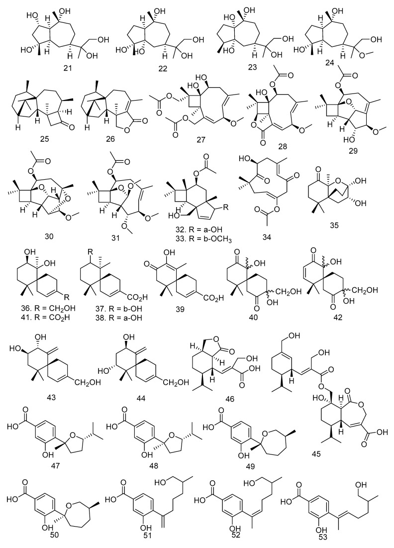
Li et al. [37] succeeded in isolating compounds from A. indica. Huang also successfully obtained five guaian-type sesquiterpenes from the fungus Xylaria sp. YM 311647. The five compounds are guaiane-2,10,11,12-tetraol (20), guaiane-2,4,10,11,12-pentaol (21), guaiane-4,5,10,11,12-pentaol (22), guaiane-1,5,10,11,12-pentaol (23), and 11-methoxyguaiane-4,10,12-triol (24) [36]. The leaves, stems, and bark of the mangrove plant Xylocarpus granatum produced the fungus Trichoderma sp. Xy24. It was fermented in PDA liquid media to isolate two new compounds of the diterpenoid type, namely (9R, 10R)-dihydroharzianone (25) and Harzianelactone (26) [37].
Caryophyllene sesquiterpenoids such as pestaloporinates A-G (27–33) and 14-acetylhumulane (34) were isolated from Pestalotiopsis sp. by an 18S rDNA sequence, which was obtained from the fresh stem bark of M. azedarach Linn and cultured in PDA [38]. In addition, six new chamigrane sesquiterpenes, merulinols A-F (35–40) and known compounds, aciicolinol C (41), aciicolinol K (42), aciicolinol F (43), and aciicolinol D (44), were obtained from the culture of an endophytic fungus XG8D isolated from the fresh leaves of a X. granatum plant in Thailand [39].
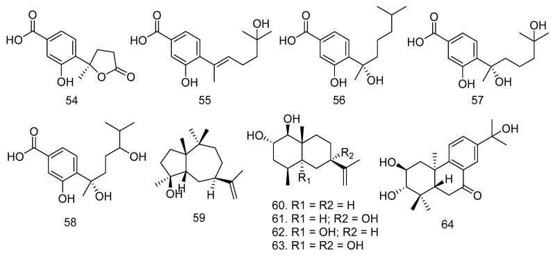
Colletotrin (45), a new sesquiterpene lactone, and one known compound, hydroheptelidic acid (46), were isolated from a rice culture of C. gloeosporioides, an endophytic fungus obtained from the stem bark of the Cameroonian medicinal plant T. monadelpha (Meliaceae) [27]. Meanwhile, seven new phenolic bisabolane sesquiterpenoids such as (7R,10S)-7,10-epoxysydonic (47), (7S,10S)-7,10-epoxysydonic (48), (7R,11S)-7,12-epoxysydonic (49), (7S,11S)-7,12-epoxysydonic (50), 7-deoxy-7,14-didehydro-12-hydroxysydonic (51), and (Z)-7-deoxy-7,8-didehydro-12-hydroxysydonic acid (52), and six known compounds, (E)-7-deoxy-7,8-didehydro-12-hydroxysydonic (53), (+)-1-hydroxyboivinianic (54), engyodontiumone I (55), (+)-sydonic (56), (+)-hydroxysydonic (57), (−)-(7S)-10-hydroxysydonic acid (58), were obtained from the culture of an endophytic fungus Aspergillus sp. xy02 isolated from the leaves of a Thai mangrove, Xylocarpus moluccensis [40].
The other sesquiterpenoid isolated from Xylaria sp. HNWSW-2, the stem of X. granatum, is guaidiol (59) [41]. In 2018, Qiu et al. [25] isolated four new eudesmane-type sesquiterpenoids, penicieudesmol A-D (60–63), from the fermentation broth of the endophytic fungus Penicillium sp. J-54 derived from mangroves. Penicillium sp. J-54 was isolated from the healthy leaves of the Ceriops tagal, which were collected in the Dong Zhai Gang Mangrove [25]. Meanwhile, a bietanetype-diterpenoid, hydroxyldecandrin G (64), was isolated from Xylaria sp. XC-16, which is obtained from leaves of T. sinensis. The structures of compounds 20–64 can be seen in Figure 2.
3.3. Steroid Compounds
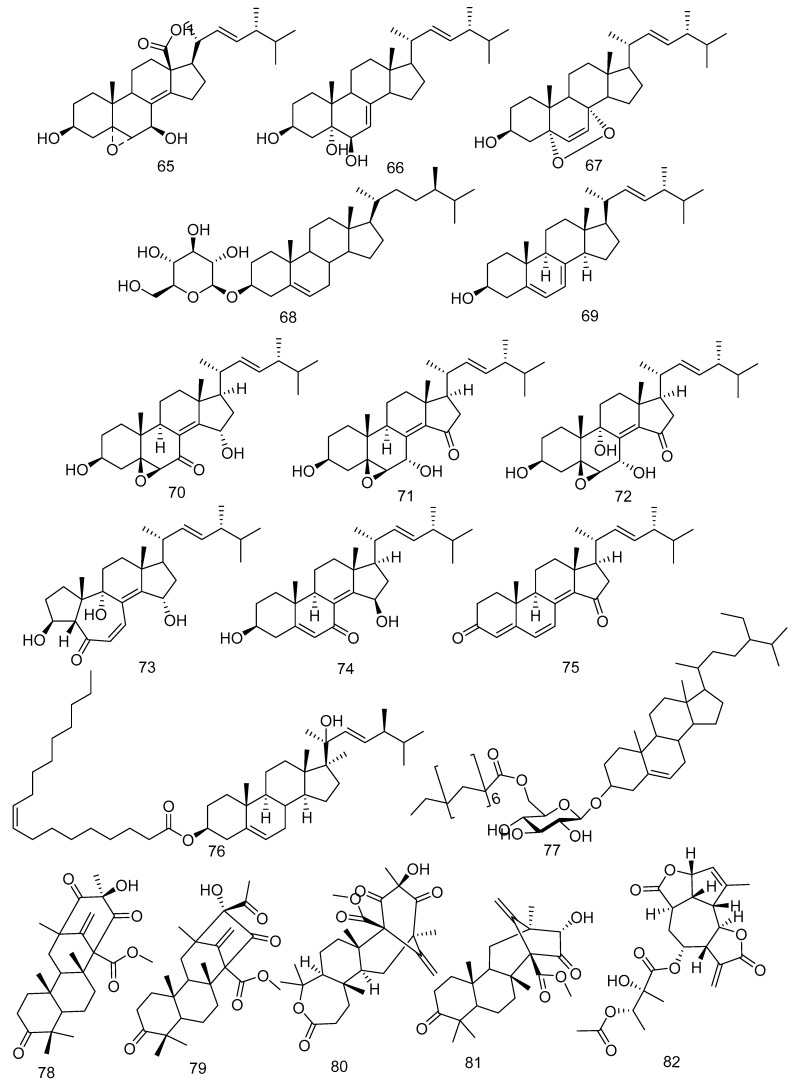
Ergokonin B (65) and cerevisterol (66) were isolated from an endophytic fungus, Fusarium sp. LN-11, from the leaves of M. azedarach [21]. Cerevisterol was also obtained from F. phaseoli [43]. In the study by Xiao et al. [34], B. dothidea KJ-1 was isolated from the stem of M. azedarach, and Sari et al. [43] stated that F. phaseoli from the root of C. macrophyllus produced ergosterol peroxide (67). β-sitosterol glucoside (68) was isolated from B. dothidea KJ-1 found in the stem of M. azedarach L. [34]. Ergosterol (69) was obtained from Eupenicillium sp. HJ002 derived from X. granatum and F. phaseoli derived from C. macrophyllus [22,43].
A new αβ-unsaturated 7-ketone sterol, 5β,6β-epoxy-3β,15α-dihydroxy-(22E,24R)-ergosta-8(14),22-dien-7-one (70), with five known sterone derivatives, 5β,6β-epoxy-3β,7α-dihydroxy(22E,24R)-ergosta-8(14),22-dien-15-one (71), 5β,6β-epoxy-3β,7α,9a-trihydroxy-(22E,24R)-ergosta-8(14),22-dien-15-one (72), 3β,9α,15a-trihydroxy-(22E,24R)-10(5→4)abeo-ergosta-6,8(14),22-trien-5-one (73), 3,15-dihydroxyl-(22E,24R)-ergosta-5,8(14),22trien-7-one (74), (22E,24R)-ergosta-4,6,8(14),22-tetraen-3,15-dione (75), were isolated from the mangrove-derived fungus Phomopsis sp. MGF222 [45]. In addition, a new ergostane-type sterol, ergost-5,22E-dien-3-oleate-20-ol (76), and one known atroside (77) were obtained from the solid brown rice culture of F. phaseoli, an endophytic fungus found in the root of C. macrophyllus [43]. The structures of the steroid compounds 65–77 can be seen in Figure 3.
Figure 3.
The steroid (65–77) and meroterpenoid (78–82) compounds were obtained from various fungi in Meliaceae Family.
3.4. Meroterpenoid Compounds
In the study conducted by Geris dos Santos and Rodrigues-Fo. [46], a Penicillium sp. isolated from the root bark of M. azedarach and cultivated over sterilized rice produced two meroterpenes, namely preaustinoid A (78) and B (79). In addition to a previous study about meroterpenoids, Fill et al. [47] reported that three known meroterpenoids, preaustinoid A1 (80), preaustinoid B2 (81), and austinolide (82), have been isolated from Penicillium brasilianum found in the root bark of Melia azedarach (Figure 3).
3.5. Polyketides
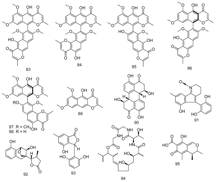
Aurasperone A (83) and fonsecinone A (84), the polyketides, were isolated from Aspergillus aculeatus, an endophytic fungus in the leaves of M. azedarach [48]. Dianhydro-aurasperone C (85), isoaurasperone A (86), fonsecinone A (87), asperpyrone A (88), and rubrofusarin B (89) were obtained from Aspergillus sp. KJ-9 fungi isolated from stembark of M. azedarach. In addition to 83–89, the Botryosphaeria dothidea KJ-1 found in the stem of this family also produced Stemphyperylenol (90) [34,44].
The Thai mangrove endophytic fungus Phomopsis sp. xy21 produced phomopsol A (91), a polyketide-derived alkaloid with a unique 3,4-dihydro-2H-indeno [1,2-b]pyridine 1-oxide motif, phomopsol B (92), which is a highly oxidized polyketide with a new 3,5-dihydro-2H-2,5-methanobenzo[e][1,4]-dioxepine moiety, and 3-(2,6-dihydroxyphenyl)-4-hydroxy- 6-methylisobenzofuran-1(3H)-one (93) [49]. Gao et al. [50] reported that Eucalactam B (94) was obtained from D. eucalyptorum KY-9 fungi found in the leaves of M. azedarach [50]. Citrinin (95) is a well-known mycotoxin produced mainly by Penicillium citrinum and several Aspergillus species. This polyketide was obtained in good yields by P. janthinellum from the host plant M. azedarach [51,71], and the structure can be found in Figure 4.
Figure 4.
The compounds of polyketides obtained from various fungi in the Meliaceae Family.
3.6. Lactones
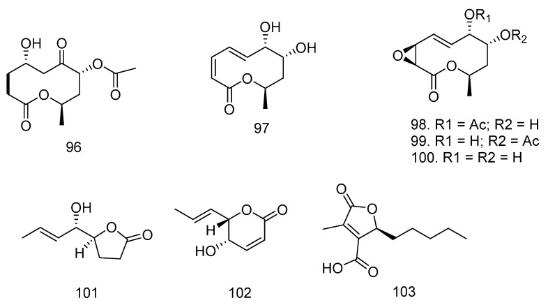
In addition to producing the azadiractin type, Wu et al. [52] reported in 2008 that the plant A. indica also produces four new lactones (8α-acetoxy-5α-hydroxy-7-oxodecan-10-olide (96), 7α,8α-dihydroxy-3,5-decadien-10-olide (97), 7α-acetoxymultiplolide A (98), and 8α-acetoxymultiplolide A (99). A known lactone, multiplolide A (100) was isolated from broth extracts of the endophytic mushroom Phomopsis sp. from the stem of A. indica. Meanwhile, in 2009, Wu et al. [52] obtained nigrosporalactone (101) and phomalactone (102) from the fermentation culture of Nigrospora sp. YB-141, an endophytic fungus isolated from A. indica. In addition to Wu, Mei et al. [22] successfully isolated a lactone compound, (R)-striatisporolide A (103), from the X. granatum Koenig-derived fungus Eupenicillium sp. HJ002 for the first time. Lactone structures can be found in Figure 5.
Figure 5.
Lactone structures isolated from Meliaceae-derived fungi.
3.7. Pyrones, Quinone, and Anthraquinones
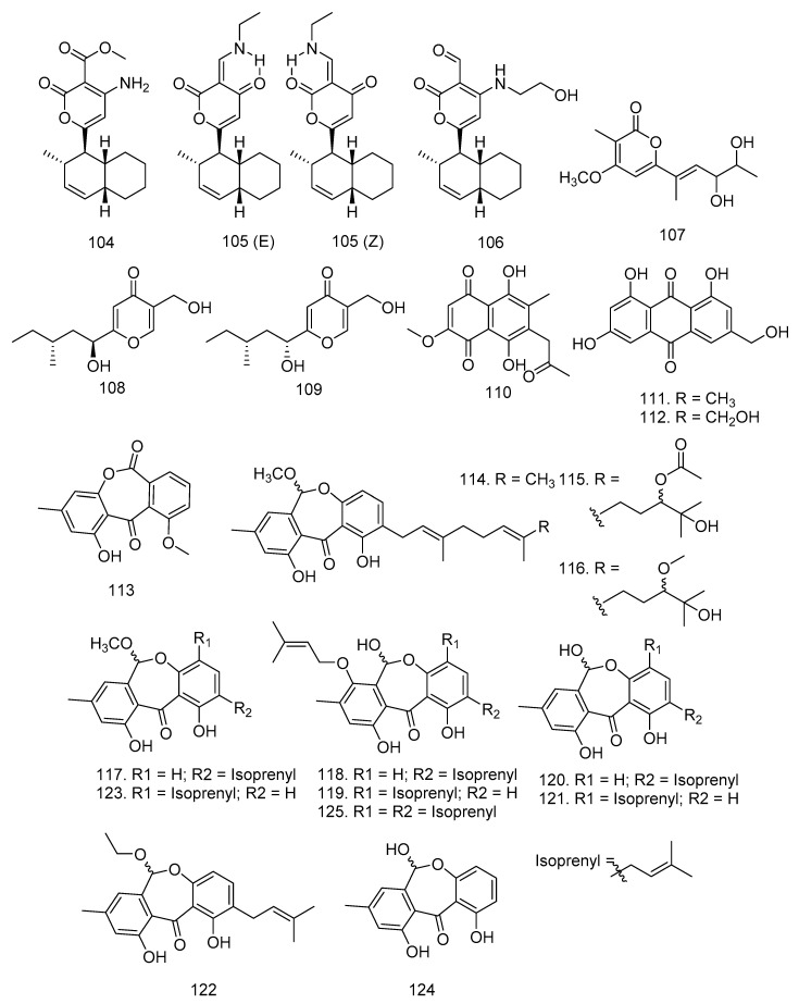
In addition to lactones, Wu et al. [52] also succeeded in proving that Nigrospora sp. YB-141, an endophytic fungus isolated from A. indica, produced two new pyrone compounds, solanapyrone N (104) and solanapyrone O (105), and one known compound, solanapyrone C (106). Meanwhile, Wang et al. [41] obtained astropyrone (107), xylaropyrone B (108), and xylaropyrone C (109) from the fermentation broth of Xylaria sp. HNWSW-2 fungus in X. granatum stem (Figure 6).
Figure 6.
Pyrones (104–109), quinone (110), and anthraquinones (111–125) were isolated from Meliaceae-derived fungi.
Endophytic fungi also produced quinone and anthraquinones. Kharwar et al. [53] isolated the quinone type, namely javanicin (110), from Chloridium sp. fungi found in A. indica root [53]. New anthraquinones such as emodin (1,6,8-trihydroxy-3-methylanthra-quinone) (111), citreorosein (1,6,8-trihydroxy-3-methylanthraquinone) (112), and janthinone (113) were obtained from P. janthinellum, isolated as an endophytic fungus from M. azedarach fruits grown on the ground for 20 days and autoclaved white corn [51,54].
Endophytic fungi Xylariaceae were isolated from fresh and healthy leaves of L. domesticum collected on tropical peatland in West Kalimantan using ITS sequencing. Internal transcribed spacer (ITS) is a piece of nonfunctional RNA located between structural ribosomal RNAs (rRNA) of a common precursor transcript, which is especially useful for elucidating relationships among congeneric species and closely related genera. Chromatographic separation of the ethyl acetate extract yielded three new arugosin-type metabolites, including arugosins O (114), P (115), and Q (116), as well as nine known compounds such as arugosin K (117), arugosin A (118), arugosin B (119), arugosin N (120), 1,6,10-trihydroxy-8-methyl-2-(3-methyl-2-butenyl)-dibenz[b,e]oxepin-11(6H)-one (121), arugosin L (122), arugosin M (123), arugosin F (124), and arugosin G (125) [54]. The structures of quinone and anthraquinone compounds are shown in Figure 6.
3.8. Xanthones, Isocoumarin, and Resorcylic Acids
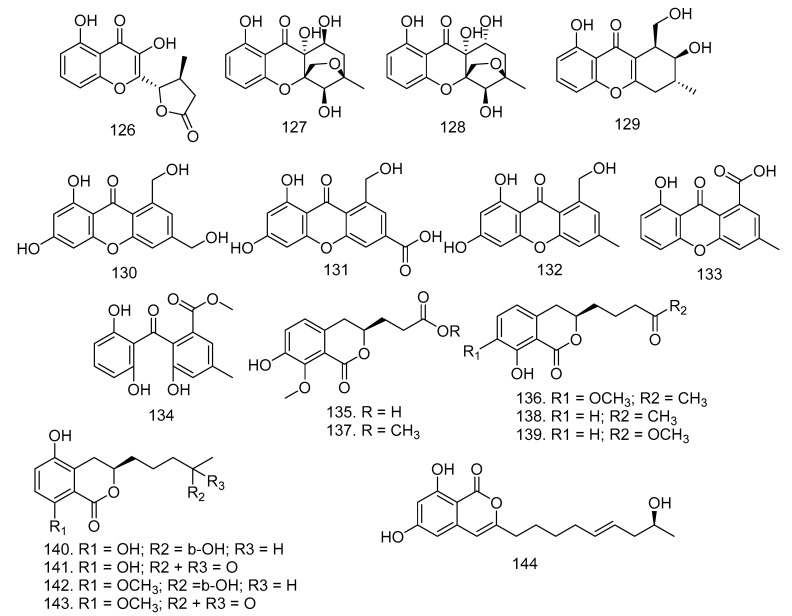
Based on the endophytic research fungi conducted by Hu et al. [55] six new xanthone-derived polyketides, phomoxanthones F-K (126–131), as well as three known xanthones, leptosphaerin E (132), mono-dictyxanthone(8-hydroxy-3-methyl-9-oxo-9H-xanthene-1-carboxylic acid) (133), and 2,2′,6′-trihydroxy-4-methyl-6-methoxy-acyl-diphenylmethanone (134), were isolated from Phomopsis sp. xy21, an endophytic fungus in the Thai mangrove X. granatum (Figure 7).
Figure 7.
Xanthones (126–134) and isocoumarins (135–144), isolated from Meliaceae-derived fungi.
In addition to xanthones, two new isocoumarins, penicimarins L-N (135–136), and seven known isocoumarins, peniisocoumarin E (137), apergilumarin A (138), penicimarin I (139), peniisocoumarin F (140), penicilloxalone B (141), penicimarin G (142), and penicimarin H (143), were successfully obtained from the endophytic fungus Penicillium sp. MGP11 in X. granatum [56]. Fusariumin (144) was isolated from Fusarium sp. LN-10 in the leaves of M. azedarach (Figure 7) [57].
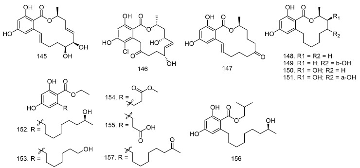
In addition to fusariumin (144), Yang et al. [57] also isolated two known resorcylic acid lactones, aigialomycin D (145), pochonin N (146), and zearalenone (147), from the cultures of Fusarium sp. LN-10. The study conducted by Sato et al. [58] reported that the endophytic fungus L. theobromae in the mangrove plant X. granatum produced nine new -resorcylic acid derivatives such as (15S)-de-O-methyllasiodiplodin (148), (13S,15S)-13-hydroxy-de-O-methyllasiodiplodin (149), (14S,15S)-14-hydroxy-de-O-methyllasiodiplodin (150), (13R,14S,15S)-13,14-dihydroxy-de-O-methyllasiodiplodin (151), ethyl (S)-2,4-dihydroxy-6-(8-hydroxynonyl)benzoate (152), ethyl 2,4-dihydroxy-6-(8-hydroxyheptyl) benzoate (153), ethyl 2,4-dihydroxy-6-(4-methoxycarbonylbutyl)benzoate (154), 3-(2-ethoxycarbonyl-3,5-dihydroxyphen-yl)propionic acid (155), (S)-2,4-dihydroxy-6-(8-hydroxynonyl)benzoate (156), and one known compound, ethyl 2,4-dihydroxy-6-(8-oxononyl)benzoate (157) (Figure 8).
Figure 8.
Compounds of resorcylic acid derivatives isolated from Endophytic fungi in Meliaceae.
3.9. Cytochalasins
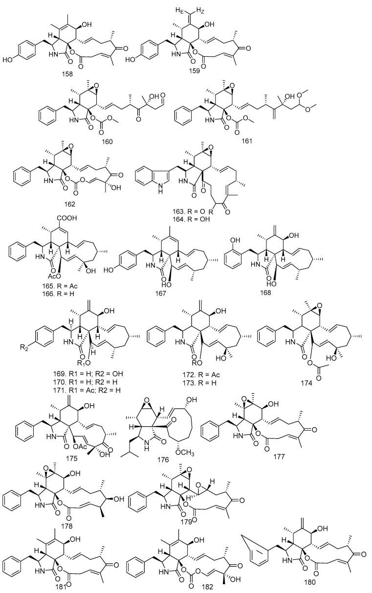
In the study by Zhang et al. [34], the fermentation extract of Xylaria sp. XC-16 from T. sinensis produced two new cytochalasins, cytochalasin Z27 (158) and cytochalasin Z28 (159), and three known compounds seco cytochalasin E (160), cytochalasin Z18 (161), and cytochalasin E (162) [24]. Chaetoglobosins C (163) and F (164) were isolated from a solid culture of the endophytic fungus B. dothidea KJ-1, collected from white cedar stems (M. azedarach L.).
Phomopsis spp. xy21 and xy22, from Thai mangrove fungal endophytes, produced four new cytochalasins, phomopsichalasins D-G (165–168), and six known ones, namely 4′-hydroxy-deacetyl-18-deoxycytochalasin H (169), deacetyl-18-deoxycytochalasin H (170), 18-deoxycytochalasin H (171), cytochalasin H (172), deacetylcytochalasin H (173), and epoxycytochalasin H (174) [59,72,73,74]. Cytochalasin D (175) was obtained from C. gloeosporioides in the stem bark of T. monadelpha [27]. Xylarisin B (176) was produced by Xylaria sp. HNWSW-2, isolated from the stem of X. granatum [41].
In 2019, Han et al. [42] conducted research on Xylaria sp. XC-16 isolated from T. sinensis leaves to yield epoxycytochalasin Z17 (177), epoxycytochalasin Z8 (178), epoxyrosellichalasin (179), 10-phenyl-[12]-cytochalasin Z16 (180), 10-phenyl-[12]-cytochalasin Z17 (181), and cytochalasin K (182). All of the cytochalasin structures can be seen in Figure 9.
Figure 9.
All of the cytochalasin structures obtained from endophytic fungi in Meliaceae.
3.10. Aromatics, Ester, Quinols
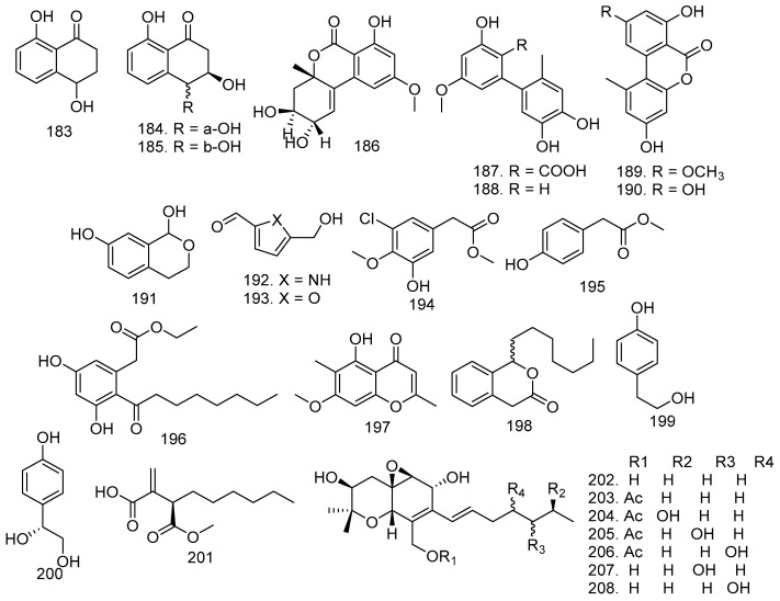
The stem bark of M. azedarach has A. fumigatus LN-4, cultured in PD liquid medium. The fungus produced 4,8-dihydroxy-1-tetralone (183), trans-3,4-dihydro-3,4,8-trihydroxynaphtalen-1(2H)-one (184), and cis-3,4-dihydro-3,4,8-trihydroxynaphtalen-1(2H)-one (185) [32]. In addition, this family also has the B. dothidea KJ-1 fungus, which produced altenuene (186), (187), djalonensone (188), alternariol (189), 5-methoxy-6-methylbiphenyl-3,4,3-triol (190), 7-hydroxy-1-isochromanone3587 (191), 5-(hydroxymethyl)-1H-pyrrole-2-carbaldehyde (192), and 5-hydroxymethylfurfural (193) (Figure 10) [34].
Figure 10.
All structures of aromatics (183–200), ester (201), and quinols (202–208) produced by endophytic fungi from the Meliaceae family.
The fungal strain Eupenicillium sp. HJ002 was obtained from the mangrove Xylocarpus granatum Koenig in the South China Sea. Extracts of this fungus yielded new aromatic compounds 3-chloro-5-hydroxy-4-methoxyphenylacetic acid methyl ester (194) and two known derivatives, 4-hydroxyphenylacetate (195) and cytosporone B (194) [22]. Apart from Xiao et al. [44], Gao et al. [50] also observed that Eucalyptorum KY-9 was isolated from the leaves of M. azaderach plant and cultured in rice. The endophytic fungus yielded eugenitol (197), cytosporone C (198), 4-hydroxyphenethyl alcohol (199), and 1-(4-hydroxyphenyl)ethane-1,2-diol (200) (Figure 10).
(R)-butanedioic acid (201) ester was produced by Eupenicillium sp. HJ002, which was isolated from X. granatum [22,50]. The leaves of T. longipes delivered P. theae fungi, which gave six new epoxyquinols, cytosporins F-K (203–208), with the known cytosporin D (202) (Figure 10) [75].
3.11. Alkaloids
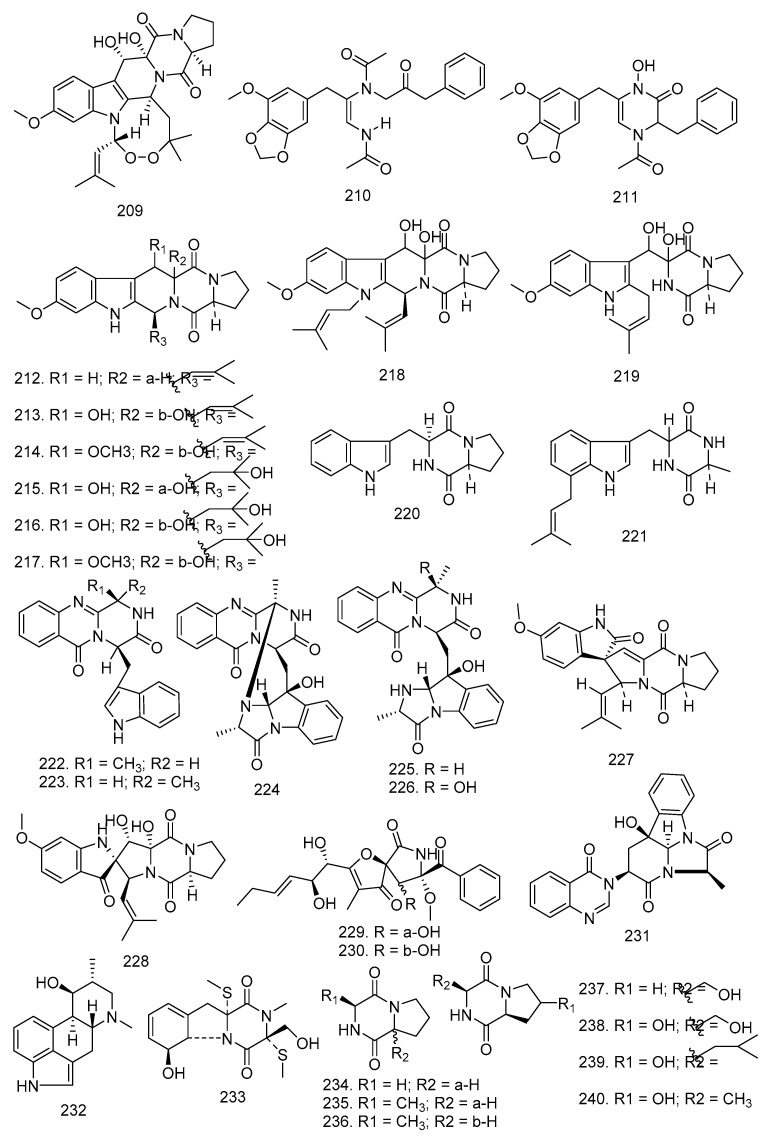
Penicillium sp. was isolated from the root bark of M. azedarach and grown on sterilized rice. The known alkaloid verruculogen (209) was obtained after chromatographic procedures. Apart from Penicillium sp., A. fumigatus LN-4 obtained from M. azedarach stem bark and cultured in PD liquid medium also yielded this compound [32,46]. In the study conducted by Li et al. [49], brasiliamide A (210) and brasiliamide B (211) were produced from P. brasilianum in the root bark of M. azaderach [47,60].
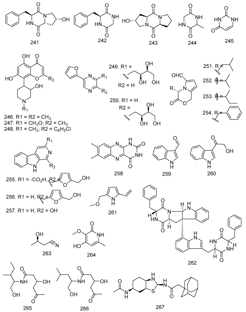
Alkaloids 212–245 were isolated from the fermentation broth of Aspergillus fumigatus LN-4, an endophytic fungus obtained from the stem bark of Melia azedarach, including 12β-hydroxy-13α-methoxyverruculogen TR-2 (217), 3-hydroxyfumiquinazoline A (226) and 32 known alkaloids such as fumitremorgin C (212), cyclotryprostatin A (213), cyclotryprostatin B (214), verrulocogen TR-2 (215), 12β-hydroxyverruculogen TR-2 (216), 12β-hydroxy-13α-methoxyverruculogen TR-2 (217), fumitremorgin B (218), tryprostatin A (219), cyclo-l-tryptophyl-l-proline (220), terezine D (221), fumiquinazoline F, G, D, A (222–225), 3-hydroxyfumiquinazoline A (226), 6-methoxyspirotryprostatin B (227), spiro [5H,10H-dipyrrolo [1,2-α:1′,2′-d]pyrazine-2-(3H),2′-[2H]indole]-3′,5,10(1′H)-trione (228), pseurotin A (229), pseurotin A1 (230), tryptoquivaline O (231), fumifaclavine B (232), bisdethiobis(methylthio)gliotoxin (233), cyclo-(Pro-Gly) (234), cyclo-(Pro-Ala) (235), cyclo-(d-Pro-l-Ala) (236), cyclo-(Pro-Ser) (237), cyclo-(Ser-trans-4-OH-Pro) (238), cyclo-(Leu-4-OH-Pro) (239), cyclo-(Ala-trans-4-OH-Pro) (240), cyclo-(cis-OH-d-Pro-l-Phe) (241), cyclo-(Gly-Phe) (242), cyclo-(Pro-tans-4-OH-Pro) (243), cyclo-(Gly-Ala) (244), and uracil (245) [32].
Further study was conducted by Kumara et al. [61] where rohitukine (246) was obtained from several endophytic fungi isolated from D. binectariferum Hook. f, such as F. proliferatum MTCC 9690, F. oxysporum MTCC 11383, F. solani MTCC 11385, F. oxysporum MTCC 11384, and G. fujikurai MTCC 11382 from the bark, leaves, fruit, and bark respectively. Apart from compound 246, rohitukine N-oxide (247) and flavopiridol (248) were also isolated from these endophytic fungi.
In 2014, Wang [62] also discovered 13 alkaloids, one of which was a new alkaloid 2-(furan-2-yl)-6-(2S,3S,4-trihydroxybutyl)pyrazine (249) and 12 known compounds such as 2-(furan-2-yl)-5-(2S,3S,4-trihydroxybutyl)pyrazine (250), (S)-4-isobutyl-3-oxo-3,4-dihydro-1-H,pyrrolo [2,1-c][1,4]oxazine-6-carbaldehyde (251), (S)-4-isopropyl-3-oxo-3,4-dihydro-1H-pyrrolo [2,1-c][1,4]oxazine-6-carbaldehyde (252), (4S)-4-(2-methylbutyl)-3-oxo-3,4-dihydro-1H-pyrrolo [2,1-c][1,4]oxazine-6-carbaldehyde (253), (S)-4-benzyl-3-oxo-3,4-dihydro-1H-pyrrolo [2,1-c][1,4]oxazine-6-carbaldehyde (254), flazin (255), perlolyrine (256), 1-hydroxy-β-carboline (257), lumichrome (258), 1H-indole-3-carboxaldehyde (259), [14,15],2-hydroxy-1-(1H-indol-3-yl), ethenone (260), and 5-(methoxymethyl)-1H-pyrrole-2-carbaldehyde (261).
Asperazine (262) was discovered by Xiao et al. [44] from Aspergillus sp. KJ-9 in the stem bark of M. azedarach. Similar endophytic fungi and plants obtained (R)-3-hydroxybutanonitrile (263), 3-hydroxy-2-methoxy-5-methylpyridine-2(1H)-one (264), 3-hydroxy-N-(1-hydroxy-3-methylpentan-2-yl)-5-oxohexanamide (265), and 3-hydroxy-N-(1-hydroxy-4-methylpentan-2-yl)-5-oxohexanamide (266) [34]. In 2016, 2S,3aR,6S,7aS)-6-acetamido-octahydro-1,3-benzothiazole-2-yl-2-(adamantan-1-yl)acetamide (267) was discovered by Mittal et al. This compound (267) was isolated from Emericella sp. derived from A. indica twig [63], and the structures can be seen in Figure 11.
Figure 11.
Alkaloids obtained from endophytic fungi in Meliaceae.
3.12. Nitro Compound, Fatty Acid, and Sugars
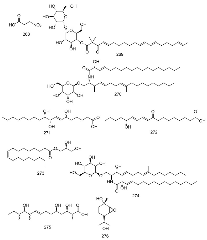
A nitro compound was isolated by Flores et al. [28] under the name 3-nitropropionic acid (268) from P. longicolla FJ 2759 fungi in the leaves of T. elegans A. Juss ssp. elegans. Furthermore, fatty acid and sugar were also obtained from endophytic fungi in Meliaceae. Fusaroside (269) was isolated from the organic extract of fermentation broths of an endophytic fungus, Fusarium sp. LN-11, in the leaves of M. azedarach. It is a unique trehalose-containing glycolipid composed of the 4-hydroxyl group of a trehalose unit attached to the carboxylic carbon of a long-chain fatty acid. Compound 269 was isolated with phalluside (270), (9R,10R,7E)-6,9,10-trihydroxyoctadec-7-enoic acid (271), porrigenic acid (272), and (9Z)-2,3-dihydroxypropyl octadeca-9-enoate (273) [21] (Figure 12).
Figure 12.
Nitro compound (268), fatty acid, and sugars (269–276) characterized by endophytic fungi in Meliaceae.
The bark of this species also contained the fungus B. dothidea KJ-1, which was cultured in rice and produced cerebroside C (274) [34]. D. eucalyptorum KY-9 produced two biosynthetically related new metabolites, eucalyptacid A (275) and phomopene (276), from the different fungus (Figure 12) [50].
4. Bioactivity of Secondary Metabolites Isolated from Endophytic Fungi
4.1. Antimicrobial
Antimicrobial activity has been discovered in several compounds isolated from endophytic fungi. This activity provides antibiotics against pathogens and microorganisms such as C. gloeosporioides, E. coli, B. subtilis, S. aureus, and B. cereus that can cause food defects. Furthermore, antimicrobial is divided into antifungal and antibacterial [76,77,78,79,80,81,82]. Helvolic acid (4) was tested against fungi such as B. cinerea, A. solani, A. alternata (Fries) Keissler, C. gloeosporioides, F. solani, F. oxysporum f. sp. niveum, F. oxysporum f. sp. vasinfectum, and G. saubinettii, and compared with two positive controls, carbendazim and hymexazole. After analyzing the comparison, helvolic acid (4) was found to be active against the fungi B. cinerea, A. solani, C. gloeosporioides, F. oxysporum f. sp. niveum, and G. saubinettii. This compound can be an antifungal [32] (Table S2).
In other studies, pycnophorin (7) was evaluated against several fungi, B. cinerea, A. solani, C. gloeosporioides, and G. saubinettii, and active against B. cinerea and A. solani. This compound (7) was also evaluated for several bacteria, such as E. coli, B. subtilis, S. aureus, and B. cereus. However, these compounds were not active against the bacteria (Table S2) [34].
Wu et al. [35] reported that compounds 8–19 had been tested for their activity against several fungi, including C. albicans YM 2005, A. niger YM 3029, P. oryzae YM 3051, F. avenaceum YM 3065, and H. compactum YM 3077, with a positive control of nystatin. The results showed that nine sesquiterpenes (8–16) were moderately active against C. albicans and H. compactum, with MIC values ranging from 32 to 256 µg/mL. Meanwhile, diterpenes (17–19) were more active, with one exhibiting the most potent inhibitory activity against C. albicans and P. oryzae, with MIC values of 16 µg/mL [35]. In 2015, Huang et al. [36] continued the research conducted by Wu et al. and evaluated five new guaiane sesquiterpenes (20–24) against the same fungi as Wu et al. The guaiane sesquiterpenes showed moderate or weak antifungal activities in a broth microdilution assay.
Seven new phenolic bisabolane sesquiterpenoids (47–53) and known analogs (54–58) were evaluated by Pan et al. [40] against S. aureus ATCC 25923. The results showed that the compounds 48, 49, 51, 53, 55, 57, and 58 have moderate antibacterial activity against S. aureus ATCC 25923 with IC50 values in the range of 31.5–41.9 μM. Compound 68 was tested against the fungi B. cinerea and A. solani, which showed strong to moderate antifungal activities against A. solani (MIC of 6.25 µM) and did not have effects against B. cinerea [34]. Minimum Inhibitory Concentration (MIC) is the lowest concentration of a compound, usually a drug, which prevents visible growth of a bacterium or bacteria or pathogenic fungus. Compound 71 was tested against the bacteria M. tenuis (Order Micrococcales, Micrococcaceae) with a MIC value of 28.2 ± 0.52 µM, while 73 has antibacterial activity against S. aureus with a MIC value of 14.6 ± 0.47 µM [45]. In the study conducted by Geris dos Santos and Rodrigues-Fo [46], preaustinoids A-B (78–79) had a bacteriostatic effect on S. aureus, P. aeruginosa, Bacillus sp. at a dosage of 250 µg/mL and on E. coli at 125 µg. Furthermore, there was a bactericidal effect on E. coli, P. aeruginosa, and Bacillus sp. at dosages of 250 µg/mL with the control such as penicillin, vancomycin, and tetracycline tested at a conc. of 25 µg/mL.
Dianhydro-aurasperone C (85), isoaurasperone A (86), fonsecinone A (87), asperpyrone A (88), and rubrofusarin B (89) have been evaluated as antifungals against G. saubinetti, M. grisea, B. cinerea, C. gloeosporioides, and A. solani. The antibacterial activity against B. cereus, S. aureus, B. subtilis, and E. coli was also evaluated. The results showed that compound 87 was active against almost all phytopathogenic fungi tested with a minimum inhibitory concentration (MIC) range of 6.25–50 µM. Compound 87 was active against all pathogenic bacteria with MIC in the range of 25–100 µM [44]. Eucalactam B (94) was tested on several fungi, such as A. solani, B. cinerea, F. solani, and G. saubinett, and was not active [50].
Citrinin (95) has a bacteriostatic effect on P. aeruginosa and B. subtilis at 62.50 and 31.25 mg/mL. It has a bactericidal effect on E. coli and P. aeruginosa at 500 and 125 mg/mL dosages, respectively. Citrinin doses of 40 g/mL suppress Leishmania mexicana (order Trypanosomatide, Trypanosomatidae) growth following a 48-hour inoculation [51]. Furthermore, four new 10-membered lactones (96–99) and one known one (100) have been evaluated against A. niger YM 3029, B. cinerea YM 3061, F. avenaceum YM 3065, F. moniliforme YM 3067, H. maydis YM 3076, P. islandicum YM 3104, and O. minus YM 3429 with control nystatin. Compound 99 demonstrated antifungal activity in the MIC value range of 31.25–500 µg/mL [26]. In 2009 Wu et al. continued testing the antifungal activity of lactone compounds (101–102) against A. niger YM 3029, B. cinerea YM 3061, P. islandicum YM 3104, and O. minus YM 3429. Similar to lactones (compounds 104–106), Wu et al. [52] also evaluated several fungi such as A. niger YM 3029, B. cinerea YM 3061, P. islandicum YM 3104, and O. minus.
The javanicin (110) was inactive or only slightly active against fungi such as Pythium ultimum, Phytophthora infestans, Botrytis cinerea, and Ceratocystis ulmi but active against Candida albicans, Escherichia coli, Bacillus sp., and Fusarium oxysporum at higher MIC values ranging from 20 to 40 µg/mL [53]. Meanwhile, compound 111 was tested for its bacteriostatic effect on P. aeruginosa and B. subtilis at 7.81 and 31.25 mg/mL, respectively. The bactericidal effect on E. coli, P. aeruginosa, and B. subtilis at 500, 62.50, and 250 mg/mL, respectively, was also analyzed. Compound 113 has no bacteriostatic effect on E. coli and B. subtilis at a dosage of 500 mg/mL and also has no bactericidal effect on P. aeruginosa at 500 mg/mL. Compound 111 was almost completely inactive against E. coli but showed promising activity against P. aeruginosa and B. subtilis [51].
Zhang et al. [24] evaluated compounds 158–162 against the fungi A. solani, B. cinerea, F. solani, and G. saubinettii. Only 159 demonstrated a strong fungicidal effect (MIC of 12.5 µM) against G. saubinetti compared to the positive control hexanol (MIC of 25 µM). Other compounds demonstrated relatively poor properties, with MIC values greater than 50 µM against the pathogens tested. Compound 163 has been evaluated against B. cinerea (200 µM), A. solani (12.5 µM), C. gloeosporioides (200 µM), and G. saubinettii (>200 µM), compared to the control carbendazim: 12.5, 1.57, 1.57, 6.25 µM; hymexazol: 200, 6.25, >200, >200 µM; toosendanin: 200, 6.25, 200, 200 µM. In addition, compound 163 has activity against A. solani [34].
Compounds 183–185 have been tested against several fungi such as B. cinerea, A. solani, A. alternata, C. gloeosporioides, F. solani, F. oxysporum f. sp. niveum, F. oxysporum f. sp. vasinfectum, G. saubinettii. From the three compounds, 183 only showed good activity against the tested fungi, namely B. cinerea (12.5 µM), A. solani (12.5 µM), A. alternata (12.5 µM), C. gloeosporioides (12.5 µM), F. solani (50 µM), F. oxysporum f. sp. niveum (25 µM), F. oxysporum f. sp. vasinfectum (50 µM), and G. saubinettii (12.5 µM) [32]. Compounds 188 and 189 were tested against the same fungi, B. cinerea and A. solani. The results showed that 188 had activity against both fungi, while 189 affected only A. solani [34].
Compounds 190 and 192 were tested against the fungi B. cinerea, A. solani, C. gloeosporioides, and G. saubinettii. They showed no activity as well as compound 190, which was tested against E. coli, B. subtilis, S. aureus, B. cereus with MIC results >100 µM [34]. Compounds 197–200 isolated by Gao et al. [50] were tested against several fungi A. solani, B. cinerea, F. solani, and G. saubinetti. The results were moderate to weak, but some active compounds, such as 198 and 200, were active against Alternaria solani (12.5 and 6.25 µM). Compounds 209 and 212–245 were isolated by Li et al. [32] and evaluated against several fungi. Furthermore, sixteen compounds demonstrated potent antifungal activity against phytopathogenic fungi (B. cinerea, A. solani, A. alternata, C. gloeosporioides, F. solani, F. oxysporum f. sp. niveum, F. oxysporum f. sp. vasinfectum, and G. saubinettii). 12β-hydroxy-13α-methoxyverruculogen TR-2 (217), fumitremorgin B (218), and verruculogen (209) demonstrated antifungal activities with MIC values of 6.25−50 μg/mL, which were comparable to the two positive controls carbendazim and hymexazol, as seen in Table S2.
Compound 262 was tested against fungi and bacteria, each giving the following MIC values: G. saubinetti, MIC 25 M; M. grisea, MIC NA; B. cinerea, MIC 50 µM; C. gloeosporioides, MIC NA. It showed no activity against B. cereus; S. aureus, MIC 50 µM; B. subtilis, MIC NA; E. coli, MIC NA. Likewise, compound 263 gave the following MIC values: G. saubinetti, MIC 12.5 µM; M. grisea, MIC 25 µM; B. cinerea, MIC NA; C. gloeosporioides, MIC 50 µM; and A. solani, MIC 25 µM [44]. Eucalyptacid A (275) showed good activity against A. solani (12.5 µM), B. cinerea (50.0 µM), F. solani (25.0 µM), and G. saubinetti (50.0 µM), compared to positive control hymexazol (MIC 6.25, 50.0, 50.0, 25.0 µM, respectively) [50].
4.2. Cytotoxic Activity
Research on cytotoxic activity in endophytic fungi has been widely reported in many compounds with different test methods. More than 140 natural products with varying levels of antitumor activity have been isolated from fungal endophytes. Alkaloids, terpenes, steroids, polyketides, quinones, isocoumarins, esters, and other secondary metabolites are prevalent. The findings can be used to develop new antitumor drugs and endophyte resources [83,84,85,86,87]. Based on the study conducted by Chokpaibon et al., [31] sesquiterpenoid compounds merulin A-C (1–3) were analyzed for their cytotoxic activity against BT474 and SW620. Compound 3 had the highest activity and was continued by 1 and 2. Compared with positive control, doxorubicin with respective IC50 values of 0.53 and 0.09 µg/mL had moderate activity.
Merulinols C-D (37–38) showed moderate activity against KATO-3 cells with IC50 values of 35.0 ± 1.20 µM and 25.3 ± 0.82 µM, respectively.[39]. Penicieduesmol B (61) has been tested against K-562 with an IC50 value of 90.1 µM (paclitaxel IC50 = 9.5 µM) [25]. Ergosterol peroxide (67) and β-sitosterol glucoside (68) were evaluated against the human colorectal HCT 116 cell line, and the results showed that the compound 67 was active with moderate activity IC50 value of 72.3 µM compared to positive control etoposide IC50 of 2.13 µM while 68 was not active [34,43]. Meanwhile, stemphyperylenol (90) indicated good activity against the HCT 116 cell line with an IC50 value of 3.13 µM, and the control was an etoposide with an IC50 of 2.13 µM [34]. Phomoxanthones G-K (127–131) were evaluated for their cytotoxicity against eight human tumor cell lines (A375, AGS, HCT-8, HCT-8/T, A549, MDA-MB-231, SMMC-7721, and A2780) using the MTT method with cisplatin as the positive control. However, none of them exhibited significant activity at 50 μM [62].
Chaetoglobosin C (163) and Chaetoglobosin F (164) were evaluated against the same cell line with an IC50 value of >100 and 26.5 µM, respectively. According to Luo et al., [59] phomopsichalasins D-G (165–168), 4′-hydroxy-deacetyl-18-deoxycytochalasin H (169), deacetyl-18-deoxycytochalasin H (170), 18-deoxycytochalasin H (171), cytochalasin H (172), deacetylcytochalasin H (173), and epoxycytochalasin H (174) were evaluated against several cells, as seen in Table S3.
Cytochalasin D (175) has cytotoxic activity against mouse lymphoma L5178Y cells with an EC50 value of 0.2 µM [72]. Compounds 186–193 were evaluated against the human colorectal HCT 116 cell line. The results showed that the compounds which have good-moderate activity after being compared to positive control etoposide IC50 value of 2.13 µM were 186 (IC50 3.13 µM), 187 (IC50 28.9 µM), 189 (IC50 33.9 µM), and 190 (IC50 73.4 µM) [34]. Compounds 256–259 have been tested against Madin-Darby canine kidney (MDCK) normal cells (MTT assay) with IC50 values of 116.3 ± 12.1 µg/mL (256), 403.2 ± 31.4 µg/mL (257), 124.1 ± 10.5 µg/mL (258), and 522.5 ± 24.5 µg/mL (259). The positive control was ribavirin with an IC50 of 744.2 ± 18.5 µg/mL [62]. Compound 274 was tested but showed no activity against the human colorectal HCT 116 cell line [34].
4.3. Antioxidant and α-Glucoside Inhibitory Activity
Natural compounds isolated from endophytic fungi in medicinal plants are a rich source of drugs with various biological activities, including antioxidant properties. According to Kharat and Mendhulkar [88], phenolic compounds are responsible for antioxidant activity. Their presence may also have contributed to the radical scavenging activity observed in this study [88,89,90,91,92,93].
Wang et al., [40] reported the antioxidant activity of (−)-(7S)-10-hydroxysydonic acid (58) based on antioxidative activity to scavenge DPPH radicals with an IC50 value of 72.1 µM. Pycnophorin (7) and 3-hydroxy-2-methoxy-5-methylpyridin-2(1H)-one (264) were tested for DPPH radical scavenging with rates of 30.9% and 22.5% at conc. of 50 µM [34]. Furthermore, altenusin (187) and 5′-methoxy-6-methylbiphenyl-3,4,3′-triol (190) have DPPH radical scavenging activity with IC50 of 17.6 ± 0.23 and 18.7 ± 0.18 µM, respectively [34].
In several studies, the antioxidant activity test of isocoumarin compounds (135–143) showed two new isocoumarins, penicimarins L-N (135–136) with IC50 28.3 and 38.9 µM, with seven known isocoumarins, peniisocoumarin E (137) (21.5 µM), apergilumarin A (138) (40.5 µM), penicimarin I (139) (IC50 > 50 µM), peniisocoumarin F (140) (IC50 > 50 µM), penicilloxalone B (141) (IC50 30.3 µM), penicimarin G (142) (4.6 µM), and penicimarin H (143) (18.6 µM). In addition to antioxidant activity, isocoumarins were also evaluated as α-glucoside inhibitory activity and showed that compounds 139, 142, and 143 have activity with IC50 values of 776.5, 683.7, and 868.7 µM, respectively. Meanwhile, the control used was acarbose with IC50 of 313.9 µM [56].
4.4. Anti-Inflammatory and Anti-Influenza
Inflammation is a condition in which catabolism takes precedence over anabolism. It can also be defined as a defense mechanism that aids in the elimination of potentially harmful factors and establishes homeostasis in the body. This causes increased blood flow to the site of inflammation due to the increased permeability of capillaries and white blood cells, resulting in symptoms such as redness, swelling, and pain [94,95]. Endophytic fungi are a valuable source of pharmacologically active metabolites, one of which is anti-inflammatory [96,97]. Liu et al. [38] reported that pestaloporinate B (28) had anti-inflammatory activity (no inhibition in LpS-induced RAW 264.7 macrophage cells) and IC50 of 19.0 µM (positive control L-NMMA IC50 = 40.5 µM).
The influenza virus is one of the most common respiratory tract pathogenic agents, causing significant mortality, morbidity, and economic loss [98]. Alkaloids contain many important chemical compounds used to develop new anti-influenza agents [99]. The alkaloid group is one of the largest groups isolated from endophytic fungi [100]. In this study, 59 alkaloids were isolated. Wang et al. [62] evaluated alkaloid compounds (249–261) against the H1N1 virus. The compounds 257–259 were active against the influenza A virus subtype H1N1 with IC50 and selectivity index (SI) values of 38.3 (±1.2)/25.0 (±3.6)/39.7 (±5.6)/45.9 (±2.1) μg/mL and 3.0/16.1/3.1/11.4, respectively. The IC50 and SI values of the positive control, ribavirin, were 23.1 (±1.7) μg/mL and 32.2, respectively.
4.5. Brine Shrimp Lethality Test (BSLT)
The Brine Shrimp Lethality Test (BSLT) determines the bioactivity of a compound derived from natural ingredients. Artemia salina (order Anostraca, Artemilidae) from larvae is widely used in environmental studies, toxicity testing, and screening of bioactive compounds from plant extracts [101]. Helvetic acid (4) had activity when tested with BSLT and showed median lethal concentration LC50 of 73.7 µg/mL with the positive control toosendanin LC50 < 1 µg/mL [32]. Pycnophorin (7) and ergosterol peroxide (67) showed 43.81% and 13.72% lethality at conc. of 100 µM [24].
Isocoumarin and resorcylic acids were evaluated against brine shrimp (Artemia salina) with mortality rates of 78.2% for fusariumin (144) at concentration 10 µg/mL, 76.7% for aigialomycin D (145) at concentration 10 µg/mL and 82.8% for pochonin N (146) at conc. 10 µg/m [57]. Zhang et al. [24] discovered cytochalasin E (162), which was evaluated against Artemia salina and showed good activity with LC50 of 2.79 µM and 100% lethality at conc. of 50 µM. Meanwhile, chaetoglobosin F (164) showed 16.76% lethality at conc. of 100 µM.
Compounds 183–185 were evaluated against brine shrimp, and 183 had the best activity compared to 184 and 185 [24]. Compound 200 showed 36% lethality at conc of 100 µM. Conversely, compounds 209 and 212–245 were evaluated, and of the 18 compounds that showed moderate lethality in brine shrimps, 218 and 209 had significant toxicities, with LC50 values of 13.6 and 15.8 g/mL, respectively [32]. Based on Yang et al. [21], 269–273 showed growth inhibitory activity against brine shrimp with mortality rates of 47.6%, 64.8%, 26.2%, 20.9%, and 18.7% at conc. 10 µg/mL and 78.2% for the positive control fusariumin. Meanwhile, compound 276 showed 36% lethality at conc. of 100 µM [50].
4.6. Allelophatic Effects on Wheat Triticum Aestivum
Allelopathy is defined as a direct or indirect harmful or beneficial effect of one plant on another through chemical compounds released into the environment [102]. It has been used as a weed control strategy for commercial herbicide-dominated programs. One of the bioactivities of endophytic fungi compounds studied was allelophatic effects on wheat Triticum aestivum (order poales, poaceae) conducted by Rawat et al. [103]. Compounds 64, 162, and 177–182 were evaluated for allelophatic effects on wheat Triticum aestivum. To some extent, all tested compounds inhibited T. aestivum shoot and root elongation with root intensity (RI) values ranging from 0.02 to 0.87 at 6.25 and 100 µM, respectively. The RI value is used as a direction to know the root growth of the individual species and for the sequence analysis. Furthermore, 179, 64, and 182 inhibited shoot elongation strongly, with IC50 values of 18.92 0.80, 23.58 0.43, and 24.02 0.51 µM, respectively. Compounds 180, 182, and 162 inhibited root elongation, with IC50 values of 17.35 0.05, 22.58 0.58, and 19.74 0.09 µM, respectively. These compounds were tested and compared with glyphosate, a commercial herbicide used as a positive control.
4.7. Antifeedant
Plant diseases caused by phytopathogens and pests cause crop loss, having a significant social and economic impact on the livelihoods of people relying on agricultural income. One of the ways to decrease the problem is to obtain antifeedant compounds. Endophytic fungi produced many compounds studied as antifeedants [104,105,106]. Li et al. studied antifungal, BSLT, and antifeedant activity in the previous study. Compounds 4, 209, 212, 218, 222, 224, 225, 226, and 228 were tested against armyworm (Mythimna separata; order Lepidoptera, Noctuidae), and each gave antifeedant index values of 7.5%, 55.0%, 15.0%, 30.0%, 10.0%, 10.0%, 45.0%, 7.5%, and 5.0% with the positive control toosendanin 97.5%. All compounds showed lower antifeedant rates than toosendanin [32].
4.8. Neuroprotective Activity
Neuroprotective activity is an action to save or regenerate the nervous system, cells, structures, and functions by preventing damage to the nervous system in neurochemical modulators. Neurological disabilities include a wide range of disorders, such as epilepsy, autism, neuromuscular disorders, brain tumors, cerebral palsy, Parkinson’s, and learning disabilities. Previous studies reported that polyketide compounds phomopsol A (91) and B (92) were tested on pC12 cells that cause Parkinson’s disease and respectively showed their cell viability results of 76% and 96% at conc. of 40.0 µM with positive control corticosterone 60% at 200.0 µM [55].
4.9. Anti-HIV Activity
Compounds phomoxanthones F, G, H, and K were tested in vitro by HIV-I virus-transfected 293 T cells. The result showed that phomoxanthone F at the concentration of 20 μM showed a weak inhibitory rate of 16.48 ± 6.67% with the positive control efavirenz, and an inhibitory rate of 88.54 ± 0.45% at the same concentration.
4.10. Phytotoxic Activity
The phytotoxic potential of plants and their compounds on other plants is increasingly being studied as a possible alternative to synthetic herbicides for controlling weeds in cropland. Apart from being an antifeedant, compounds from endophytic fungi were also tested for their phytotoxic activity such as compounds 148, 150, 153, and 162 against Digitaria ciliaris, with the most significant result being compound 150.
4.11. Enhanced Root Elongation Activity
The activity of a compound can be influenced by the surrounding environment. Compounds 152, 153, 154, 156, and 157 were tested for their activity on root elongation and the results showed that all the tested compounds increased root elongation activity. This is enough to warrant further studies on their mode of action and their role in chemical ecology, including allelopathy.
5. Conclusions
Meliaceae plant endophytes have been studied thus far for secondary metabolites. All endophytes use extra-cellular hydrolysis to defend against host attacks, invade pathogens, or obtain the host’s nutrients. Previous studies found that several of the same compounds were isolated from the host and its endophytes. Endophytic fungi are one of the microorganisms frequently found in plants. A comprehensive review of secondary metabolites (triterpenoids, sesquiterpenoids, alkaloids, flavonoids, quinones, and other compounds) isolated from Meliaceae endophytes and their pharmacological activities has provided information about species identification, compound isolation, and their vital role in medicine, which can be seen from their pharmacological activity. Endophytic fungi isolated from Meliaceae contain secondary metabolites with antioxidant, antibacterial, antifungal, anti-influenza, cytotoxic, and BSLT toxicity properties. This review provides sufficient evidence that endophytic fungi are potential bioactive plants. Hopefully, this review will contribute to the advancement of endophytic research, particularly in plants of the Meliaceae family.
Acknowledgments
The authors are grateful to Universitas Padjadjaran for providing the Academic Leadership Grant and for supporting all research facilities.
Supplementary Materials
The following supporting information can be downloaded at: https://www.mdpi.com/article/10.3390/molecules28020778/s1, Table S1: Secondary Metabolites Derived from Endophytic fungi-Meliaceae; Table S2: Antimicrobial Activity of Compounds that have been evaluated against several fungi and bacteria; Table S3: Cytotoxic Activity of compounds (165–174) against Several Cells.
Author Contributions
Conceptualization, Y.M. and S.E.S., methodology, S.E.S. and Y.M., validation, U.S.; formal analysis, S.E.S., resources, Y.M. and S.E.S., data curation, S.E.S., writing–original draft preparation, Y.M. and S.E.S., writing–review and editing, Y.M., S.E.S. and U.S., visualization, S.E.S., supervision, U.S., project administration, U.S., funding acquisition, U.S. All authors have read and agreed to the published version of the manuscript.
Institutional Review Board Statement
Not applicable.
Informed Consent Statement
Not applicable.
Data Availability Statement
This study did not report any data.
Conflicts of Interest
The authors declared no conflict of interest.
Funding Statement
This study was funded by Universitas Padjadjaran from Academic Leadership Grant No: 1959/UN6.3.1/pT.00/2022 to Unang Supratman.
Footnotes
Disclaimer/Publisher’s Note: The statements, opinions and data contained in all publications are solely those of the individual author(s) and contributor(s) and not of MDPI and/or the editor(s). MDPI and/or the editor(s) disclaim responsibility for any injury to people or property resulting from any ideas, methods, instructions or products referred to in the content.
References
- 1.Yadav R., Pednekar A., Avalaskar A., Rathi M., Rewachandani Y. A comprehensive review on Meliaceae family. World J. Pharm. Sci. 2015;3:1572–1577. [Google Scholar]
- 2.Sinaga S.E., Mayanti T., Naini A.A., Harneti D., Nurlelasari N., Maharani R., Farabi K., Supratman U., Fajriah S., Azmi M.N. 1035 Sesquiterpenoids from the Stem Bark of Lansium domesticum Corr. Cv. Kokossan and Their Cytotoxic Activity against MCF-7 Breast Cancer Cell Lines. J. Pharm. Pharmacol. 2022;22:1035–1042. doi: 10.22146/ijc.72742. [DOI] [Google Scholar]
- 3.Olatunji T.L., Odebunmi C.A., Adetunji A.E. Biological activities of limonoids in the Genus Khaya (Meliaceae): A review. Futur. J. Pharm. Sci. 2021;7:74. doi: 10.1186/s43094-021-00197-4. [DOI] [Google Scholar]
- 4.Suzuki T., Ariefta N.R., Koseki T., Furuno H., Kwon E., Momma H., Harneti D., Maharani R., Supratman U., Kimura K., et al. New polyketides, paralactonic acids A–E produced by Paraconiothyrium sp. SW-B-1, an endophytic fungus associated with a seaweed, Chondrus ocellatus Holmes. Fitoterapia. 2019;132:75–81. doi: 10.1016/j.fitote.2018.11.017. [DOI] [PubMed] [Google Scholar]
- 5.Shiono Y., Sasaki T., Shibuya F., Yasuda Y., Koseki T., Supratman U. Isolation of a Phomoxanthone A Derivative, a New Metabolite of Tetrahydroxanthone, from a Phomopsis sp. Isolated from the Mangrove, Rhizhopora mucronata. Nat. Prod. Commun. 2013;8:1735–1737. doi: 10.1177/1934578X1300801220. [DOI] [PubMed] [Google Scholar]
- 6.Sun Y., Wang Q. Endophytic fungal community in stems and leaves of plants from desert areas in China. Mycol. Prog. 2012;11:781–790. doi: 10.1007/s11557-011-0790-x. [DOI] [Google Scholar]
- 7.Sofian F.F., Suzuki T., Supratman U., Harneti D., Maharani R., Salam S., Abdullah F.F., Yosida J., Ito Y., Koseki T., et al. The 2,3-epoxy naphtoquinol produced by endophyte Arthrinium marii M-211. Nat. Prod. Res. 2021;9:1–7. doi: 10.1080/14786419.2021.1998899. [DOI] [PubMed] [Google Scholar]
- 8.Strobel G., Daisy B. Bioprospecting for Microbial Endophytes and Their Natural Products. Microbiol. Mol. Biol. Rev. 2003;67:491–502. doi: 10.1128/MMBR.67.4.491-502.2003. [DOI] [PMC free article] [PubMed] [Google Scholar]
- 9.Tan R.X., Zou W.X. Endophytes: A rich source of functional metabolites. Nat. Prod. Res. 2001;18:448–459. doi: 10.1039/b100918o. [DOI] [PubMed] [Google Scholar]
- 10.De Battista J.P., Bacon C.W., Severson R., Plattner R.D., Bouton J.H. Indole Acetic Acid Production by the Fungal Endophyte of Tall Fescue (AJ) Agron. 1990;880:878–880. doi: 10.2134/agronj1990.00021962008200050006x. [DOI] [Google Scholar]
- 11.Khan S.A., Hamayun M., Khan A.L., Shinwari Z.K. Isolation of plant growth promoting endophytic fungi from dicots inhabiting coastal sand dunes of korea. Pak. J. Bot. 2012;44:1453–1460. [Google Scholar]
- 12.Alvin A., Miller K.I., Neilan B.A. Exploring the potential of endophytes from medicinal plants as sources of antimycobacterial compounds. Microbiol. Res. 2014;169:483–495. doi: 10.1016/j.micres.2013.12.009. [DOI] [PMC free article] [PubMed] [Google Scholar]
- 13.Nicoletti R., Fiorentino A. Plant Bioactive Metabolites and Drugs Produced by Endophytic Fungi of Spermatophyta. Agriculture. 2015;5:918–970. doi: 10.3390/agriculture5040918. [DOI] [Google Scholar]
- 14.Kusari S., Spiteller M. Are we ready for industrial production of bioactive plant secondary. Nat. Prod. Rep. 2015;28:1203–1207. doi: 10.1039/c1np00030f. [DOI] [PubMed] [Google Scholar]
- 15.Khan A.L., Hussain J., Al-harrasi A., Al-rawahi A., Lee J., Latif A., Hussain J., Al-harrasi A., Al-rawahi A. Endophytic fungi: Resource for gibberellins and crop abiotic stress resistance. Crit. Rev. Biotechnol. 2015;35:62–74. doi: 10.3109/07388551.2013.800018. [DOI] [PubMed] [Google Scholar]
- 16.Mabberley D.J. Florae Malesianae PraecursoresLXVII Meliaceae (Divers Genera) Blumea. 1985;31:129–152. [Google Scholar]
- 17.van der Nat J.M., van der Sluis W.G., de Silva K.T.D., Labadie R.P. Ethnopharmacognostical survey of Azadirachta indica A. Juss (Meliaceae) J. Ethnopharmacol. 1991;35:1–24. doi: 10.1016/0378-8741(91)90131-V. [DOI] [PubMed] [Google Scholar]
- 18.Mayanti T., Sinaga S.E., Supratman U. Phytochemistry and biological activity of Lansium domesticum Corr. species: A review. J. Pharm. Pharmacol. 2022;74:1568–1587. doi: 10.1093/jpp/rgac057. [DOI] [PubMed] [Google Scholar]
- 19.Samaranada V.A., Wijekumar P.J., Nirmani D., Samarakoon A.W., Perera P.K., Wijekumar J. The phytochemical constituents and pharmacological properties of Munronia pinnata: A review. Int. J. Herb. Med. 2021;9:85–91. [Google Scholar]
- 20.Ogbuewu I.P., Odoemenam Y.U., Obikaonu H.O., Opara M.N., Emenalom O.O., Uchegbu M.C., Okoli I.C., Esonu B.O., Iloeje M.U. The growing importance of neem (Azadirachta indica A. Juss) in agriculture, industry, medicine and environment: A review. Res. J. Med. Plant. 2011;5:230–245. doi: 10.3923/rjmp.2011.230.245. [DOI] [Google Scholar]
- 21.Yang S.X., Wang H.P., Gao J.M., Zhang Q., Laatsch H., Kuang Y. Fusaroside, a unique glycolipid from Fusarium sp., an endophytic fungus isolated from Melia azedarach. Org. Biomol. Chem. 2012;10:819–824. doi: 10.1039/C1OB06426F. [DOI] [PubMed] [Google Scholar]
- 22.Mei R.Q., Nong X.H., Wang B., Sun X.P., Huang G.L., Luo Y.P., Zheng C.J., Chen G.Y. A new phenol derivative isolated from mangrove-derived fungus Eupenicillium sp. HJ002. Nat. Prod. Res. 2021;35:4051–4057. doi: 10.1080/14786419.2020.1712388. [DOI] [PubMed] [Google Scholar]
- 23.Geris Dos Santos R.M., Rodrigues-Fo E., Caldas Rocha W., Simas Teixeira M.F. Endophytic fungi from Melia azedarach. World J. Microbiol. Biotechnol. 2003;19:767–770. doi: 10.1023/A:1026000731189. [DOI] [Google Scholar]
- 24.Zhang Q., Xiao J., Sun Q.Q., Qin J.C., Pescitelli G., Gao J.M. Characterization of cytochalasins from the endophytic Xylaria sp. and their biological functions. J. Agric. Food Chem. 2014;62:10962–10969. doi: 10.1021/jf503846z. [DOI] [PubMed] [Google Scholar]
- 25.Qiu L., Wang P., Liao G., Zeng Y., Cai C., Kong F., Guo Z., Proksch P., Dai H., Mei W. New eudesmane-type sesquiterpenoids from the mangrove-derived endophytic fungus Penicillium sp. J-54. Mar. Drugs. 2018;16:108. doi: 10.3390/md16040108. [DOI] [PMC free article] [PubMed] [Google Scholar]
- 26.Wu S.H., Chen Y.W., Shao S.C., Wang L.D., Li Z.Y., Yang L.Y., Li S.L., Huang R. Ten-membered lactones from Phomopsis sp., an endophytic fungus of Azadirachta indica. J. Nat. Prod. 2008;71:731–734. doi: 10.1021/np070624j. [DOI] [PubMed] [Google Scholar]
- 27.Kemda P.N., Akone S.H., Tontsa A.T., Zhen L., Müller W.E.G., Proksch P., Nkengfack A.E. Colletotrin: A sesquiterpene lactone from the endophytic fungus Colletotrichum gloeosporioides associated with Trichilia monadelpha. Z. Für Nat. B. 2017;72:697–703. doi: 10.1515/znb-2017-0058. [DOI] [Google Scholar]
- 28.Flores A.C., Pamphile J.A., Sarragiotto M.H., Clemente E. Production of 3-nitropropionic acid by endophytic fungus Phomopsis longicolla isolated from Trichilia elegans A. JUSS ssp. elegans and evaluation of biological activity. World J. Microbiol. Biotechnol. 2013;29:923–932. doi: 10.1007/s11274-013-1251-2. [DOI] [PubMed] [Google Scholar]
- 29.Mohana Kumara P., Zuehlke S., Priti V., Ramesha B.T., Shweta S., Ravikanth G., Vasudeva R., Santhoshkumar T.R., Spiteller M., Uma Shaanker R. Fusarium proliferatum, an endophytic fungus from Dysoxylum binectariferum Hook.f, produces rohitukine, a chromane alkaloid possessing anti-cancer activity. Antonie Van Leeuwenhoek. 2012;101:323–329. doi: 10.1007/s10482-011-9638-2. [DOI] [PubMed] [Google Scholar]
- 30.Value N. Lansium domesticum—A Fruit with Multi-Benefits: Traditional Uses, Phytochemicals, Nutritional Value, and Bioactivities. Nutrients. 2022;147:1531. doi: 10.3390/nu14071531. [DOI] [PMC free article] [PubMed] [Google Scholar]
- 31.Chokpaiboon S., Sommit D., Teerawatananond T., Muangsin N., Bunyapaiboonsri T., Pudhom K. Cytotoxic nor-chamigrane and chamigrane endoperoxides from a basidiomycetous fungus. J. Nat. Prod. 2010;73:1005–1007. doi: 10.1021/np100103j. [DOI] [PubMed] [Google Scholar]
- 32.Li X., Zhang Q., Zhang A., Gao J. Metabolites from Aspergillus an endophytic fungus associated with Melia azedarach, and Their Antifungal, Antifeedant and Toxic Activities. J. Agric. Food Chem. 2012;60:3424–3431. doi: 10.1021/jf300146n. [DOI] [PubMed] [Google Scholar]
- 33.Kusari S., Verma V.C., Lamshoeft M., Spiteller M. An endophytic fungus from Azadirachta indica A. Juss. that produces azadirachtin. World J. Microbiol. Biotechnol. 2012;28:1287–1294. doi: 10.1007/s11274-011-0876-2. [DOI] [PubMed] [Google Scholar]
- 34.Xiao J., Zhang Q., Gao Y.Q., Tang J.J., Zhang A.L., Gao J.M. Secondary metabolites from the endophytic Botryosphaeria dothidea of Melia azedarach and their antifungal, antibacterial, antioxidant, and cytotoxic activities. J. Agric. Food Chem. 2014;62:3584–3590. doi: 10.1021/jf500054f. [DOI] [PubMed] [Google Scholar]
- 35.Wu S.H., He J., Li X.N., Huang R., Song F., Chen Y.W., Miao C.P. Guaiane sesquiterpenes and isopimarane diterpenes from an endophytic fungus Xylaria sp. Phytochemistry. 2014;105:197–204. doi: 10.1016/j.phytochem.2014.04.016. [DOI] [PubMed] [Google Scholar]
- 36.Huang R., Xie X.S., Fang X.W., Ma K.X., Wu S.H. Five new guaiane sesquiterpenes from the endophytic fungus Xylaria sp. YM 311647 of Azadirachta indica. Chem. Biodivers. 2015;12:1281–1286. doi: 10.1002/cbdv.201400405. [DOI] [PubMed] [Google Scholar]
- 37.Zhang M., Liu J.M., Zhao J.L., Li N., Chen R.D., Xie K.B., Zhang W.J., Feng K.P., Yan Z., Wang N., et al. Two new diterpenoids from the endophytic fungus Trichoderma sp. Xy24 isolated from mangrove plant Xylocarpus granatum. Chin. Chem. Lett. 2016;27:957–960. doi: 10.1016/j.cclet.2016.02.008. [DOI] [Google Scholar]
- 38.Liu Y., Yang M.H., Wang X.B., Li T.X., Kong L.Y. Caryophyllene sesquiterpenoids from the endophytic fungus, Pestalotiopsis sp. Fitoterapia. 2016;109:119–124. doi: 10.1016/j.fitote.2015.12.003. [DOI] [PubMed] [Google Scholar]
- 39.Choodej S., Teerawatananond T., Mitsunaga T., Pudhom K. Chamigrane sesquiterpenes from a basidiomycetous endophytic fungus XG8D associated with Thai mangrove Xylocarpus granatum. Mar. Drugs. 2016;14:132. doi: 10.3390/md14070132. [DOI] [PMC free article] [PubMed] [Google Scholar]
- 40.Wang P., Yu J.H., Zhu K., Wang Y., Cheng Z.Q., Jiang C.S., Dai J.G., Wu J., Zhang H. Phenolic bisabolane sesquiterpenoids from a Thai mangrove endophytic fungus, Aspergillus sp. xy02. Fitoterapia. 2018;127:322–327. doi: 10.1016/j.fitote.2018.02.031. [DOI] [PubMed] [Google Scholar]
- 41.Wang P., Cui Y., Cai C.H., Kong F.D., Chen H.Q., Zhou L.M., Song X.M., Mei W.L., Dai H.F. A new cytochalasin derivative from the mangrove-derived endophytic fungus Xylaria sp. HNWSW-2. J. Asian Nat. Prod. Res. 2018;20:1002–1007. doi: 10.1080/10286020.2018.1497610. [DOI] [PubMed] [Google Scholar]
- 42.Han W.B., Zhai Y.J., Gao Y., Zhou H.Y., Xiao J., Pescitelli G., Gao J.M. Cytochalasins and an Abietane-Type Diterpenoid with Allelopathic Activities from the Endophytic Fungus Xylaria Species. J. Agric. Food Chem. 2019;67:3643–3650. doi: 10.1021/acs.jafc.9b00273. [DOI] [PubMed] [Google Scholar]
- 43.Sari A.P., Nurlelasari, Azhari A., Harneti D., Maharani R., Mayanti T., Farabi K., Darwati, Supratman U., Fajriah S., et al. New Ergostane-Type Sterol Produced by an Endophytic Fungus Fusarium phaseoli Isolated from Chisocheton macrophyllus (Meliaceae) Rec. Nat. Prod. 2022;16:614–621. doi: 10.25135/rnp.334.2203.2387. [DOI] [Google Scholar]
- 44.Xiao J., Zhang Q., Gao Y.Q., Shi X.W., Gao J.M. Antifungal and antibacterial metabolites from an endophytic Aspergillus sp. associated with Melia azedarach. Nat. Prod. Res. 2014;28:1388–1392. doi: 10.1080/14786419.2014.904308. [DOI] [PubMed] [Google Scholar]
- 45.Zhu X.C., Huang G.L., Mei R.Q., Wang B., Sun X.P., Luo Y.P., Xu J., Zheng C.J. One new α,β-unsaturated 7-ketone sterol from the mangrove-derived fungus Phomopsis sp. MGF222. Nat. Prod. Res. 2021;35:3970–3976. doi: 10.1080/14786419.2020.1752210. [DOI] [PubMed] [Google Scholar]
- 46.Geris dos Santos R.M., Rodrigues-Fo E. Meroterpenes from Penicillium sp. found in association with Melia azedarach. Phytochemistry. 2002;61:907–912. doi: 10.1016/S0031-9422(02)00379-5. [DOI] [PubMed] [Google Scholar]
- 47.Fill T.P., dos Santos R.M.G., Barisson A., Rodrigues-Filho E., Souza A.Q.L. Co-Production of bisphenylpropanoid amides and meroterpenes by an endophytic Penicillium brasilianum found in the root bark of Melia azedarach. Z. Für Nat. C. 2009;64:355–360. doi: 10.1515/znc-2009-5-609. [DOI] [PubMed] [Google Scholar]
- 48.Campos F.R., Barison A., Daolio C., Ferreira A.G., Rodrigues-Fo E. Complete 1H and 13C NMR assignments of aurasperone A and fonsecinone A, two bis-naphthopyrones produced by Aspergillus aculeatus. Magn. Reson. Chem. 2005;43:962–965. doi: 10.1002/mrc.1654. [DOI] [PubMed] [Google Scholar]
- 49.Li W.S., Hu H.B., Huang Z.H., Yan R.J., Tian L.W., Wu J. Phomopsols A and B from the Mangrove Endophytic Fungus Phomopsis sp. xy21: Structures, Neuroprotective Effects, and Biogenetic Relationships. Org. Lett. 2019;21:7919–7922. doi: 10.1021/acs.orglett.9b02906. [DOI] [PubMed] [Google Scholar]
- 50.Gao Y.Q., Du S.T., Xiao J., Wang D.C., Han W.B., Zhang Q., Gao J.M. Isolation and Characterization of Antifungal Metabolites from the Melia Azedarach-Associated Fungus Diaporthe Eucalyptorum. J. Agric. Food Chem. 2020;68:2418–2425. doi: 10.1021/acs.jafc.9b07825. [DOI] [PubMed] [Google Scholar]
- 51.do R., Marinho A.M., Rodrigues-Filho E., Moitinho M.D.L.R., Santos L. Biologically Active Polyketides Produced by Penicillium janthinellum Isolated as an Endophytic Fungus from Fruits of Melia azedarach. J. Braz. Chem. Soc. 2005;16:280–283. [Google Scholar]
- 52.Wu S.H., Chen Y.W., Shao S.C., Wang L.D., Yu Y., Li Z.Y., Yang L.Y., Li S.L., Huang R. Two new solanapyrone analogues from the endophytic fungus Nigrospora sp. YB-141 of Azadirachta indica. Chem. Biodivers. 2009;6:79–85. doi: 10.1002/cbdv.200700421. [DOI] [PubMed] [Google Scholar]
- 53.Kharwar R.N., Verma V.C., Kumar A., Gond S.K., Harper J.K., Hess W.M., Lobkovosky E., Ma C., Ren Y., Strobel G.A. Javanicin, an antibacterial naphthaquinone from an endophytic fungus of neem, Chloridium sp. Curr. Microbiol. 2009;58:233–238. doi: 10.1007/s00284-008-9313-7. [DOI] [PubMed] [Google Scholar]
- 54.Rudiyansyah, Alimuddin A.H., Masriani, Muharini R., Liu Z., Lin W., Hartmann R., Proksch P. Arugosins O-Q, new fungal metabolites from the fungus Xylariaceae sp. isolated from leaves of Lansium domesticum (Meliaceae) Nat. Prod. Commun. 2019;14:125–128. doi: 10.1177/1934578X1901400133. [DOI] [Google Scholar]
- 55.Hu H.B., Luo Y.F., Wang P., Wang W.J., Wu J. Xanthone-derived polyketides from the Thai mangrove endophytic fungus Phomopsis sp. xy21. Fitoterapia. 2018;131:265–271. doi: 10.1016/j.fitote.2018.11.004. [DOI] [PubMed] [Google Scholar]
- 56.Mei R.Q., Wang B., Zeng W.N., Huang G.L., Chen G.Y., Zheng C.J. Bioactive isocoumarins isolated from a mangrove-derived fungus Penicillium sp. MGP11. Nat. Prod. Res. 2022;36:1260–1265. doi: 10.1080/14786419.2021.1873981. [DOI] [PubMed] [Google Scholar]
- 57.Yang S.X., Gao J.M., Zhang Q., Laatsch H. Toxic polyketides produced by Fusarium sp., an endophytic fungus isolated from Melia azedarach. Bioorganic Med. Chem. Lett. 2011;21:1887–1889. doi: 10.1016/j.bmcl.2010.12.043. [DOI] [PubMed] [Google Scholar]
- 58.Sato S., Sofian F.F., Suehiro W., Harneti D., Maharani R., Supratman U., Abdullah F.F., Salam S., Koseki T., Shiono Y. β-Resorcylic Acid Derivatives, with Their Phytotoxic Activities, from the Endophytic Fungus Lasiodiplodia theobromae in the Mangrove Plant Xylocarpus granatum. Chem. Biodivers. 2021;18:1–14. doi: 10.1002/cbdv.202000928. [DOI] [PubMed] [Google Scholar]
- 59.Luo Y.F., Zhang M., Dai J.G., Pedpradab P., Wang W.J., Wu J. Cytochalasins from mangrove endophytic fungi Phomopsis spp. xy21 and xy22. Phytochem. Lett. 2016;17:162–166. doi: 10.1016/j.phytol.2016.07.027. [DOI] [Google Scholar]
- 60.Fill T.P., Asenha H.B.R., Marques A.S., Ferreira A.G., Rodrigues-Fo E. Time course production of indole alkaloids by an endophytic strain of Penicillium brasilianum cultivated in rice. Nat. Prod. Res. 2013;27:967–974. doi: 10.1080/14786419.2012.701210. [DOI] [PubMed] [Google Scholar]
- 61.Kumara P.M., Soujanya K.N., Ravikanth G., Vasudeva R., Ganeshaiah K.N., Shaanker R.U. Rohitukine, a chromone alkaloid and a precursor of flavopiridol, is produced by endophytic fungi isolated from Dysoxylum binectariferum Hook.f and Amoora rohituka (Roxb). Wight & Arn. Phytomedicine. 2014;21:541–546. doi: 10.1016/j.phymed.2013.09.019. [DOI] [PubMed] [Google Scholar]
- 62.Wang P., Kong F., Wei J., Wang Y., Wang W., Hong K., Zhu W. Alkaloids from the mangrove-derived actinomycete Jishengella endophytica 161111. Mar. Drugs. 2014;12:477–490. doi: 10.3390/md12010477. [DOI] [PMC free article] [PubMed] [Google Scholar]
- 63.Mittal S., Shrivastava D., Govil S., Kumar S., Bisen P.S. A Novel Anticandidal Compound Containing Sulfur from Endophytic Fungus Emericella sp. Nat. Prod. J. 2016;6:188–193. doi: 10.2174/2210315506666160513160732. [DOI] [Google Scholar]
- 64.Liang Y., Li X.M., Cui C.M., Li C.S., Sun H., Wang B.G. Sesquiterpene and acetogenin derivatives from the marine red alga Laurencia okamurai. Mar. Drugs. 2012;10:2817–2825. doi: 10.3390/md10122817. [DOI] [PMC free article] [PubMed] [Google Scholar]
- 65.Dorta E., Díaz-Marrero A.R., Cueto M., D’Croz L., Maté J.L., Darias J. Chamigrenelactone, a polyoxygenated sesquiterpene with a novel structural type and devoid of halogen from Laurencia obtusa. Tetrahedron Lett. 2004;45:7065–7068. doi: 10.1016/j.tetlet.2004.07.125. [DOI] [Google Scholar]
- 66.Suzuki M., Daitoh M., Vairappan C.S., Tsuyoshi A., Masuda M. Novel halogenated metabolites from the Malaysian Laurencia pannosa. J. Nat. Prod. 2001;64:597–602. doi: 10.1021/np0006317. [DOI] [PubMed] [Google Scholar]
- 67.Guella G., Öztunç A., Mancini I., Pietra F. Stereochemical features of sesquiterpene metabolites as a distinctive trait of red seaweeds in the genus Laurencia. Tetrahedron Lett. 1997;38:8261–8264. doi: 10.1016/S0040-4039(97)10162-9. [DOI] [Google Scholar]
- 68.Davyt D., Fernandez R., Suescun L., Mombrú A.W., Saldaña J., Domínguez L., Coll J., Fujii M.T., Manta E. New sesquiterpene derivatives from the red alga Laurencia scoparia. Isolation, structure determination, and anthelmintic activity. J. Nat. Prod. 2001;64:1552–1555. doi: 10.1021/np0102307. [DOI] [PubMed] [Google Scholar]
- 69.Juagdan E.G., Kalidindi R., Scheuer P. Two new chamigranes from an Hawaiian red alga, Laurencia cartilaginea. Tetrahedron. 1997;53:521–528. doi: 10.1016/S0040-4020(96)01002-2. [DOI] [Google Scholar]
- 70.De Carvalho L.R., Fujii M.T., Roque N.F., Kato M.J., Lago J.H.G. Aldingenin A, new brominated sesquiterpene from red algae Laurencia aldingensis. Tetrahedron Lett. 2003;44:2637–2640. doi: 10.1016/S0040-4039(03)00379-4. [DOI] [Google Scholar]
- 71.Vrabcheva T., Usleber E., Dietrich R., Märtlbauer E. Co-occurrence of ochratoxin A and citrinin in cereals from bulgarian villages with a history of Balkan endemic nephropathy. J. Agric. Food Chem. 2000;48:2483–2488. doi: 10.1021/jf990891y. [DOI] [PubMed] [Google Scholar]
- 72.Beno M.A., Christoph G.G., Cox R.H., Wells J.M., Cole R.J., Kirksey J.W. Structure of a New [11]Cytochalasin, Cytochalasin H or Kodo-cytochalasin-1. J. Am. Chem. Soc. 1977;99:4123–4130. doi: 10.1021/ja00454a035. [DOI] [PubMed] [Google Scholar]
- 73.Chen H., Daletos G., Okoye F., Lai D., Dai H., Proksch P. A new cytotoxic cytochalasin from the endophytic fungus Trichoderma harzianum. Nat. Prod. Commun. 2015;10:585–587. doi: 10.1177/1934578X1501000412. [DOI] [PubMed] [Google Scholar]
- 74.Elsässer B., Krohn K., Flörke U., Root N., Aust H.J., Draeger S., Schulz B., Antus S., Kurtán T. X-ray structure determination, absolute configuration and biological activity of Phomoxanthone A. Eur. J. Org. Chem. 2005;2005:4563–4570. doi: 10.1002/ejoc.200500265. [DOI] [Google Scholar]
- 75.Akone S.H., El Amrani M., Lin W., Lai D., Proksch P. Cytosporins F-K, new epoxyquinols from the endophytic fungus Pestalotiopsis theae. Tetrahedron Lett. 2013;54:6751–6754. doi: 10.1016/j.tetlet.2013.10.005. [DOI] [Google Scholar]
- 76.Nischitha R., Shivanna M.B. Antimicrobial activity and metabolite profiling of endophytic fungi in Digitaria bicornis (Lam) Roem. and Schult. and Paspalidium flavidum (Retz.) A. Camus. 3 Biotech. 2021;11:1–15. doi: 10.1007/s13205-020-02590-x. [DOI] [PMC free article] [PubMed] [Google Scholar]
- 77.de Carvalho C.R., Maia M.Q., Sobral M., Pereira G.M.D., da Silva K., Vital M.J.S., Zilli J.É., Rosa C.A., Rosa L.H. Diversity and antimicrobial activity of culturable endophytic fungi associated with the neotropical ethnomedicinal plants Copaifera langsdorffii and Copaifera pubiflora. S. Afr. J. Bot. 2021;142:305–315. doi: 10.1016/j.sajb.2021.06.021. [DOI] [Google Scholar]
- 78.Fouda A., Hassan S.E.D., Eid A.M., Abdel-Rahman M.A., Hamza M.F. Light enhanced the antimicrobial, anticancer, and catalytic activities of selenium nanoparticles fabricated by endophytic fungal strain, Penicillium crustosum EP-1. Sci. Rep. 2022;12:11834. doi: 10.1038/s41598-022-15903-2. [DOI] [PMC free article] [PubMed] [Google Scholar]
- 79.Sette L.D., Passarini M.R.Z., Delarmelina C., Salati F., Duarte M.C.T. Molecular characterization and antimicrobial activity of endophytic fungi from coffee plants. World J. Microbiol. Biotechnol. 2006;22:1185–1195. doi: 10.1007/s11274-006-9160-2. [DOI] [Google Scholar]
- 80.Ramos H.P., Braun G.H., Pupo M.T., Said S. Antimicrobial activity from endophytic fungi Arthrinium state of Apiospora montagnei Sacc. and Papulaspora immersa. Braz. Arch. Biol. Technol. 2010;53:629–632. doi: 10.1590/S1516-89132010000300017. [DOI] [Google Scholar]
- 81.Vaz A.B.M., Mota R.C., Bomfim M.R.Q., Vieira M.L.A., Zani C.L., Rosa C.A., Rosa L.H. Antimicrobial activity of endophytic fungi associated with Orchidaceae in Brazil. Can. J. Microbiol. 2009;55:1381–1391. doi: 10.1139/W09-101. [DOI] [PubMed] [Google Scholar]
- 82.Phongpaichit S., Rungjindamai N., Rukachaisirikul V., Sakayaroj J. Antimicrobial activity in cultures of endophytic fungi isolated from Garcinia species. FEMS Immunol. Med. Microbiol. 2006;48:367–372. doi: 10.1111/j.1574-695X.2006.00155.x. [DOI] [PubMed] [Google Scholar]
- 83.Sang X.N., Chen S.F., Tang M.X., Wang H.F., An X., Lu X.J., Zhao D., Wang Y.B., Bai J., Hua H.M., et al. α-Pyrone derivatives with cytotoxic activities, from the endophytic fungus Phoma sp. YN02-P-3. Bioorganic Med. Chem. Lett. 2017;27:3723–3725. doi: 10.1016/j.bmcl.2017.06.079. [DOI] [PubMed] [Google Scholar]
- 84.Wang L.-W., Zhang Y.-L., Lin F.-C., Hu Y.-Z., Zhang C.-L. Natural Products with Antitumor Activity from Endophytic Fungi. Mini-Rev. Med. Chem. 2011;11:1056–1074. doi: 10.2174/138955711797247716. [DOI] [PubMed] [Google Scholar]
- 85.Li S.J., Zhang X., Wang X.H., Zhao C.Q. Novel natural compounds from endophytic fungi with anticancer activity. Eur. J. Med. Chem. 2018;156:316–343. doi: 10.1016/j.ejmech.2018.07.015. [DOI] [PubMed] [Google Scholar]
- 86.Uzma F., Mohan C.D., Hashem A., Konappa N.M., Rangappa S., Kamath P.V., Singh B.P., Mudili V., Gupta V.K., Siddaiah C.N., et al. Endophytic fungi-alternative sources of cytotoxic compounds: A review. Front. Pharmacol. 2018;9:309. doi: 10.3389/fphar.2018.00309. [DOI] [PMC free article] [PubMed] [Google Scholar]
- 87.Janakiraman V., Govindarajan K., Magesh C.R. Biosynthesis of Silver Nanoparticles from Endophytic Fungi, and its Cytotoxic Activity. Bionanoscience. 2019;9:573–579. doi: 10.1007/s12668-019-00631-1. [DOI] [Google Scholar]
- 88.Kharat S.N., Mendhulkar V.D. synthesis, characterization and studies on antioxidant activity of silver nanoparticles using Elephantopus scaber leaf extract. Mater. Sci. Eng. C. 2016;62:719–724. doi: 10.1016/j.msec.2016.02.024. [DOI] [PubMed] [Google Scholar]
- 89.Huang W.Y., Cai Y.Z., Xing J., Corke H., Sun M. A potential antioxidant resource: Endophytic fungi from medicinal plants. Econ. Bot. 2007;61:14–30. doi: 10.1663/0013-0001(2007)61[14:APAREF]2.0.CO;2. [DOI] [Google Scholar]
- 90.Praptiwi, Raunsai M., Wulansari D., Fathoni A., Agusta A. Antibacterial and antioxidant activities of endophytic fungi extracts of medicinal plants from Central Sulawesi. J. Appl. Pharm. Sci. 2018;8:069–074. doi: 10.7324/JAPS.2018.8811. [DOI] [Google Scholar]
- 91.Yadav M., Yadav A., Yadav J.P. In Vitro antioxidant activity and total phenolic content of endophytic fungi isolated from Eugenia jambolana Lam. Asian Pac. J. Trop. Med. 2014;7:S256–S261. doi: 10.1016/S1995-7645(14)60242-X. [DOI] [PubMed] [Google Scholar]
- 92.Manjunath Hulikere M., Joshi C.G. Characterization, antioxidant and antimicrobial activity of silver nanoparticles synthesized using marine endophytic fungus—Cladosporium cladosporioides. Process. Biochem. 2019;82:199–204. doi: 10.1016/j.procbio.2019.04.011. [DOI] [Google Scholar]
- 93.Abuzaid H., Amin E., Moawad A., Ramadan U., Abdelmohsen, Hetta M., Mohammedl R. Liquid Chromatography High-Resolution Mass Spectrometry Analysis, Phytochemical and Biological Study of Two Aizoaceae Plants Plants: A New Kaempferol Derivative from Trianthema portulacastrum L. Pharmacogn. Res. 2020;10:24–30. [Google Scholar]
- 94.Stankov S.V. Definition of Inflammation, Causes of Inflammation and Possible Anti-inflammatory Strategies. Open Inflamm. J. 2012;5:1–9. doi: 10.2174/1875041901205010001. [DOI] [Google Scholar]
- 95.Widiyastuti Y., Sholikhah I.Y.M., Haryanti S. Cytotoxic activities of ethanolic and dichloromethane extract of leaves, stems, and flowers of Jarong [Stachytarpheta jamaicensis (L.) Vahl.] on HeLa and T47D cancer cell line. AIP Conf. Proc. 2019;2202:020101. doi: 10.1063/1.5141714. [DOI] [Google Scholar]
- 96.Chen S., Ding M., Liu W., Huang X., Liu Z., Lu Y., Liu H., She Z. Anti-inflammatory meroterpenoids from the mangrove endophytic fungus Talaromyces amestolkiae YX1. Phytochemistry. 2018;146:8–15. doi: 10.1016/j.phytochem.2017.11.011. [DOI] [PubMed] [Google Scholar]
- 97.Debbab A., Aly A.H., Proksch P. Mangrove derived fungal endophytes—A chemical and biological perception. Fungal Divers. 2013;61:1–27. doi: 10.1007/s13225-013-0243-8. [DOI] [Google Scholar]
- 98.Stephenson I., Nicholson K.G., Gluck R., Mischler R. Safety and antigenicity of whole virus and subunit influenza A/Hong Kong/1073. Lancet. 2003;362:1959–1966. doi: 10.1016/S0140-6736(03)15014-3. [DOI] [PubMed] [Google Scholar]
- 99.Moradi M.T., Karimi A., Lorigooini Z. Alkaloids as the natural anti-influenza virus agents: A systematic review. Toxin Rev. 2018;37:11–18. doi: 10.1080/15569543.2017.1323338. [DOI] [Google Scholar]
- 100.Zheng R., Li S., Zhang X., Zhao C. Biological activities of some new secondary metabolites isolated from endophytic fungi: A review study. Int. J. Mol. Sci. 2021;22:959. doi: 10.3390/ijms22020959. [DOI] [PMC free article] [PubMed] [Google Scholar]
- 101.Simorangkir M., Nainggolan B., Juwitaningsih T., Silaban S. The Toxicity of n-Hexane, Ethyl Acetate and Ethanol Extracts of SarangBanua (Clerodendrumfragrans Vent Willd) Leaves by Brine Shrimp Lethality Test (BSLT) Method. J. Phys. Conf. Ser. 2021;1811:012053. doi: 10.1088/1742-6596/1811/1/012053. [DOI] [Google Scholar]
- 102.Koul O. Phytochemicals and insect control: An antifeedant approach. CRC. Crit. Rev. Plant Sci. 2008;27:1–24. doi: 10.1080/07352680802053908. [DOI] [Google Scholar]
- 103.Rawat L.S., Narwal S.S., Kadiyan H.S., Maikhuri R.K., Negi V.S., Pharswan D.S. Allelopathic effects of sunflower on seed germination and seedling growth of Trianthema portulacastrum. Allelopath. J. 2012;30:11–22. [Google Scholar]
- 104.Yin H.Y., Yang X.Q., Wang D.L., De Zhao T., Wang C.F., Yang Y.B., Ding Z.T. Antifeedant and antiphytopathogenic metabolites from co-culture of endophyte Irpex lacteus, phytopathogen Nigrospora oryzae, and entomopathogen Beauveria bassiana. Fitoterapia. 2021;148:104781. doi: 10.1016/j.fitote.2020.104781. [DOI] [PubMed] [Google Scholar]
- 105.Ownley B.H., Gwinn K.D., Vega F.E. Endophytic fungal entomopathogens with activity against plant pathogens: Ecology and evolution. BioControl. 2010;55:113–128. doi: 10.1007/s10526-009-9241-x. [DOI] [Google Scholar]
- 106.d’Errico G., Aloj V., Flematti G.R., Sivasithamparam K., Worth C.M., Lombardi N., Ritieni A., Marra R., Lorito M., Vinale F. Metabolites of a Drechslera sp. endophyte with potential as biocontrol and bioremediation agent. Nat. Prod. Res. 2021;35:4508–4516. doi: 10.1080/14786419.2020.1737058. [DOI] [PubMed] [Google Scholar]
Associated Data
This section collects any data citations, data availability statements, or supplementary materials included in this article.
Supplementary Materials
Data Availability Statement
This study did not report any data.


