Abstract
Currently, probiotic bacteria with not transferable antibiotic resistance represent a sustainable strategy for the treatment and prevention of enterotoxigenic Escherichia coli (ETEC) in farm animals. Lactiplantibacillus plantarum is among the most versatile species used in the food industry, either as starter cultures or probiotics. In the present work, the immunobiotic potential of L. plantarum CRL681 and CRL1506 was studied to evaluate their capability to improve the resistance to ETEC infection. In vitro studies using porcine intestinal epithelial (PIE) cells and in vivo experiments in mice were undertaken. Expression analysis indicated that both strains were able to trigger IL-6 and IL-8 expression in PIE cells in steady-state conditions. Furthermore, mice orally treated with these strains had significantly improved levels of IFN-γ and TNF-α in the intestine as well as enhanced activity of peritoneal macrophages. The ability of CRL681 and CRL1506 to beneficially modulate intestinal immunity was further evidenced in ETEC-challenge experiments. In vitro, the CRL1506 and CRL681 strains modulated the expression of inflammatory cytokines (IL-6) and chemokines (IL-8, CCL2, CXCL5 and CXCL9) in ETEC-stimulated PIE cells. In vivo experiments demonstrated the ability of both strains to beneficially regulate the immune response against this pathogen. Moreover, the oral treatment of mice with lactic acid bacteria (LAB) strains significantly reduced ETEC counts in jejunum and ileum and prevented the spread of the pathogen to the spleen and liver. Additionally, LAB treated-mice had improved levels of intestinal IL-10 both at steady state and after the challenge with ETEC. The protective effect against ETEC infection was not observed for the non-immunomodulatory TL2677 strain. Furthermore, the study showed that L. plantarum CRL1506 was more efficient than the CRL681 strain to modulate mucosal immunity highlighting the strain specific character of this probiotic activity. Our results suggest that the improved intestinal epithelial defenses and innate immunity induced by L. plantarum CRL1506 and CRL681 would increase the clearance of ETEC and at the same time, protect the host against detrimental inflammation. These constitute valuable features for future probiotic products able to improve the resistance to ETEC infection.
Keywords: probiotics, lactic acid bacteria, Lactiplantibacillus plantarum, intestinal immune response, enterotoxigenic Escherichia coli
1. Introduction
Enterotoxigenic Escherichia coli (ETEC) is one of the most common causes of human diarrhea in developing countries [1] These bacteria can also affect farm animals. For example, it is the main bacterial etiologic agent of post-weaning diarrhea in pigs [2]. ETEC-associated disease causes huge economic losses in the global swine industry due to high morbidity and mortality, substantial veterinary costs, and stunted growth of animals [3]. Symptoms of ETEC infection include watery diarrhea with associated depression, loss of appetite, and dehydration. ETEC strains adhere to small intestinal epithelial cells (IECs) through flexible fimbriae present on their surface, which mediate recognition and adherence to the corresponding receptors [4]. ETECs expressing F4 fimbriae are the most prevalent strains in pigs [3]. After colonization, porcine ETEC strains produce one or more thermolabile (LT) and/or thermostable enterotoxins (ST) [2], which activate a flow of electrolytes towards the intestinal lumen, creating a hypertonic environment. Consequently, water moves from the epithelial cells into the intestinal lumen causing hypersecretory diarrhea. In addition to enterotoxins damage, the lipopolysaccharide (LPS) from the cell wall can induce intestinal damage through the stimulation of the inflammatory response [5]. The innate immune response of IECs is initiated when the pathogen-associated molecular pattern (PAMPs) such as LPS, binds to specialized pattern recognition receptors, including membrane-bound Toll-like receptors (TLRs). This interaction activates the signaling pathway for nuclear factor kappa B (NF-κB) and mitogen-activated protein kinase (MAPK) [6], leading to the transcriptional expression of various pro-inflammatory chemokines, cytokines, and antimicrobial peptides that trigger the recruitment and activation of inflammatory cells [7,8]. The TLR4-mediated production of inflammatory cytokines (TNF-α, IL-6, IL-17), chemokines chemoattractant for neutrophils (IL-8, CXCL5), monocytes (CCL2, CCL8) and lymphocytes (CXCL9, CXCL10, CXCL11) can contribute to intestinal tissue damage in pigs during ETEC infection if it is not regulated properly [5,9].
Currently, the treatment and prevention of ETEC in animals are based on the use of antimicrobials. This has led to the emergence of antibiotic resistant bacterial strains around the world [10]. Emerging clones carry multiple resistance genes, have high dissemination capacity and high pathogenicity [11]. Therefore, it is necessary to develop alternatives that help to reduce the impact of ETEC infection and eliminate the need for antimicrobial treatment. In this sense, probiotics represent an attractive strategy since they are selected from bacteria with not transferable antibiotic resistance, and they do not have the deleterious effect of antibiotics on the intestinal microbiota [12]. Research has clearly demonstrated the protective effects of probiotic microorganisms against pathogenic E. coli. Earlier studies performed with the probiotic strains Bifidobacterium lactis HN019 [13]. or Lacticaseibacillus rhamnosus HN001 [14]. demonstrated that their preventive administration to mice significantly improved the resistance against E. coli O157:H7 infection This effect was associated with an enhancement of intestinal immunity. Furthermore, by using a piglet model it was shown that B. lactis HN019 administration increased feed conversion efficiency during weaning and that this effect was associated with a reduction of the severity of weanling diarrhea [15]. A multispecies probiotic formulation was also evaluated in its capacity to diminish the severity of post-weaning diarrhea caused by ETEC on newly weaned pigs [16]. The multispecies probiotics improved growth performance by preserving the intestinal mucosa integrity and diminishing intestinal inflammatory factors like TNF-α. Similarly, the administration of Pediococcus acidilactici to weaned pigs was shown to increase resistance to ETEC challenge by modulating the expression of intestinal cytokines [17]. The probiotic treatment significantly reduced the attachment of ETEC to the intestinal mucosa in pigs and differentially regulated the expression of IL-6 and TNF-α. Our research group has experience identifying probiotic strains capable of beneficially regulating the intestinal immune system, increasing the resistance to ETEC infection [5,18,19]. These immunomodulatory probiotic strains, referred as immunobiotics, were selected by in vitro assays based on the porcine intestinal epitheliocyte cell line (PIE cells) developed by our group [20]. In previous studies, we have shown that TLR4 is strongly expressed in this cell line and that PIE cells can increase the expression of proinflammatory chemokines and cytokines in response to LPS stimulation [21]. In addition, by using this in vitro model we were able to select immunobiotic lactobacilli capable of modulating cytokines and chemokines expression caused by ETEC or LPS challenges [19,20].
Lactiplantibacillus plantarum can survive in a wide range of environmental niches including the gastrointestinal tract, easily colonizing the intestine of humans and other mammals [22,23]. In addition, many strains of this bacterial species have shown to possess beneficial properties for the host, including their ability to beneficially modulate the immune system [24]. Due to these properties, L. plantarum is considered one of the most widely used bacterial strains in the food industry both as a starter culture and as a probiotic [25]. Previously, we performed in vitro studies in PIE cells and in vivo studies in mice with different L. plantarum strains and we demonstrated that those lactic acid bacteria (LAB) possess a differential ability to modulate the respiratory and intestinal innate antiviral immune responses [26]. Of note, L. plantarum CRL1506 showed a remarkable capacity to beneficially regulate the mucosal antiviral immune response triggered by the activation of TLR3 [26,27,28]. The CRL1506 strain improves the production of type I interferons (IFNs) and antiviral factors and differentially regulates the expressions of inflammatory cytokines and chemokines in epithelial cells from the intestinal [26] and respiratory [28] tracts. Furthermore, in vivo studies in mice demonstrated that the oral treatment with L. plantarum CRL1506 can modulate the TLR3-mediated intestinal damage through its ability to reduce the expression of IL-15 in the intestinal epithelium and regulate the function of CD3+NK1.1+CD8αα+ intraepithelial lymphocytes [26,29]. Strains like L. plantarum TL2677 do not have those immunomodulatory capacities [26,30]. On the other hand, L. plantarum CRL681 has a proven technological potential as starter and bioprotective culture for meat and meat products. The CRL681 strain has remarkable acidogenic [31] and proteolytic [32,33,34] activities. In addition. L. plantarum CRL681 has bioprotective potential due to the high inhibitory activity toward Escherichia coli O157:H7 [35].
In the present work we aimed to deepen the characterization of the immunomodulatory properties of L. plantarum CRL1506 and CRL681 particularly focused on their ability to enhance intestinal immune responses and the resistance against ETEC. Therefore, we conducted in vitro studies in PIE cells to evaluate their capacity to modulate the innate immune response on the intestinal mucosa before and after ETEC challenge. In addition, we performed experiments in mice as preliminary studies to demonstrate in vivo the protective potential of CRL1506 and CRL681 strains against ETEC infection and to provide the scientific basis for carrying out future in vivo studies in pigs.
2. Materials and Methods
2.1. Bacterial Strains and Culture Conditions
Lactiplantibacillus plantarum CRL1506 was originally isolated from goat milk and CRL681 from fermented sausage. Both strains were obtained from the CERELA culture collection (Tucumán Argentina). L. plantarum TL2766, originally isolated from human feces, was included in the experiments as a non-immunomodulatory control strain. The TL2766 strain was obtained from the Meiji dairy culture collection (Tokyo, Japan).
For the experiments of this work, all Lactiplantibacillus strains were activated from frozen stock and grown on Mann-Rogosa Sharpe Agar (MRS Difco) at 37 °C. After 24 h of incubation, a single colony was transferred to MRS broth (MRS Difco) and was cultured at 37 °C for 24 h. Bacterial cells were then washed three times with phosphate-buffered saline (PBS) and adjusted to appropriate concentrations for in vitro and in vivo experiments using a microscope and a Petroff-Hausser counting chamber. They were stored at −80 °C until use [21].
Enterotoxigenic Escherichia coli (ETEC) strain 987P (O9: H-: 987 pilus +: heat stable toxin +) was obtained from the National Institute of Animal Health (Tsukuba, Japan) [21,36]. ETEC cells were cultured on blood agar (5% sheep blood) for 24 h at 37 °C, transferred to tryptic soy broth (TSB; Becton, Dickinson and Company, Franklin Lakes, NJ, USA) and cultured 20 h at 37 °C with shaking. After incubation, the bacterial subcultures were centrifuged at 5000× g for 10 min at 4 °C and washed with PBS (pH 7.2). Finally, the ETEC cells were suspended in Dulbecco’s Modified Eagle’s Medium (DMEM) for the experimental challenge with PIE cells. Live human ETEC O9, F4 pilus +, STp + kanamycin resistant strain was used for in vivo experiments as described below.
2.2. Porcine Intestinal Epitheliocyte Cells
PIE cells are untransformed intestinal cultured cells. They were originally derived from intestinal epithelium isolated from a non-suckling newborn pig [20]. PIE cells were maintained in DMEM (Invitrogen Corporation, Carlsbad, CA, USA) supplemented with 10% fetal bovine serum, 100 mg/mL penicillin and 100 U/mL streptomycin at 37 °C in a 5% CO2 atmosphere [21,36]. PIE cells were cultured in 250 mL flasks (1.0 × 10⁶ cells) for 5 days changing the culture medium every 1–2 days. After reaching 80–90% confluence, cells were subcultured in 24-well flasks for immunostimulation assays as described below.
2.3. Immunomodulatory Assay in PIE Cells
Twelve-well type I collagen coated plates (Iwaki, Tokyo, Japan) were used to seed the PIE cells (3 × 10⁴ cells/well) and they were cultured for 3 days. The medium was then replaced and the lactobacilli (108 cells/mL) were added. They were shaken in a microplate mixer and co-cultured for 48 h at 37 °C in a 5% CO2 atmosphere. Each well was then vigorously washed with medium at least 3 times to remove bacteria. The gene expression of inflammatory cytokines (IL-6 and IL-8) and chemokines (CCL2, CCL8, CXCL5, CXCL9, CXCL10, and CXCL11), as well as negative regulators of TLR4 signaling (SIGIRR, Tollip, A20, Bcl-3, IRAK-M, and MKP-1), were studied without any inflammatory challenge (basal levels) or after a thermostable ETEC PAMPs challenge (5 × 10⁷ cells/mL) for 12 h using RT-PCR as described below.
2.4. Quantitative Expression Analysis by RT-PCR
We performed two-step real-time quantitative PCR to characterize the expression of selected genes in PIE cells as described before [13,32]. The primers used in this study were described previously [18,21,37]. The PCR cycling conditions were 2 min at 50 °C, followed by 2 min at 95 °C, and then 40 cycles of 15 s at 95 °C, 30 s at 60 °C, and 30 s at 72 °C. The reaction mixtures contained 5 μL of sample cDNA and 15 μL of master mix, including the sense and antisense primers. Expression of β-actin was used to normalize cDNA levels for differences in total cDNA levels in the samples. In ETEC challenge experiments, a relative index was calculated after normalization with β-actin and results were expressed as normalized fold expression based on challenged control PIE cells set as 1.0.
2.5. ETEC Challenge in Mice
This study was carried out in strict accordance with the recommendations of the Guide for the Care and Use of Laboratory Animals of the CERELA, Guide for Animal Experimentation. Five-week-old female BALB/c mice were obtained from the closed colony maintained at CERELA (Tucumán, Argentina). They were housed in plastic cages with controlled room temperature (22 ± 2 °C temperature, 55 ± 2% humidity) and mice were fed ad libitum with a conventional balanced diet. Researchers and personnel specialized in animal care and handling at CERELA ensured animal welfare. The health and behavior of the animals were monitored twice a day. The tests for each parameter studied were carried out in 5–6 mice per group. Animals were euthanized immediately after the time point was reached using xylazine and ketamine. No signs of discomfort or pain were observed and there were no deaths before the mice reached the end points.
L. plantarum CRL1506, CRL681 or TL2677 were administered orally to different groups of mice for 5 consecutive days at a dose of 108 cells/mouse/day. On the sixth day, the lactobacilli-treated groups and the untreated control mice were orally inoculated with 200 mL of a bacterial suspension containing human ETEC O9, F4 pilus +, STp + kanamycin resistant strain (1 × 10⁹ cells) diluted with 0.1 M carbonate buffer (pH 9.0). Two days after ETEC inoculation, the mice were sacrificed to collect jejunum, ileum, spleen, and liver samples. The collected tissues were weighed and homogenized in BHI broth. Homogenates were plated on MAC agar plates containing kanamycin for ETEC counts. The results were expressed as logarithm of colony forming units (CFU) per gram of organ.
2.6. Ex Vivo Peritoneal Macrophage Phagocytosis Assay
Peritoneal macrophages were collected aseptically from mice as previously described [18,38]. Briefly, the inner skin was exposed, and cold PBS supplemented with 10% fetal calf serum by carefully injection into the peritoneal cavity. The liquid was collected, and the macrophages were washed twice with PBS containing bovine serum albumin (BSA). Cells were adjusted to a concentration of 1 × 10⁶ per ml. Phagocytosis was assessed using heat-treated Saccharomyces boulardi. For this purpose, mixtures of opsonized yeast in autologous mouse serum (10%) were added to 0.2 mL of macrophage suspension. Samples were incubated for 30 min at 37 °C. The percentage of phagocytosis was expressed as the percentage of phagocytic macrophages in 200 cells counted using a light microscope.
2.7. Bactericidal Activity of Peritoneal Macrophages
The bactericidal activity (oxidative blast) of peritoneal macrophages was measured using the nitro blue tetrazolium reduction test (NBT, Sigma-Aldrich, St. Louis, MO, USA) as previously described [18,38]. Briefly, peritoneal macrophages were obtained as described above and 200 µL of these cells were incubated with 120 µL of NBT reagent. Samples were incubated first at 37 °C for 10 min and then 10 min at room temperature. Then, NBT was added and incubated at 37 °C for 20 min. In the presence of oxidative metabolites, NBT (yellow) is reduced to formazan, which forms a blue precipitate. Finally, the samples were examined with a light microscope for blue precipitates. At random, 100 cells were counted and the percentage of NBT positive (+) cells was determined.
2.8. Cytokine Concentrations
Concentrations of cytokines were determined in blood and intestinal samples from lactobacilli-treated and control mice. Blood samples were obtained by cardiac puncture under anesthesia. Intestinal fluid samples were obtained as previously described (Indo et al., 2021). TNF-α, IL-6, IL-10, IFN-γ, chemokine KC (or CXCL1) and monocyte chemoattractant protein 1 (MCP-1) concentrations were measured with enzyme-linked immunosorbent assay (ELISA) kits following the manufacturer’s recommendations (R&D Systems, Minneapolis, MN, USA).
2.9. Statistical Analysis
Experiments were performed in triplicate and results expressed as the mean ± SD. For the comparison of two groups, the Student’s t-test was used after the verification of normal distribution. For the comparison of more than two groups, a one-way analysis of variance (ANOVA) was performed followed by and Tukey’s test. In all cases, a level of significance of p < 0.05 was considered.
3. Results
3.1. Effect of L. plantarum Strains on the Expression of Cytokines and the Negative Regulators of the TLR4 Signaling in PIE Cells
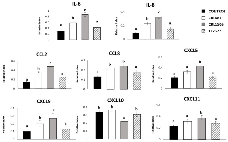
We evaluated whether L. plantarum CRL1506, CRL681 or TL2766 could modify the cytokine expression profile of PIE cells and whether the immunomodulatory property was strain specific. For this purpose, we comparatively analyzed the mRNA levels of IL-6 and the chemokines IL-8, CCL2, CCL8, CXCL5, CXCL9, CXCL10, and CXCL11 in PIE cells stimulated with these three bacterial strains (Figure 1). Stimulation of PIE cells with strains CRL1506 or CRL681 increased the expression of IL-6, IL-8, CCL2, CCL8 and CXCL9, while no significant differences were found in the levels of these inflammatory cytokines and chemokines between PIE cells treated with L. plantarum TL2677 and the control group. Only the strain CRL1506 was able to increase CXCL9 and CXCL11 levels compared to the control group, while CXCL10 levels decreased significantly in PIE cells treated with this Lactiplantibacillus strain. There was no significant difference in the levels of this chemokine among the other groups (Figure 1).
Figure 1.
Effect of Lactiplantibacillus plantarum CRL681, CRL1506 or TL2677 on the expression of cytokines and chemokines in porcine intestinal epithelial (PIE) cells. PIE cells were stimulated with CRL681, CRL1506 or TL2677 strains for 48 h. Untreated cells were used as controls. The expression of cytokines (IL-6) and chemokines (CCL2, CXCL5, CXCL8, CXCL9, CXCL10, CXCL11, and CCL8) were studied at 48 h after lactobacilli stimulation (basal). The results represent three independent experiments. Results are expressed as mean ± SD. Bold letters indicate significant differences when compared to the control group (p < 0.05).
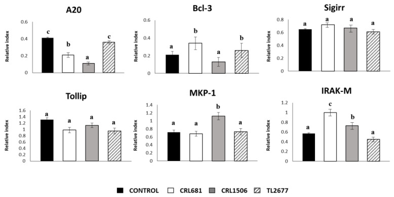
In addition, we evaluated the influence of the three L. plantarum strains on the expression of the negative regulators of the TLR4 signaling in PIE cells (Figure 2). No significant differences were observed in the expression of SIGIRR and Tollip when untreated control PIE cells and those treated with the different L. plantarum strains were compared. In addition, L. plantarum CRL681 and CRL1506 were able to reduce the expression of A20 in PIE cells and both strains increased the expression of Bcl-3 and IRAK-M. L. plantarum CRL1506 was the only strain capable of increasing the expression of MKP-1 compared to the control group (Figure 2).
Figure 2.
Effect of Lactiplantibacillus plantarum CRL681, CRL1506 or TL2677 on the expression of negative regulators of the Toll-like receptor (TLR) signaling pathway in porcine intestinal epithelial (PIE) cells. PIE cells were stimulated with CRL681, CRL1506 or TL2677 strains for 48 h. Untreated cells were used as controls. The expression of negative regulators of the TLR signaling pathway were studied at 48 h after lactobacilli stimulation (basal). The results represent three independent experiments. Results are expressed as mean ± SD. Bold letters indicate significant differences when compared to the control group (p < 0.05).
3.2. Effect of L. plantarum Strains on ETEC-Activated Innate Immune Response in PIE Cells
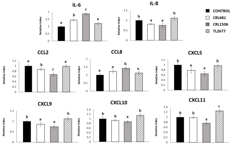
To study modulation of cytokines and chemokines in the context of inflammation, PIE cells were treated with L. plantarum CRL681, CRL1506 or TL2677. Then, cells were challenged with thermostable ETEC PAMPs. These molecular patterns can trigger TLR4 activation in this cell line, as we described previously [18,36]. Non-Lactiplantibacillus treated PIE cells challenged with ETEC were used as controls. The ETEC PAMPs significantly increased the expression of all inflammatory cytokines and chemokines in all experimental groups as shown in Figure 3, when compared to basal levels. The mRNA expression levels of IL-6 were significantly higher in cells treated with CRL681 and CRL1506 strains. On the other hand, the expressions of IL-8, CCL2, CXCL5 and CXCL9 were lower in PIE cells treated with these strains compared to controls. Interestingly, only L. plantarum CRL1506 enhanced the expression of CCL8 (Figure 3). L. plantarum TL2677 increased the expression of CXCL11 after exposure to ETEC, while strain CRL1506 reduced the expression of this chemokine (Figure 3). No significant differences were observed in the levels of CXCL10 between the groups treated with lactobacilli and the control group after ETEC challenge (Figure 3).
Figure 3.
Effect of Lactiplantibacillus plantarum CRL681, CRL1506 or TL2677 on the expression of cytokines and chemokines in porcine intestinal epithelial (PIE) cells after Enterotoxigenic Escherichia coli (ETEC) challenge. PIE cells were pre-treated with CRL681, CRL1506 or TL2677strains for 48 h and then stimulated with heat-stable ETEC pathogen-associated molecular patterns (PAMPs). Non-lactobacilli treated cells were used as controls. The expression of cytokines (IL-6) and chemokines (CCL2, CXCL5, CXCL8, CXCL9, CXCL10, CXCL11, and CCL8) were studied at 12 h after heat-stable ETEC PAMPs challenge. The results represent three independent experiments. Results are expressed as mean ± SD. Bold letters indicate significant differences when compared to the control group (p < 0.05).
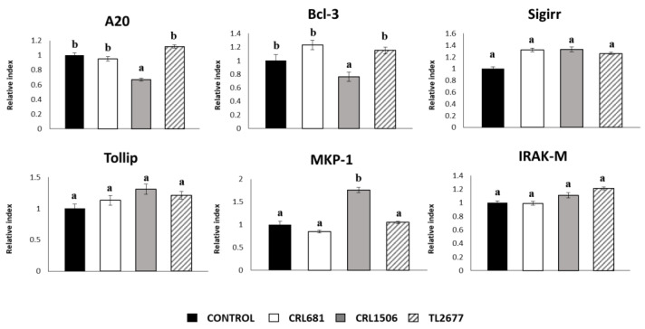
When the negative regulators of the TLR signaling pathway were investigated after exposure to ETEC PAMPs, it was found that only L. plantarum CRL1506 reduced the expressions of A20 and Bcl-3 (Figure 4). This strain also increased the levels of MKP-1 in ETEC-challenged PIE cells. No significant differences were observed in the levels of the other mediators analyzed in this study (Figure 4).
Figure 4.
Effect of Lactiplantibacillus plantarum CRL681, CRL1506 or TL2677 on the expression of negative regulators of the Toll-like receptor (TLR) signaling pathway in porcine intestinal epithelial (PIE) cells after Enterotoxigenic Escherichia coli (ETEC) challenge. PIE cells were pre-treated with CRL681, CRL1506 or TL2677 strains for 48 h and then stimulated with heat-stable ETEC pathogen-associated molecular patterns (PAMPs). Non-lactobacilli treated cells were used as controls. The expression of negative regulators of the TLR signaling pathway were studied at 12 h after heat-stable ETEC PAMPs challenge. The results represent three independent experiments. Results are expressed as mean ± SD. Bold letters indicate significant differences when compared to the control group (p < 0.05).
3.3. L. plantarum CRL681 and CRL1506 Modulate Intestinal Immunity In Vivo
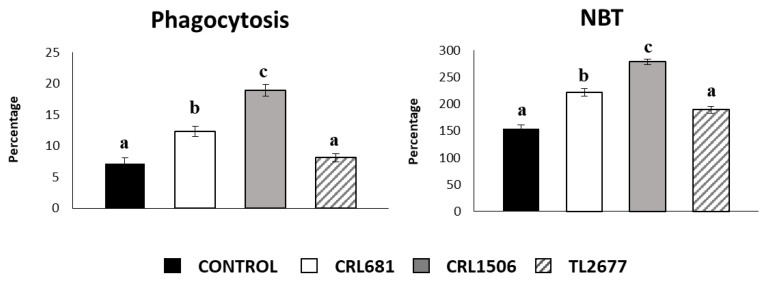
In addition, the ability of the different L. plantarum strains to stimulate macrophages was evaluated. For this purpose, ex vivo analysis of the phagocytic and bactericidal activity of the peritoneal macrophages were carried out. L. plantarum strains CRL681 and CRL1506 significantly increased the phagocytic activity of peritoneal macrophages, while this effect was absent in the strain TL2677 (Figure 5). In addition, a significant difference in the percentage of phagocytosis between the strains CRL681 and CRL1506 was observed, the latter strain showing higher phagocytic activity (Figure 5). To study the activation of the respiratory burst in peritoneal macrophages, we used the NBT method as previously described [18]. The treatment with the CRL1506 strain was more effective to enhance the percentage of NBT+ cells in the macrophage population obtained from the peritoneal cavity than the treatment with the CRL681 strain (Figure 5). Of note, L. plantarum TL2677 did not induce significant changes compared to the control group (Figure 5).
Figure 5.
Effect of Lactiplantibacillus plantarum CRL681, CRL1506 or TL2677 on peritoneal macrophages activities. Mice were orally treated with L. plantarum CRL681, CRL1506 or TL2677 (108 cells/mouse per day for 5 consecutive days). Untreated mice were used as controls. One day after the last lactobacilli administration, phagocytic and bactericidal (oxidative burst) activities of peritoneal macrophages were determined. The results represent three independent experiments. Results are expressed as mean ± SD. Bold letters indicate significant differences when compared to the control group (p < 0.05).
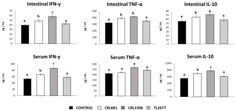
We also analyzed the cytokine concentrations in the intestinal fluid and serum obtained from mice treated with lactobacilli to determine the local and systemic effects induced by the L. plantarum strains (Figure 6). Both L. plantarum CRL681 and CRL1506 increased the levels of intestinal and serum IFN-γ. The concentrations of this cytokine found in the group stimulated with CRL1506 was higher than the CRL681 group (Figure 6). In addition, L. plantarum CRL681 and CRL1506 increased the level of intestinal TNF-α, while no differences were observed for serum TNF-α between the groups. Increased levels of IL-10 were found in both the intestinal fluid and serum of CRL1506- and CRL681-treated mice. However, serum levels of this immunoregulatory cytokine were significantly higher in mice treated with L. plantarum CRL1506 compared to those that received the CRL681 strain (Figure 6). No significant differences were observed between mice treated with L. plantarum TL2677 and control animals when the concentrations of intestinal and serum cytokines were analyzed (Figure 6).
Figure 6.
Effect of Lactiplantibacillus plantarum CRL681, CRL1506 or TL2677 on intestinal and serum cytokines of adult immunocompetent mice after enterotoxigenic Escherichia coli (ETEC) challenge. Mice were orally treated with L. plantarum CRL681, CRL1506 or TL2677 (108 cells/mouse per day for 5 consecutive days) and then challenged orally with ETEC F4 strain (109 cells). Mice with no lactobacilli treatment and challenged with ETEC were used as controls. Two days after the challenge, the concentrations of tumor necrosis factor (TNF)-α, interferon (IFN)-γ, and interleukin (IL)-10 in intestinal fluid and serum were determined by ELISA. The results represent three independent experiments. Results are expressed as mean ± SD. Bold letters indicate significant differences when compared to the control group (p < 0.05).
3.4. L. plantarum CRL681 and CRL1506 prevent the Spread of ETEC and Modulate the Expression of Intestinal Cytokines after Bacterial Challenge
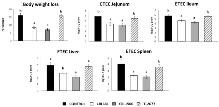
In order to evaluate the effect of L. plantarum strains on resistance to ETEC infection, body weight loss and bacterial counts in the jejunum, ileum, liver and spleen of infected mice were determined two days after the challenge. We evaluated body weight loss to study the general health state of mice (Figure 7). The infection with ETEC significantly increased the body weight loss of mice we described previously [37]. Of note, mice treated with the CRL1506 and CRL681 strains had significantly lower percentages of body weight loss than controls. L. plantarum CRL681 and CRL1506 were also able to significantly reduce ETEC counts in jejunum and ileum compared to controls (Figure 7). Furthermore, both lactobacilli treatments prevented the spread of the pathogen to the spleen and liver. There were no significant differences in body weight loss and the pathogen’s counts observed in the different organs of the mice treated with TL2677 and the control group (Figure 7).
Figure 7.
Immunomodulatory effect of Lactiplantibacillus plantarum CRL681, CRL1506 or TL2677 in mice in response to enterotoxigenic Escherichia coli (ETEC) challenge. Mice were orally treated with L. plantarum CRL681, CRL1506 or TL2677 (108 cells/mouse per day for 5 consecutive days) and then challenged orally with ETEC F4 strain (109 cells). Mice with no lactobacilli treatment and challenged with ETEC were used as controls. ETEC counts in jejunum, ileum, liver, and spleen were determined two days after the challenge. Values are means ± SD. Bold letters indicate significant differences when compared to the ETEC control group (p < 0.05).
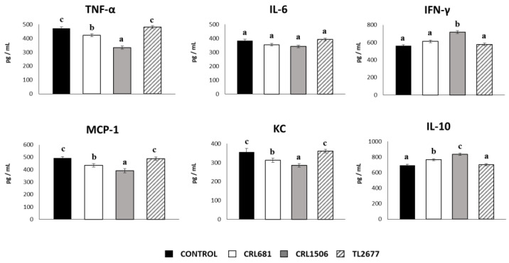
The levels of TNF-α, IL-6, IFN-γ, MCP-1, KC and IL-10 were also quantified in the gut mucosa of mice treated with lactobacilli and challenged with ETEC. Both L. plantarum CRL681 and CRL1506 significantly reduced the intestinal levels of TNF-α, KC and MCP-1 in comparison with the controls, being the CRL1506 strain the most effective to induce the decrease of these cytokines (Figure 8). Mice treated with strains CRL681 or CRL1506 showed higher intestinal IL-10 concentrations than controls, while only CRL1506 increased IFN-γ levels compared to the control group (Figure 8). No significant differences were observed in IL-6 levels between the studied groups. In addition, no differences were detected in intestinal cytokines levels between the group treated with L. plantarum TL2677 and the control group (Figure 8).
Figure 8.
Immunomodulatory effect of Lactiplantibacillus plantarum CRL681, CRL1506 or TL2677 in mice in response to enterotoxigenic Escherichia coli (ETEC) challenge. Mice were orally treated with L. plantarum CRL681, CRL1506 or TL2677 (108 cells/mouse per day for 5 consecutive days) and then challenged orally with ETEC F4 strain (109 cells). Mice with no lactobacilli treatment and challenged with ETEC were used as controls. The intestinal levels of tumor necrosis factor (TNF)-α, interferon (IFN)-γ, interleukin (IL)-6, IL-10, chemokine KC (or CXCL1), and monocyte chemoattractant protein 1 (MCP-1) were determined two days after the challenge with ETEC. Values are means ± SD. Bold letters indicate significant differences when compared to the ETEC control group (p < 0.05).
4. Discussion
LAB of various species can be used as probiotics for animals with the aim to control pathogenic microorganisms and improve natural defense mechanisms, reducing health problems and therefore increasing productivity [39,40]. L. plantarum is among the most versatile species used for decades in the food industry either as starter cultures or probiotics [25]. They are applied as starter cultures to produce cheeses, sausages, olives and a wide variety of fermented foods and beverages, contributing to their organoleptic properties, flavor and texture. This outstanding versatility and metabolic activity can be explained by its big genome (2.91 to 3.7 Mbp in length) compared to other LAB such as Latilactobacillus curvatus or Lactobacillus sakei which account for less than 1.9 Mbp [41,42] This feature contributes to its survival capability in a wide range of environmental niches including plants, fermented foods, and the gastrointestinal tract humans and other mammals [22,23]. The increasing significance as probiotic of this species are mainly linked to health promotion in humans and animals [24,43,44,45,46,47,48,49]. However, their beneficial effects are strain dependent and not universal. Therefore, the probiotic properties need to be characterized on a strain level.
In this context, the immunobiotic potential of two L. plantarum strains with different origins and probiotic/technological properties was studied and compared to evaluate their capability to improve the resistance to ETEC infection. L. plantarum CRL681, originally isolated from fermented sausages, has an efficient acidogenic activity that guarantees safety and texture development during ripening [31,44]. In addition, detailed peptidomic studies confirmed its peptidogenic ability and its capacity to increase free amino acid contents in meat or fermented-meat models [31,32,33]. The CRL681 strain is capable of degrading biogenic amines in vitro and lacks the ability to produce them [40]. Moreover, this strain has remarkable bioprotective potential due to the high inhibitory activity toward E. coli O157:H7 [35]. On the other hand, L. plantarum CRL1506, originally isolated from goat milk, has demonstrated to possess remarkable immunomodulatory activities in the context of antiviral immunity [26,27,28,29,46], although its capacity to modulate antibacterial immune responses in the intestinal tract has not been explored in depth.
In the present study, we demonstrated that both CRL681 and CRL1506 strains can modulate the intestinal immune response in steady-state conditions. L. plantarum CRL681 and CRL1506 triggered the expression of IL-6 and IL-8 in porcine IECs. Furthermore, mice orally treated with these strains had significantly improved levels of IFN-γ and TNF-α in the intestine as well as enhanced activity of peritoneal macrophages. Studies evaluating the effect of lactobacilli strains with the capacity to reduce the severity of intestinal infections found that the most remarkable effect was the increase in the intestinal levels of TNF-α, IFN-γ, IL-1β, IL-6, and IL-12 for the mice treated with the probiotic strains, as well as the phagocytic activity of intestinal and peritoneal macrophages [47,48]. In vitro and ex vivo studies in a primary culture of IECs demonstrated that probiotic lactobacilli interact with these cells and induce release of IL-6 [47,49]. This cytokine was shown to regulate the survival and proliferation of IECs as well as to stimulate immune cells in the intestinal mucosa [50]. Autophagy in IECs is involved in the homeostatic control of cell death and differentiation. It was shown that IECs have a high basal level of autophagy that is regulated by TLR-mediated IL-8 production [51]. On the other hand, probiotic bacteria can stimulate macrophages by increasing their phagocytic activities and their capacity to produce cytokines like IFN-γ and TNF-α. This macrophage activity is essential for the protection against infections [52]. Thus, our results suggest that both CRL681 and CRL1506 strains have the capacity to interact with IECs and macrophages in the gut, reinforcing the epithelial defenses and stimulating immune responses. In fact, the ability of L. plantarum CRL681 and CRL1506 to beneficially modulate intestinal immunity was put into greater evidence in the experiments in which challenges with ETEC were performed.
In this work, using the in vitro PIE cell system we observed that L. plantarum CRL681 and CRL1506 differentially modulated the innate immune responses of porcine IECs triggered by ETEC challenge. The CRL1506 and CRL681 strains modulated the expression of inflammatory cytokines (IL-6) and chemokines (IL-8, CCL2, CXCL5 and CXCL9) in ETEC-stimulated PIE cells. Furthermore, our studies in the mice model of ETEC infection demonstrated in vivo the ability of L. plantarum CRL681 and CRL1506 to beneficially regulate the immune response against this pathogen. In our hands, the oral treatment of mice with CRL681 or CRL1506 strains significantly reduced body weight loss as well as ETEC counts in jejunum and ileum and prevented the spread of the pathogen to the spleen and liver. This protective effect could be related to a differential regulation of cytokines and chemokines in the intestinal mucosa, particularly in IECs. Our results are in line with previous works demonstrating that probiotic microorganisms can regulate the immune response against ETEC in both mice models [13,14] and pigs [15,16,17] as well as in IECs [53,54]. Lacticaseibacillus rhamnosus GG and Bifidobacterium animalis MB5 protected human Caco-2 cells from the ETEC K88-associated inflammation by reducing IL-1β and TNF-α and enhancing TGF-β1 expression [53,55]. Enterococcus faecium HDRsEf1 has been shown to have positive effects on piglet diarrhea, and studies performed in the porcine IPEC-J2 cell line demonstrated that this effect is partially related to its ability to reduce the expression of IL-8 after ETEC challenge, protecting IECs from the acute inflammatory response [54]. In addition, it was shown that the porcine IECs line IPEC-1 treated with L. plantarum CGMCC 1258 had reduced expressions of IL-1α, IL-6, IL-8 and TNF-α after the challenge with ETEC K88. This effect was associated with the ability of the CGMCC 1258 strain to regulate MAPK and NF-κB signaling pathways [56]. Our previous studies in PIE cells showed a reduction in the activation of NF-κB and MAPK signaling pathways and in the expression of some inflammatory cytokines and chemokines in ETEC-challenged PIE cells, preventively stimulated with Lactobacillus jensenii TL2937 [21], or Bifidobacterium breve M-16V [36].
Then, our results suggest that the improved intestinal epithelial defenses and innate immunity induced by L. plantarum CRL681 and CRL1506 would increase the clearance of ETEC and at the same time, protect the host against detrimental inflammation. The activation of TLR4 in the intestinal mucosa induce the production of cytokines to stimulate the recruitment and activation of inflammatory cells. Although this mechanism is a key primary line of host defense, prolonged or dysregulated proinflammatory response may lead to tissue damage and dysfunction [57]. Thus, the reduction of the intestinal levels of TNF-α, MCP-1 and KC induced by the CRL681 and CRL1506 strains could indicate a better control of inflammation and its detrimental effects. Furthermore, it was observed that mice treated with L. plantarum CRL681 or CRL1506 had improved levels of intestinal IL-10 both at steady state and after the challenge with ETEC. Studies performed in healthy adult volunteers challenged with ETEC demonstrated that higher pre-challenge concentrations of IL-10 were associated with protection from ETEC diarrhea [9]. In addition, in agreement with our results it was reported that the treatment of mice with L. plantarum CCFM1143 was able to alleviate diarrhea caused by ETEC infection, and that this beneficial effect was associated with reductions of TNF-α and improvements of IL-10 [58]. Furthermore, in line with the relevant role of IL-10 in controlling TNF-α production, our studies showed that in baseline determinations the levels of this regulatory cytokine increased only 1.4 times in animals treated with CRL1506 or CRL681 strains compared to controls and was not able to decrease the levels of the inflammatory cytokine. In contrast, in the ETEC-challenge experiments, IL-10 was augmented more than 10-fold compared to baseline values and 1.6-fold between CRL1506 and CRL681 versus controls.
We have previously demonstrated that the beneficial effects of immunobiotic bacteria in the context of TLR4-triggered inflammation are mediated by a differential modulation of negative regulators of the TLR signaling pathway [21,36]. Then, we also evaluated here the ability of L. plantarum CRL681 or CRL1506 to modulate the expression of SIGIRR, Tollip, A20, Bcl-3, IRAK-M, and MKP-1 in porcine IECs. We found that both strains upregulated the expression of IRAK-M and reduced A20 in PIE cells without ETEC challenge. In addition, CRL1506 and CRL681 increased the expressions of MKP-1 and Bcl-3, respectively. The TLR negative regulators play important roles in the maintenance of intestinal hemostasis as well as in the control of immune responses against pathogens. IRAK-M exerts its regulatory effect by acting on the TRAF6/IRAK-1 complex, diminishing the activation of NF-κB and MAPK pathways [59]. It was described that LPS challenge induce the expression of IRAK-M, and that the tolerance to TLR4 activation is diminished in IRAK-M-deficient cells [60]. On the other hand, it was reported that A20 [61,62] and Bcl-3 [63] regulate TLR4 signaling pathway by inducing the inhibition of NF-κB activation while MKP-1 inactivates the MAPK p38 signaling pathway [64]. Thus, it is tempting to speculate that lactobacilli would induce a differential expression of negative regulators of the TLR4 pathway in the intestinal epithelium, and when the challenged with ETEC is induced, this signaling pathway would be differentially regulated allowing an efficient induction of an antibacterial state in the gut, and the protection against the inflammatory-mediated damage.
Of note, the immunostimulatory activity of the CRL1506 and CRL681 strains were not achieved by L. plantarum TL2677 in porcine IECs. In addition, the protective effect of L. plantarum CRL681 and CRL1506 against ETEC infection was not observed for the TL2677 strain in mice, highlighting that the immunomodulatory effects are strain specific. Furthermore, the analysis of the immunomodulatory activities in steady state conditions as well as the immune responses triggered by ETEC challenge showed that L. plantarum CRL1506 is more efficient than CRL681 strain to modulate mucosal immunity. In line with our results, it was shown that different strains had individual capacities to regulate TLR4-NF-κB signaling pathway and the expression of IL-8 in HEK cells as well as to regulate trans-epithelial electrical resistance and tight junction integrity in LPS-challenged Caco-2 monolayers [65]. The different abilities of the L. plantarum strains studied here to influence the immune response against ETEC could be associated with their distinct capacity to modulate negative regulators of TLR. While the TL2677 did not modulate the expression of TLR negative regulators, L. plantarum CRL1506 was more efficient than the CRL681 strain to upregulate IRAK-M expression and diminish A20. Moreover, when the TLR negative regulators were evaluated in PIE cells challenged with ETEC, only cells treated with L. plantarum CRL1506 showed differences in the expressions of A20, Bcl-3 and MKP-1. Interestingly, despite the more notable changes in the expression of immune factors both in vitro and in vivo induced by the CRL1506 strain, no significant differences in ETEC counts were observed when compared to mice treated with L. plantarum CRL681. This may be due to the fact that in this work only one post-infection point was studied. Perhaps a study of the kinetics of ETEC clearance could find differences between the two treatments. In addition, a further study to evaluate whether a combination of both L. plantarum CRL1506 and CRL681 could improve immunity more effectively than individual strains and induce a more efficient clearance of ETEC in mice intestine would be of value. Another important point for future studies is to find the bacterial molecules and immune receptors involved in the induction of TLR negative regulators in the intestinal epithelium by L. plantarum, which also explain the differences between the strains. Our recent comparative genomic study showed a great variability in the predicted surface proteins of L. plantarum strains, including CRL1506 and CRL681 [30]. These results suggest that the surface molecules could be involved in their differential ability to modulate the intestinal innate immune response against ETEC.
5. Conclusions
Our results demonstrated that the improved intestinal epithelial defenses and innate immunity induced by L. plantarum CRL1506 and CRL681 would increase the clearance of ETEC and at the same time, the differential expression of negative regulators of the TLR4 pathway in the intestinal epithelium allowing the establishment of an antibacterial state in the gut, and the protection against the inflammatory-mediated damage. These findings constitute valuable features for a future probiotic culture for animal feed able to improve the resistance to ETEC infection. The preliminary studies carried out in this work using porcine IECs and mice provide the scientific basis for carrying out in vivo studies in pigs to reliably demonstrate the protective potential of CRL1506 and CRL681 strains against ETEC infection in the porcine host.
Author Contributions
A.B. carried out the experiments, analyzed the results, contributed to the discussion, and wrote the paper. J.V. conceived the idea of the project, carried out the experiments, analyzed the results, contributed to the discussion, wrote the paper, and participated in funding acquisition. L.A. carried out the experiments, analysed the results and contributed to the discussion. M.T. contributed to the discussion and wrote the paper. M.E. carried out the experiments, analyzed the results, analysed the results and contributed to the discussion. K.F. carried out the experiments, analyzed the results, contributed to the discussion and wrote the paper. S.Q.-V. contributed to the discussion and wrote the paper. S.F. coordinate, analyzed and discussed the results, wrote the paper, and participated in funding acquisition. H.K. coordinate, analyzed and discussed the results, wrote the paper, and participated in funding acquisition. All authors have read and agreed to the published version of the manuscript.
Institutional Review Board Statement
Experiments with mice were performed in accordance with the guide for the care and use of laboratory animals and were approved by the CERELA-CONICET Animal Care and Ethics Committee under the BIOT-CRL/19 protocol.
Informed Consent Statement
Not applicable.
Data Availability Statement
Data are contained within this article.
Conflicts of Interest
The authors declare that the research was conducted in the absence of any commercial or financial relationships that could be construed as a potential conflict of interest.
Funding Statement
This study was supported by the research program on development of innovative technology grants (JPJ007097) from the Project of the Bio-oriented Technology Research Advancement Institution (BRAIN), and by a Grant-in-Aid for Scientific Research (A) (19H00965) from the Japan Society for the Promotion of Science (JSPS), and by Japan Racing Association (JRA) Livestock Industry Promotion Project, and by the Association for Research on Lactic Acid Bacteria to HK. This study was also supported by ANPCyT–FONCyT Grant PICT-2016-0410 to JV, Grant PICT-2018-02249; PICT-2018-0664 to SF and by JSPS Core-to-Core Program, A. Advanced Research Networks entitled Establishment of international agricultural immunology research-core for a quantum improvement in food safety, and by AMED Grant Number JP21zf0127001. Mikado Tomokiyo was supported by JST SPRING, Grant Number JPMJSP2114. Kohtaro Fukuyama was supported by JST, the establishment of university fellowships towards the creation of science technology innovation, Grant Number JPMJFS2102.
Footnotes
Disclaimer/Publisher’s Note: The statements, opinions and data contained in all publications are solely those of the individual author(s) and contributor(s) and not of MDPI and/or the editor(s). MDPI and/or the editor(s) disclaim responsibility for any injury to people or property resulting from any ideas, methods, instructions or products referred to in the content.
References
- 1.Khalil I.A., Troeger C., Blacker B.F., Rao P.C., Brown A., Atherly D.E., Brewer T.G., Engmann C.M., Houpt E.R., Kang G., et al. Morbidity and Mortality Due to Shigella and Enterotoxigenic Escherichia coli Diarrhoea: The Global Burden of Disease Study 1990–2016. Lancet Infect. Dis. 2018;18:1229–1240. doi: 10.1016/S1473-3099(18)30475-4. [DOI] [PMC free article] [PubMed] [Google Scholar]
- 2.Laird T.J., Abraham S., Jordan D., Pluske J.R., Hampson D.J., Trott D.J., O’Dea M. Porcine Enterotoxigenic Escherichia coli: Antimicrobial Resistance and Development of Microbial-Based Alternative Control Strategies. Vet. Microbiol. 2021;258:109117. doi: 10.1016/j.vetmic.2021.109117. [DOI] [PubMed] [Google Scholar]
- 3.Wu Q., Cui D., Chao X., Chen P., Liu J., Wang Y., Su T., Li M., Xu R., Zhu Y., et al. Transcriptome Analysis Identifies Strategies Targeting Immune Response-Related Pathways to Control Enterotoxigenic Escherichia coli Infection in Porcine Intestinal Epithelial Cells. Front. Vet. Sci. 2021;8:677897. doi: 10.3389/fvets.2021.677897. [DOI] [PMC free article] [PubMed] [Google Scholar]
- 4.Dubreuil J.D., Isaacson R.E., Schifferli D.M. Animal Enterotoxigenic Escherichia coli. EcoSal Plus. 2016;7:10. doi: 10.1128/ecosalplus.ESP-0006-2016. [DOI] [PMC free article] [PubMed] [Google Scholar]
- 5.Kobayashi H., Albarracin L., Sato N., Kanmani P., Kober A., Ikeda-Ohtsubo W., Suda Y., Nochi T., Aso H., Makino S., et al. Modulation of Porcine Intestinal Epitheliocytes Immunetranscriptome Response by Lactobacillus jensenii TL2937. Benef. Microbes. 2016;7:769–782. doi: 10.3920/BM2016.0095. [DOI] [PubMed] [Google Scholar]
- 6.Hu M.M., Shu H.B. Cytoplasmic Mechanisms of Recognition and Defense of Microbial Nucleic Acids. Annu. Rev. Cell Dev. Biol. 2018;34:357–379. doi: 10.1146/annurev-cellbio-100617-062903. [DOI] [PubMed] [Google Scholar]
- 7.Afonina I.S., Zhong Z., Karin M., Beyaert R. Limiting Inflammation-the Negative Regulation of NF-ΚB and the NLRP3 Inflammasome. Nat. Immunol. 2017;18:861–869. doi: 10.1038/ni.3772. [DOI] [PubMed] [Google Scholar]
- 8.Zhou M., Xu W., Wang J., Yan J., Shi Y., Zhang C., Ge W., Wu J., Du P., Chen Y. Boosting MTOR-Dependent Autophagy via Upstream TLR4-MyD88-MAPK Signalling and Downstream NF-ΚB Pathway Quenches Intestinal Inflammation and Oxidative Stress Injury. EBioMedicine. 2018;35:345–360. doi: 10.1016/j.ebiom.2018.08.035. [DOI] [PMC free article] [PubMed] [Google Scholar]
- 9.Brubaker J., Zhang X., Bourgeois A.L., Harro C., Sack D.A., Chakraborty S. Intestinal and Systemic Inflammation Induced by Symptomatic and Asymptomatic Enterotoxigenic E. coli Infection and Impact on Intestinal Colonization and ETEC Specific Immune Responses in an Experimental Human Challenge Model. Gut Microbes. 2021;13:1–13. doi: 10.1080/19490976.2021.1891852. [DOI] [PMC free article] [PubMed] [Google Scholar]
- 10.Jiang F., Wu Z., Zheng Y., Frana T.S., Sahin O., Zhang Q., Li G. Genotypes and Antimicrobial Susceptibility Profiles of Hemolytic Escherichia coli from Diarrheic Piglets. Foodborne Pathog. Dis. 2019;16:94–103. doi: 10.1089/fpd.2018.2480. [DOI] [PubMed] [Google Scholar]
- 11.de Lagarde M., Vanier G., Arsenault J., Fairbrother J.M. High Risk Clone: A Proposal of Criteria Adapted to the One Health Context with Application to Enterotoxigenic Escherichia coli in the Pig Population. Antibiotics. 2021;10:244. doi: 10.3390/antibiotics10030244. [DOI] [PMC free article] [PubMed] [Google Scholar]
- 12.Dubreuil J.D. Enterotoxigenic Escherichia coli and Probiotics in Swine: What the Bleep Do We Know? Biosci. Microbiota Food Health. 2017;36:75–90. doi: 10.12938/bmfh.16-030. [DOI] [PMC free article] [PubMed] [Google Scholar]
- 13.Shu Q., Gill H.S. A dietary probiotic (Bifidobacterium lactis HN019) reduces the severity of Escherichia coli O157:H7 infection in mice. Med. Microbiol. Immunol. 2001;189:147–152. doi: 10.1007/s430-001-8021-9. [DOI] [PubMed] [Google Scholar]
- 14.Shu Q., Gill H.S. Immune protection mediated by the probiotic Lactobacillus rhamnosus HN001 (DR20) against Escherichia coli O157:H7 infection in mice. FEMS Immunol. Med. Microbiol. 2002;34:59–64. doi: 10.1016/S0928-8244(02)00340-1. [DOI] [PubMed] [Google Scholar]
- 15.Shu Q., Qu F., Gill H.S. Probiotic treatment using Bifidobacterium lactis HN019 reduces weanling diarrhea associated with rotavirus and Escherichia coli infection in a piglet model. J. Pediatr. Gastroenterol. Nutr. 2001;33:171–177. doi: 10.1097/00005176-200108000-00014. [DOI] [PubMed] [Google Scholar]
- 16.Sun Y., Duarte M.E., Kim S.W. Dietary inclusion of multispecies probiotics to reduce the severity of post-weaning diarrhea caused by Escherichia coli F18+ in pigs. Anim. Nutr. 2021;7:326–333. doi: 10.1016/j.aninu.2020.08.012. [DOI] [PMC free article] [PubMed] [Google Scholar]
- 17.Daudelin J.F., Lessard M., Beaudoin F., Nadeau E., Bissonnette N., Boutin Y., Brousseau J.P., Lauzon K., Fairbrother J.M. Administration of probiotics influences F4 (K88)-positive enterotoxigenic Escherichia coli attachment and intestinal cytokine expression in weaned pigs. Veter Res. 2011;42:69. doi: 10.1186/1297-9716-42-69. [DOI] [PMC free article] [PubMed] [Google Scholar]
- 18.Garcia-Castillo V., Komatsu R., Clua P., Indo Y., Takagi M., Salva S., Islam M.A., Alvarez S., Takahashi H., Garcia-Cancino A., et al. Evaluation of the Immunomodulatory Activities of the Probiotic Strain Lactobacillus fermentum UCO-979C. Front. Immunol. 2019;10:1376. doi: 10.3389/fimmu.2019.01376. [DOI] [PMC free article] [PubMed] [Google Scholar]
- 19.Villena J., Kitazawa H. Modulation of Intestinal TLR4-Inflammatory Signaling Pathways by Probiotic Microorganisms: Lessons Learned from Lactobacillus jensenii TL2937. Front. Immunol. 2014;4:512. doi: 10.3389/fimmu.2013.00512. [DOI] [PMC free article] [PubMed] [Google Scholar]
- 20.Moue M., Tohno M., Shimazu T., Kido T., Aso H., Saito T., Kitazawa H. Toll-like Receptor 4 and Cytokine Expression Involved in Functional Immune Response in an Originally Established Porcine Intestinal Epitheliocyte Cell Line. Biochim. Biophys. Acta Gen. Subj. 2008;1780:134–144. doi: 10.1016/j.bbagen.2007.11.006. [DOI] [PubMed] [Google Scholar]
- 21.Shimazu T., Villena J., Tohno M., Fujie H., Hosoya S., Shimosato T., Aso H., Suda Y., Kawai Y., Saito T., et al. Immunobiotic Lactobacillus jensenii Elicits Anti-Inflammatory Activity in Porcine Intestinal Epithelial Cells by Modulating Negative Regulators of the Toll-like Receptor Signaling Pathway. Infect. Immun. 2012;80:276–288. doi: 10.1128/IAI.05729-11. [DOI] [PMC free article] [PubMed] [Google Scholar]
- 22.de Vries M.C., Vaughan E.E., Kleerebezem M., de Vos W.M. Lactobacillus plantarum-Survival, Functional and Potential Probiotic Properties in the Human Intestinal Tract. Int. Dairy J. 2006;16:1018–1028. doi: 10.1016/j.idairyj.2005.09.003. [DOI] [Google Scholar]
- 23.Siezen R.J., Tzeneva V.A., Castioni A., Wels M., Phan H.T.K., Rademaker J.L.W., Starrenburg M.J.C., Kleerebezem M., van Hylckama Vlieg J.E.T. Phenotypic and Genomic Diversity of Lactobacillus plantarum Strains Isolated from Various Environmental Niches. Environ. Microbiol. 2010;12:758–773. doi: 10.1111/j.1462-2920.2009.02119.x. [DOI] [PubMed] [Google Scholar]
- 24.Villena J., Li C., Vizoso-Pinto M.G., Sacur J., Ren L., Kitazawa H. Lactiplantibacillus plantarum as a Potential Adjuvant and Delivery System for the Development of SARS-CoV-2 Oral Vaccines. Microorganisms. 2021;9:683. doi: 10.3390/microorganisms9040683. [DOI] [PMC free article] [PubMed] [Google Scholar]
- 25.Behera S.S., Ray R.C., Zdolec N. Lactobacillus plantarum with Functional Properties: An Approach to Increase Safety and Shelf-Life of Fermented Foods. Biomed Res. Int. 2018;2018:9361614. doi: 10.1155/2018/9361614. [DOI] [PMC free article] [PubMed] [Google Scholar]
- 26.Albarracin L., Garcia-Castillo V., Masumizu Y., Indo Y., Islam M.A., Suda Y., Garcia-Cancino A., Aso H., Takahashi H., Kitazawa H., et al. Efficient Selection of New Immunobiotic Strains With Antiviral Effects in Local and Distal Mucosal Sites by Using Porcine Intestinal Epitheliocytes. Front. Immunol. 2020;11:543. doi: 10.3389/fimmu.2020.00543. [DOI] [PMC free article] [PubMed] [Google Scholar]
- 27.Mizuno H., Arce L., Tomotsune K., Albarracin L., Funabashi R., Vera D., Islam M.A., Vizoso-Pinto M.G., Takahashi H., Sasaki Y., et al. Lipoteichoic Acid Is Involved in the Ability of the Immunobiotic Strain Lactobacillus plantarum CRL1506 to Modulate the Intestinal Antiviral Innate Immunity Triggered by TLR3 Activation. Front. Immunol. 2020;11:571. doi: 10.3389/fimmu.2020.00571. [DOI] [PMC free article] [PubMed] [Google Scholar]
- 28.Islam M.A., Albarracin L., Tomokiyo M., Valdez J.C., Sacur J., Vizoso-Pinto M.G., Andrade B.G.N., Cuadrat R.R.C., Kitazawa H., Villena J. Immunobiotic Lactobacilli Improve Resistance of Respiratory Epithelial Cells to SARS-CoV-2 Infection. Pathogens. 2021;10:1197. doi: 10.3390/pathogens10091197. [DOI] [PMC free article] [PubMed] [Google Scholar]
- 29.Tada A., Zelaya H., Clua P., Salva S., Alvarez S., Kitazawa H., Villena J. Immunobiotic Lactobacillus Strains Reduce Small Intestinal Injury Induced by Intraepithelial Lymphocytes after Toll-like Receptor 3 Activation. Inflamm. Res. 2016;65:771–783. doi: 10.1007/s00011-016-0957-7. [DOI] [PubMed] [Google Scholar]
- 30.Albarracin L., Tonetti F.R., Fukuyama K., Suda Y., Zhou B., Baillo A.A., Fadda S., Saavedra L., Kurata S., Hebert E.M., et al. Genomic Characterization of Lactiplantibacillus plantarum Strains Possessing Differential Antiviral Immunomodulatory Activities. Bacteria. 2022;1:136–160. doi: 10.3390/bacteria1030012. [DOI] [Google Scholar]
- 31.Fadda S., Vildoza M.J., Vignolo G. The acidogenic metabolism of Lactobacillus plantarum CRL 681 improves sarcoplasmic protein hydrolysis during meat fermentation. J. Muscle Foods. 2010;21:545–556. doi: 10.1111/j.1745-4573.2009.00202.x. [DOI] [Google Scholar]
- 32.Fadda S., Chambon C., Champomier-Vergès M.C., Talon R., Vignolo G. Lactobacillus Role during Conditioning of Refrigerated and Vacuum-Packaged Argentinean Meat. Meat Sci. 2008;79:603–610. doi: 10.1016/j.meatsci.2007.04.003. [DOI] [PubMed] [Google Scholar]
- 33.de Almeida M.A., Saldaña E., da Silva Pinto J.S., Palacios J., Contreras-Castillo C.J., Sentandreu M.A., Fadda S.G. A Peptidomic Approach of Meat Protein Degradation in a Low-Sodium Fermented Sausage Model Using Autochthonous Starter Cultures. Food Res. Int. 2018;109:368–379. doi: 10.1016/j.foodres.2018.04.042. [DOI] [PubMed] [Google Scholar]
- 34.Fadda S., López C., Vignolo G. Role of Lactic Acid Bacteria during Meat Conditioning and Fermentation: Peptides Generated as Sensorial and Hygienic Biomarkers. Meat Sci. 2010;86:66–79. doi: 10.1016/j.meatsci.2010.04.023. [DOI] [PubMed] [Google Scholar]
- 35.Orihuel A., Terán L., Renaut J., Vignolo G.M., de Almeida A.M., Saavedra M.L., Fadda S. Differential Proteomic Analysis of Lactic Acid Bacteria- Escherichia coli O157:H7 Interaction and Its Contribution to Bioprotection Strategies in Meat. Front. Microbiol. 2018;9:1083. doi: 10.3389/fmicb.2018.01083. [DOI] [PMC free article] [PubMed] [Google Scholar]
- 36.Tomosada Y., Villena J., Murata K., Chiba E., Shimazu T., Aso H., Iwabuchi N., Xiao J.Z., Saito T., Kitazawa H. Immunoregulatory Effect of Bifidobacteria Strains in Porcine Intestinal Epithelial Cells through Modulation of Ubiquitin-Editing Enzyme A20 Expression. PLoS One. 2013;8:e59259. doi: 10.1371/journal.pone.0059259. [DOI] [PMC free article] [PubMed] [Google Scholar]
- 37.Indo Y., Kitahara S., Tomokiyo M., Araki S., Islam M.A., Zhou B., Albarracin L., Miyazaki A., Ikeda-Ohtsubo W., Nochi T., et al. Ligilactobacillus Salivarius Strains Isolated From the Porcine Gut Modulate Innate Immune Responses in Epithelial Cells and Improve Protection Against Intestinal Viral-Bacterial Superinfection. Front. Immunol. 2021;12:1. doi: 10.3389/fimmu.2021.652923. [DOI] [PMC free article] [PubMed] [Google Scholar]
- 38.Marranzino G., Villena J., Salva S., Alvarez S. Stimulation of Macrophages by Immunobiotic Lactobacillus Strains: Influence beyond the Intestinal Tract. Microbiol. Immunol. 2012;56:771–781. doi: 10.1111/j.1348-0421.2012.00495.x. [DOI] [PubMed] [Google Scholar]
- 39.Adetoye A., Pinloche E., Adeniyi B.A., Ayeni F.A. Characterization and Anti-Salmonella Activities of Lactic Acid Bacteria Isolated from Cattle Faeces. BMC Microbiol. 2018;18:96. doi: 10.1186/s12866-018-1248-y. [DOI] [PMC free article] [PubMed] [Google Scholar]
- 40.Markowiak P., Ślizewska K. The Role of Probiotics, Prebiotics and Synbiotics in Animal Nutrition. Gut Pathog. 2018;10:21. doi: 10.1186/s13099-018-0250-0. [DOI] [PMC free article] [PubMed] [Google Scholar]
- 41.Chaillou S., Champomier-Vergès M.C., Cornet M., Crutz-Le Coq A.M., Dudez A.M., Martin V., Beaufils S., Darbon-Rongère E., Bossy R., Loux V., et al. The Complete Genome Sequence of the Meat-Borne Lactic Acid Bacterium Lactobacillus sakei 23K. Nat. Biotechnol. 2005;23:1527–1533. doi: 10.1038/nbt1160. [DOI] [PubMed] [Google Scholar]
- 42.Hebert E.M., Saavedra L., Taranto M.P., Mozzi F., Magni C., Nader M.E.F., de Valdez G.F., Sesma F., Vignolo G., Raya R.R. Genome Sequence of the Bacteriocin-Producing Lactobacillus curvatus Strain CRL705. J. Bacteriol. 2012;194:538–539. doi: 10.1128/JB.06416-11. [DOI] [PMC free article] [PubMed] [Google Scholar]
- 43.Corsetti A., Valmorri S. Lactic Acid Bacteria|Lactobacillus spp.: Lactobacillus plantarum. Encycl. Dairy Sci. Second. Ed. 2011;3:111–118. doi: 10.1016/B978-0-12-374407-4.00263-6. [DOI] [Google Scholar]
- 44.Vignolo G.M., de Ruiz Holgado A.P., Oliver G. Acid Production and Proteolytic Activity of Lactobacillus Strains Isolated from Dry Sausages. J. Food Prot. 1988;51:481–484. doi: 10.4315/0362-028X-51.6.481. [DOI] [PubMed] [Google Scholar]
- 45.Fadda S., Vignolo G., Oliver G. Tyramine Degradation and Tyramine/Histamine Production by Lactic Acid Bacteria and Kocuria Strains. Biotechnol. Lett. 2001;23:2015–2019. doi: 10.1023/A:1013783030276. [DOI] [Google Scholar]
- 46.Albarracin L., Kobayashi H., Iida H., Sato N., Nochi T., Aso H., Salva S., Alvarez S., Kitazawa H., Villena J. Transcriptomic Analysis of the Innate Antiviral Immune Response in Porcine Intestinal Epithelial Cells: Influence of Immunobiotic Lactobacilli. Front. Immunol. 2017;8:57. doi: 10.3389/fimmu.2017.00057. [DOI] [PMC free article] [PubMed] [Google Scholar]
- 47.Maldonado Galdeano C., de Moreno De Leblanc A., Vinderola G., Bibas Bonet M.E., Perdigón G. Proposed Model: Mechanisms of Immunomodulation Induced by Probiotic Bacteria. Clin. Vaccine Immunol. 2007;14:485–492. doi: 10.1128/CVI.00406-06. [DOI] [PMC free article] [PubMed] [Google Scholar]
- 48.Salva S., Villena J., Alvarez S. Immunomodulatory Activity of Lactobacillus rhamnosus Strains Isolated from Goat Milk: Impact on Intestinal and Respiratory Infections. Int. J. Food Microbiol. 2010;141:82–89. doi: 10.1016/j.ijfoodmicro.2010.03.013. [DOI] [PubMed] [Google Scholar]
- 49.Villena J., Chiba E., Vizoso-Pinto M.G., Tomosada Y., Takahashi T., Ishizuka T., Aso H., Salva S., Alvarez S., Kitazawa H. Immunobiotic Lactobacillus rhamnosus Strains Differentially Modulate Antiviral Immune Response in Porcine Intestinal Epithelial and Antigen Presenting Cells. BMC Microbiol. 2014;14:126. doi: 10.1186/1471-2180-14-126. [DOI] [PMC free article] [PubMed] [Google Scholar]
- 50.Grivennikov S., Karin E., Terzic J., Mucida D., Yu G.Y., Vallabhapurapu S., Scheller J., Rose-John S., Cheroutre H., Eckmann L., et al. IL-6 and Stat3 Are Required for Survival of Intestinal Epithelial Cells and Development of Colitis-Associated Cancer. Cancer Cell. 2009;15:103–113. doi: 10.1016/j.ccr.2009.01.001. [DOI] [PMC free article] [PubMed] [Google Scholar]
- 51.Li Y.Y., Ishihara S., Aziz M.M., Oka A., Kusunoki R., Tada Y., Yuki T., Amano Y., Ansary M.U., Kinoshita Y. Autophagy Is Required for Toll-like Receptor-Mediated Interleukin-8 Production in Intestinal Epithelial Cells. Int. J. Mol. Med. 2011;27:337–344. doi: 10.3892/IJMM.2011.596. [DOI] [PubMed] [Google Scholar]
- 52.Elean M., Albarracin L., Fukuyama K., Zhou B., Tomokiyo M., Kitahara S., Araki S., Suda Y., Saavedra L., Villena J., et al. Lactobacillus delbrueckii CRL 581 Differentially Modulates TLR3-Triggered Antiviral Innate Immune Response in Intestinal Epithelial Cells and Macrophages. Microorganisms. 2021;9:2449. doi: 10.3390/microorganisms9122449. [DOI] [PMC free article] [PubMed] [Google Scholar]
- 53.Roselli M., Finamore A., Britti M.S., Mengheri E. Probiotic Bacteria Bifidobacterium animalis MB5 and Lactobacillus rhamnosus GG Protect Intestinal Caco-2 Cells from the Inflammation-Associated Response Induced by Enterotoxigenic Escherichia coli K88. Br. J. Nutr. 2006;95:1177–1184. doi: 10.1079/BJN20051681. [DOI] [PubMed] [Google Scholar]
- 54.Tian Z., Liu X., Dai R., Xiao Y., Wang X., Bi D., Shi D. Enterococcus faecium HDRsEf1 Protects the Intestinal Epithelium and Attenuates ETEC-Induced IL-8 Secretion in Enterocytes. Mediat. Inflamm. 2016;6:7474306. doi: 10.1155/2016/7474306. [DOI] [PMC free article] [PubMed] [Google Scholar]
- 55.Roselli M., Finamore A., Britti M.S., Konstantinov S.R., Smidt H., de Vos W.M., Mengheri E. The Novel Porcine Lactobacillus sobrius Strain Protects Intestinal Cells from Enterotoxigenic Escherichia coli K88 Infection and Prevents Membrane Barrier Damage. J. Nutr. 2007;137:2709–2716. doi: 10.1093/jn/137.12.2709. [DOI] [PubMed] [Google Scholar]
- 56.Yang J., Qiu Y., Hu S., Zhu C., Wang L., Wen X., Yang X., Jiang Z. Lactobacillus plantarum Inhibited the Inflammatory Response Induced by Enterotoxigenic Escherichia coli K88 via Modulating MAPK and NF-ΚB Signalling in Intestinal Porcine Epithelial Cells. J. Appl. Microbiol. 2021;130:1684–1694. doi: 10.1111/jam.14835. [DOI] [PubMed] [Google Scholar]
- 57.Kessel A., Toubi E., Pavlotzky E., Mogilner J., Coran A.G., Lurie M., Karry R., Sukhotnik I. Treatment with Glutamine Is Associated with Down-Regulation of Toll-like Receptor-4 and Myeloid Differentiation Factor 88 Expression and Decrease in Intestinal Mucosal Injury Caused by Lipopolysaccharide Endotoxaemia in a Rat. Clin. Exp. Immunol. 2008;151:341–347. doi: 10.1111/j.1365-2249.2007.03571.x. [DOI] [PMC free article] [PubMed] [Google Scholar]
- 58.Yue Y., He Z., Zhou Y., Ross R.P., Stanton C., Zhao J., Zhang H., Yang B., Chen W. Lactobacillus plantarum Relieves Diarrhea Caused by Enterotoxin-Producing Escherichia coli through Inflammation Modulation and Gut Microbiota Regulation. Food Funct. 2020;11:10362–10374. doi: 10.1039/D0FO02670K. [DOI] [PubMed] [Google Scholar]
- 59.Hubbard L.L.N., Moore B.B. IRAK-M Regulation and Function in Host Defense and Immune Homeostasis. Infect. Dis. Rep. 2010;2:22–29. doi: 10.4081/idr.2010.e9. [DOI] [PMC free article] [PubMed] [Google Scholar]
- 60.Escoll P., del Fresno C., García L., Vallés G., Lendínez M.J., Arnalich F., López-Collazo E. Rapid up-Regulation of IRAK-M Expression Following a Second Endotoxin Challenge in Human Monocytes and in Monocytes Isolated from Septic Patients. Biochem. Biophys. Res. Commun. 2003;311:465–472. doi: 10.1016/j.bbrc.2003.10.019. [DOI] [PubMed] [Google Scholar]
- 61.Ning S., Pagano J.S. The A20 Deubiquitinase Activity Negatively Regulates LMP1 Activation of IRF7. J. Virol. 2010;84:6130–6138. doi: 10.1128/JVI.00364-10. [DOI] [PMC free article] [PubMed] [Google Scholar]
- 62.Vereecke L., Sze M., Mc Guire C., Rogiers B., Chu Y., Schmidt-Supprian M., Pasparakis M., Beyaert R., van Loo G. Enterocyte-Specific A20 Deficiency Sensitizes to Tumor Necrosis Factor-Induced Toxicity and Experimental Colitis. J. Exp. Med. 2010;207:1513–1523. doi: 10.1084/jem.20092474. [DOI] [PMC free article] [PubMed] [Google Scholar]
- 63.Wessells J., Baer M., Young H.A., Claudio E., Brown K., Siebenlist U., Johnson P.F. BCL-3 and NF-KappaB P50 Attenuate Lipopolysaccharide-Induced Inflammatory Responses in Macrophages. J. Biol. Chem. 2004;279:49995–50003. doi: 10.1074/jbc.M404246200. [DOI] [PubMed] [Google Scholar]
- 64.Wang J., Ford H.R., Grishin A.V. NF-KappaB-Mediated Expression of MAPK Phosphatase-1 Is an Early Step in Desensitization to TLR Ligands in Enterocytes. Mucosal Immunol. 2010;3:523–534. doi: 10.1038/mi.2010.35. [DOI] [PubMed] [Google Scholar]
- 65.Stephens M., von der Weid P.Y. Lipopolysaccharides Modulate Intestinal Epithelial Permeability and Inflammation in a Species-Specific Manner. Gut Microbes. 2020;11:421–432. doi: 10.1080/19490976.2019.1629235. [DOI] [PMC free article] [PubMed] [Google Scholar]
Associated Data
This section collects any data citations, data availability statements, or supplementary materials included in this article.
Data Availability Statement
Data are contained within this article.


