Abstract
Enterobacteriaceae represent one of the main families of Gram-negative bacilli responsible for serious urinary tract infections (UTIs). The present study aimed to define the resistance profile and the virulence of Enterobacteriaceae strains isolated in urinary tract infections in Benin. A total of 390 urine samples were collected from patients with UTIs, and Enterobacteriaceae strains were isolated according to standard microbiology methods. The API 20E gallery was used for biochemical identification. All the isolated strains were subjected to antimicrobial susceptibility testing using the disc diffusion method. Extended-spectrum beta-lactamase (ESBL) production was investigated using a double-disc synergy test (DDST), and biofilm production was quantified using the microplate method. Multiplex PCR was used to detect uro-virulence genes, namely: PapG, IronB, Sfa, iucD, Hly, FocG, Sat, FyuA and Cnf, using commercially designed primers. More than 26% (103/390) of our samples were contaminated by Enterobacteriaceae strains at different levels. Thus, E. coli (31.07%, 32/103), Serratia marcescens (11.65%, 12/103), Klebsiella ornithinolytica (8.74%, 9/103), Serratia fonticola (7.77%, 8/103) and Enterobacter cloacae (6.80%, 7/103) were identified. Among the isolated strains, 39.81% (41/103) were biofilm-forming, while 5.83% (6/103) were ESBL-producing. Isolates were most resistant to erythromycin, cefixime, ceftriaxone and ampicillin (≥90%) followed by ciprofloxacin, gentamycin, doxycycline and levofloxacin (≥50%), and least resistant to imipenem (27.18%). In regard to virulence genes, Sfa was the most detected (28.15%), followed by IronB (22.23%), iucD (21.36%), Cnf (15.53%), PapG (9.71%), FocG (8.74%), Sat (6.79%), FyuA (5.82%) and Hyl (2.91%). These data may help improve the diagnosis of uropathogenic strains of Enterobacteriaceae, but also in designing effective strategies and measures for the prevention and management of severe, recurrent, or complicated urinary tract infections in Benin.
Keywords: urinary tract infections, Enterobacteriaceae, resistance, biofilm, ESBL, virulence, Benin
1. Introduction
Urinary tract infections (UTIs) affect nearly 250 million people yearly and represent approximately 40% of infections worldwide. They account for 10–20% of nosocomial infections [1,2]. UTIs are associated with considerable morbidity and a large spectrum of clinical symptoms, ranging from asymptomatic bacteriuria to cystitis or septic shock, that can lead to life-threatening multiple-organ failure [3]. Most UTIs have a bacterial origin, and the most frequent cause of the infection is Enterobacteriaceae [4]. The most commonly encountered Enterobacteriaceae are Escherichia coli, Klebsiella pneumoniae and Enterobacter cloacae [5].
The ability of Enterobacteriaceae to invade and persist in the uroepithelium depends on several virulence factors and their ability to form biofilms [6]. Biofilm-forming bacteria are a common cause of recurrent and severe urinary tract infections and are generally multidrug-resistant bacteria [7]. In addition to the formation of biofilms, resistance to empirical antimicrobial treatments has increased in recent years [8], especially in Gram-negative bacteria [9]. Several studies have shown an increase in antimicrobial resistance to the most commonly used antibiotics, such as ciprofloxacin and trimethoprim-sulfamethoxazole, in strains of Enterobacteriaceae isolated from urinary tract infections [10,11]. Recent studies in Africa and Europe reported a substantial increase in Gram-negative bacteria from ESBL-producing urinary tract infections [12,13]. Indeed, the spread of ESBL-producing bacteria has made the empirical treatment of infections more difficult and has promoted resistance to beta-lactam antibiotics such as penicillin, cephalosporins and sometimes even carbapenems [14].
The pathogenicity of Enterobacteriaceae in urinary tract infections increases with the presence of virulence factors. Indeed, Enterobacteriaceae strains harbor several virulence genes associated with serious or recurrent urinary tract infections [15]. Among these genes, P fimbriae (pap), S-fimbriae (sfa), hemolysins (hly), cytotoxic-necrotizing-factor (cnf1) and Aerobactin (iucD) are the most relevant [16]. While pap and sfa genes are well known to promote docking, factors associated with the colonization of the host [17], the hly, cnf1 and fyuA genes, are mainly associated with intracellular survival, iron acquisition, immune system leakage, the inflammatory response and host tissue damage [18,19].
Effective management and treatment of urinary tract infections require an in-depth understanding of antimicrobial resistance, virulence genes and biofilm formation in strains of Enterobacteriaceae isolated from urinary tract infections [20,21]. Understanding the link between biofilm formation, the presence of virulence genes and the distribution of antimicrobial resistance in strains of Enterobacteriaceae implicated in urinary tract infections will also allow for more effective prevention and management strategies [20]. Thus, the present study analyzed resistance profiles and virulence factors associated with Enterobacteriaceae-related urinary tract infections in Benin.
2. Materials and Methods
2.1. Urine Sample Collection
The sample size was determined using the Schwartz [22] formula , with n = required sample size, t = 95% confidence level (typical value of 1.96), p = the prevalence of urinary tract infections (11.7%) and m = 5%. The urine samples (390) used in this study included samples from hospitalized patients and outpatients with clinical symptoms suggestive of a possible urinary tract infection and were obtained before the start of any antimicrobial treatment. Samples from patients under antibiotic therapy were not taken into account in our study. Sample collection was performed between March 2021 and March 2022 in 9 hospitals, namely the Natitingou Area Hospital (n = 75), Djougou Area Hospital (n = 10), Ménontin Area Hospital (n = 30), Departmental Hospital and University Centers of Borgou/Alibori (n = 90), Departmental Hospital Centers of Zou/Colline (n = 50), Departmental Hospital Centers of Mono/Couffo (n = 70), Departmental Hospital and University Centers of Ouémé/Plateau (n = 25), Clinique bon Samaritan of Porto-Novo (20) and Clinique Senalia (n = 20).
2.2. Isolation and Identification of Enterobacteriaceae Strains
Once collected, urine samples were cultured on different media, including blood agar, Eosin Methylene Blue (EMB) agar and nutrient agar, and incubated at 37 °C for 24 h. After the incubation time, suspected colonies were stained using the Gram staining method. In addition, the shapes, colors and arrangements of the colonies were observed [23]. The identification of Enterobacteriaceae species was performed using 23 biochemical tests (0- nitrophenyl-fi-D-galactosidase, arginine di-hydrolase, lysine and ornithine decarboxylase, citrate utilization, hydrogen sulfide, urease, tryptophan deaminase, indole, Voges–Proskauer, gelatin liquefaction, fermentation of glucose, mannitol, inositol, sorbitol, rhamnose, sucrose, melibiose, amygdalin and arabinose, nitrate reduction and nitrogen gas production, and catalase production) on an API 20E (BioMerieux SA, Marcy-l’Etoile, France) strip.
2.3. Antibiotic Susceptibility of Isolates
The susceptibility of isolated Enterobacteriaceae to 12 antibiotics was tested using the disc diffusion method on Mueller Hinton agar medium, in accordance with the recommendations of the Antibiogram Committee of the French Society of Microbiology [24]. The bacterial suspension was standardized using the McFarland 0.5 control. The antibiotics studied were ampicillin (AMP, 10 μg), cefuroxime (CXM, 30 μg), amoxicillin + clavulanic acid (AMC, 30 μg), gentamicin (G, 10 μg), ciprofloxacin (CIP, 5 μg), ceftriaxone (CRO, 30 μg), cefixime (CFM, 5μg), levofloxacin (LEV, 5 μg), sulfonamide (SSS, 300 μg), erythromycin (E, 15 μg), imipenem (IPM, 10 μg) and doxycycline (DO, 30 μg).
2.4. ESBL Production Detection Tests
The extended-spectrum beta-lactamase (ESBL) production test was carried out with 3rd generation cephalosporins, namely cefuroxime (CXM) and ceftriaxone (CRO) in the presence of amoxicillin + clavulanic acid (AMC) placed in the center of two cephalosporin discs. The result was considered positive if potentiation of the corkscrew-shaped zone of inhibition between the discs of CXM and AMC, and that of AMC and CRO, was observed [25].
2.5. Detection of the Bacterial Ability to Form Biofilm
The in vitro ability of isolated Enterobacteriaceae to form biofilm was determined using the method previously described by Christensen et al. [26]. Briefly, a 48-well microplate was inoculated with 10 μL of 18 h bacterial suspension to which 150 μL of Brain–Heart Infusion (BHI) was added. The microplates were incubated for 24 h at 37 °C, and then, the wells were washed three times with 0.2 mL of sterile physiological water in order to eliminate free bacteria. The biofilms formed by the adhesion of the sessile organisms to the microplate wells were stained with crystal violet (0.1%) for 10 min [27]. After drying in the open air, the appearance of a visible film on the walls of the microplates and the bottom of the walls was considered an indication of biofilm production.
2.6. Molecular Identification of Virulence Factors
DNA extraction was performed using the method of Rasmussen and Morrissey [28] and multiplex PCR was used for virulence gene detection. The amplification was carried in a 20 µL mix containing 2.0 µL of buffer (10×), 0.4 µL of MgCl2 (25 mM), 0.2 µL of dNTPs (10 mM), 1 µL of primer F (10 mM), 1 µL of primer R (10 mM), 0.2 µL of Taq DNA polymerase, and DNA (5 µL) under initial denaturation conditions of 94 °C for 5 min, followed by 30 cycles of denaturation (60 s at 94 °C), annealing (60 s at 53 °C) and elongations (60 s at 72 °C), followed by final elongation at 72 °C for 10 min. After amplification, PCR products were migrated on 1.2% agarose gel for about 30 min at 100 V. A 100 bp molecular weight marker (Gene Ruler) was used. The primers sequences [29] used to target the detection of virulence factors are shown in Table 1.
Table 1.
Sequences of the primers used for target genes.
| Screened Gene | Primer | Primer Sequences (5′------->3′) | Expected Sizes (bp) |
|---|---|---|---|
| cnf1 | Cnf1 | 5′-aagatggagtttcctatgcaggag-3′ | 498 |
| Cnf2 | 5′-cattcagagtcctgccctcattatt-3′ | ||
| sat | SAT F | 5′-ggtattgatatctccggtgaac-3′ | 779 |
| SAT R | 5′-atagccgcctgacatcagtaat-3′ | ||
| papG II/III | pF f | 5′-ctgtaattacggaagtgatttctg-3′ | 1070 |
| pG r | 5′-actatccggctccggataaaccat-3 | ||
| iucD | iucD f | 5′-aaaactgacatcggatggc-3′ | 253 |
| iucD r | 5′-gtatttgtggcaacgcagaa-3′ | ||
| fyuA | FyuA f’ | 5′-tgattaaccccgcgacgggaa-3′ | 880 |
| FyuA r’ | 5′-cgcagtaggcacgatgttgta-3 | ||
| focG | FocG f | 5′-cagcacaggcagtggatacga-3′ | 360 |
| FocG r | 5′-gaatgtcgcctgcccattgct-3′ | ||
| sfaS | SfaS f | 5′-gtggatacgacgattactgtg-3′ | 240 |
| SfaS r | 5′-ccgccagcattccctgtattc-3′ | ||
| iroN | IRON1 | 5′-tattcgtggtatggggccgga-3′ | 547 |
| IRON2 | 5′-gcccgcatagatattcccctg-3′ | ||
| hlyA | Hly f | 5′-aacaasgataagcactgttctggct-3′ | 1177 |
| Hly r | 5′-accatataagcggtcattcccrtca-3′ |
Cnf1: cytotoxic necrotizing factor type 1, SAT: secreted autotransporter toxin, Sfas: S-fimbriae adhesin, Pap G: adhesin P-fimbria, Hly: hemolysin RTX, Foc G: F1C fimbrial adhesin, Fyu A: yersiniabactin outer membrane receptor, IUCD: aerobactin biosynthesis receptor, IronB: salmochelin outer membrane receptor.
2.7. Data Analysis
Means and standard deviations were calculated from the experimental results using an Excel 2013 spreadsheet. Graph Pad Prism 8 software was used to determine significant differences at the 5% threshold (p ˂ 0.05) between the calculated means. In addition, resistant and biofilm-forming species possessing virulence genes were subjected to principal component analysis (PCA) using R 4.2.1 software to determine the correlation between resistant, biofilm-forming and virulence genes.
3. Results
3.1. Sociodemographic Characteristics of Patients
The age of the majority of the patients (22.23%) included in the study ranges from 21 to 30 years (Table 2). The average age was 41 years old and the maximum was 76 years old. Positive samples for Enterobacteriaceae were mainly seen in female patients (64.08%). The sex ratio of M/F was 0.56.
Table 2.
Breakdown of patients according to sex and age.
| Parameter | Variable | Percentage (%) |
|---|---|---|
| Sex | M | 35.92 |
| F | 64.08 | |
| Age | 10–20 years | 11.65 |
| 21–30 years | 22.23 | |
| 31–40 years | 19.42 | |
| 41–50 years | 6.8 | |
| 51–60 years | 19.42 | |
| 61–70 years | 16.5 | |
| 71–80 years | 3.88 |
3.2. Enterobacteriaceae Strains’ Isolation Frequency
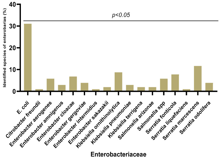
Out of the 390 urine samples collected, 103 (26.41%) were contaminated with Enterobacteriaceae strains. Remarkable diversity was observed among these Enterobacteriaceae strains (Figure 1). A total of 18 species of Enterobacteriaceae were identified with predominance of Escherichia coli (32/103, 31.07%), followed by Serratia marcescens (12/103, 11.65%) and Klebsiella ornithinolytica (9/103, 8.74%). The less represented species were Serratia liquefaciens, Citrobacter freundii, Enterobacter intermidius and Klebsiella pneumoniae (1/103, 0.97%). There was non-significant variation (p < 0.05) in the different species according to sex.
Figure 1.
Different Enterobacteriaceae species isolated from urinary tract infections.
3.3. Antibiotic Susceptibility of Enterobacteriaceae Strains
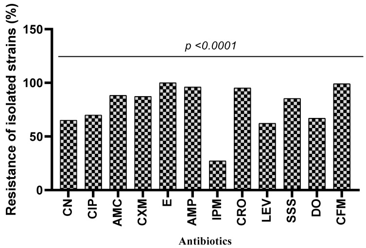
The antibiotic resistance of Enterobacteriaceae isolates is shown in Figure 2. The results showed variable (between 27.18% and 100%) susceptibility of isolated Enterobacteriaceae strains to the tested antibiotics. Indeed, the highest strain resistance rates were observed with antibiotics such as erythromycin (100%), cefixime (99.02%), ampicillin (96.11%) and amoxicillin/clavulanic acid (88.35%). However, a low resistance rate was observed with imipenem (27.18%). In addition, the results indicated that all Enterobacter sakazakii strains are resistant to gentamicin, ciprofloxacin, augmentin, cefuroxime, erythromycin, ceftriaxone, levofloxacin, sulfonamide, doxycycline and cefixime. All the isolated Escherichia coli were resistant to erythromycin, ampicillin, ceftriaxone and cefixime. In contrast, all Citrobacter freundii and Enterobacter intermidius were sensitive to gentamicin, doxycycline and imipenem (Table 3). The analysis of variance showed that there was a significant difference between the resistance of species and antibiotics (p < 0.0001).
Figure 2.
Resistance profile of isolated Enterobacteriaceae strains to antibiotics. CN: gentamicin, CIP: ciprofloxacin, AMC: amoxicillin/clavulanic acid, CXM: cefuroxime, E: erythromycin, AMP: ampicillin, IPM: imipenem, CRO: ceftriaxone, LEV: levofloxacin, SSS: sulfonamide, DO: doxycycline, CFM: cefixime.
Table 3.
Resistance to antibiotics by isolated species of Enterobacteriaceae.
| Species | Antibiotics | |||||||||||
|---|---|---|---|---|---|---|---|---|---|---|---|---|
| CN | CIP | AMC | CXM | E | AMP | IPM | CRO | LEV | SSS | DO | CFM | |
| Escherichia coli | 61.29% | 70.96% | 96.77% | 80.64% | 100% | 100% | 22.58% | 100% | 58.06% | 90.32% | 58.06% | 100% |
| Citrobacter freundii | 0% | 100% | 0% | 100% | 100% | 100% | 0% | 100% | 100% | 100% | 0% | 100% |
| Enterobacter aerogenes | 66.66% | 66.66% | 100% | 100% | 100% | 100% | 33.33% | 100% | 66.66% | 100% | 66.66% | 83.33% |
| Enterobacter amnigenus | 66.66% | 33.33% | 66.66% | 100% | 100% | 100% | 66.66% | 100% | 33.33% | 100% | 33.33% | 100% |
| Enterobacter cloacae | 85.71% | 71.42% | 85.71% | 85.71% | 100% | 100% | 42.85% | 85.71% | 57.14% | 100% | 100% | 100% |
| Enterobacter gergoviae | 75% | 25% | 50% | 75% | 100% | 100% | 25% | 100% | 75% | 100% | 75% | 100% |
| Enterobacter intermidius | 0% | 0% | 100% | 100% | 100% | 100% | 0% | 100% | 0% | 0% | 0% | 100% |
| Enterobacter sakazakii | 100% | 100% | 100% | 100% | 100% | 50% | 50% | 100% | 100% | 100% | 100% | 100% |
| Klebsiella ornithinolytica | 66.66% | 88.88% | 88.88% | 88.88% | 100% | 100% | 33.33% | 88.88% | 66.66% | 88.88% | 77.77% | 100% |
| Klebsiella pneumoniae | 50% | 100% | 100% | 50% | 100% | 50% | 50% | 100% | 50% | 0% | 50% | 100% |
| Klebsiella ssp | 100% | 100% | 100% | 100% | 100% | 100% | 0% | 100% | 100% | 100% | 100% | 100% |
| Klebsiella terrigena | 100% | 100% | 100% | 100% | 100% | 100% | 0% | 100% | 50% | 100% | 100% | 100% |
| Salmonalla spp | 50% | 66.66% | 100% | 83.33% | 100% | 100% | 50% | 100% | 33.33% | 100% | 83.33% | 100% |
| Salmonella arizonae | 100% | 100% | 100% | 100% | 100% | 100% | 0% | 100% | 50% | 50% | 50% | 100% |
| Serratia fonticola | 37.50% | 62.50% | 75% | 100% | 100% | 75% | 12.50% | 100% | 37.50% | 75% | 75% | 100% |
| Serratia liquefaciens | 100% | 100% | 0 | 100% | 100% | 100% | 0 | 100% | 100% | 100% | 0 | 100% |
| Serratia marcescens | 66.66% | 66.66% | 91.66% | 91.66% | 100% | 100% | 25% | 75% | 91.66% | 75% | 66.66% | 100% |
| Serratia odolifera | 75% | 75% | 75% | 75% | 100% | 100% | 0% | 100% | 75% | 50% | 50% | 100% |
CN: gentamicin, CIP: ciprofloxacin, AMC: amoxicillin/clavulanic acid, CXM: cefuroxime, E: erythromycin, AMP: ampicillin, IPM: imipenem, CRO: ceftriaxone, LEV: levofloxacin, SSS: sulfonamide, DO: doxycycline, CFM: cefixime.
3.4. ESBL Production by Isolated Enterobacteriaceae Strains
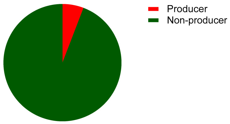
Of the 103 bacterial isolates, 6% were ESBL producers (Figure 3).
Figure 3.
Proportion of ESBL producers among the enterobacterial strains isolated from urinary tract infections in Benin.
3.5. Biofilm Formation Capability
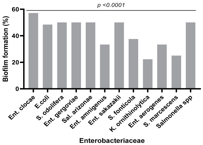
The biofilm formation test revealed that 40% of the strains were biofilm-forming. In regard to the species, isolated Enterobacter clocae strains were the most efficient at biofilm formation (57%) followed by Serratia odolifera (50%), Enterobacter gergoviae (50%), Salmonella arizonae (50%), Enterobacter sakazakii (50%), Salmonella spp. (50%) and Escherichia coli (48.38%) (Figure 4).
Figure 4.
Biofilm production rate by bacterial species.
3.6. Detection of Virulence Genes
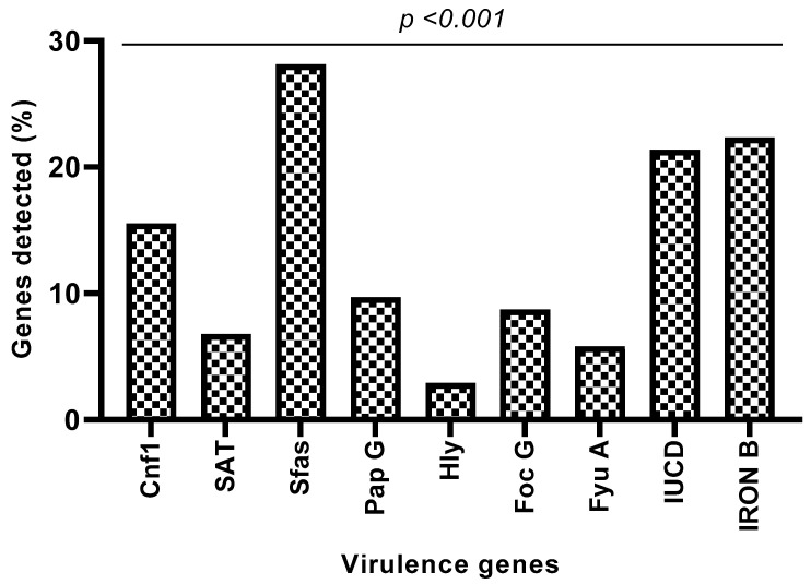
Virulence genes were detected in 64.07% of Enterobacteriaceae isolates. Thus, genes encoding for S-frimbria adhesin (28.15%), salmochelin outer membrane receptor (22.23%), aerobactin biosynthesis receptor (21.36%), cytotoxic necrotizing factor type 1 (15.53%), adhesin P-fimbria (PapG) (9.71%), F1C fimbrial adhesin (8.74%), secreted autotransporter toxin (6.79%), yersiniabactin outer membrane receptor (5.82%) and hemolysin RTX (2.91%) were found in various proportions (Figure 5).
Figure 5.
Proportion of genes sought in all strains. Cnf1: cytotoxic necrotizing factor type 1, SAT: secreted autotransporter toxin, Sfas: S-fimbriae adhesin, Pap G: adhesin P-fimbria, Hly: hemolysin RTX, Foc G: F1C fimbrial adhesin, Fyu A: yersiniabactin outer membrane receptor, IUCD: aerobactin biosynthesis receptor, IronB: salmochelin outer membrane receptor.
Considering the presence of nine targeted virulence genes by species, Escherichia coli isolates harbored eight, namely Sfas (25.8%), IronB (32.25%), iucD (35.48%), Cnfl (19.35%), PapG (9.67%), FocG (16.12%), Sat (6.45%) and FyuA (12.9%), in different proportions. Among Serratia marcescens isolates, six virulence genes (Sfas: 41.66%, IronB: 25%, iucD: 16.66%, Cnfl: 33.33%, PapG: 25% and Sat: 16.66%) were reported to be present. Only one gene was found among the isolates of Citrobacter freundii, Enterobacter gergoviae and Serratia odolifera (Table 4). The analysis of variance showed a significant difference between the presence of the virulence factors of a species (p < 0.0173) and a highly significant difference between the species and the virulence genes (p < 0.0039).
Table 4.
Proportion of genes recorded by species.
| Species | Frequency of Virulence Genes (%) | ||||||||
|---|---|---|---|---|---|---|---|---|---|
| Cnf1 | SAT | Sfas | Pap G | Hly | Foc G | Fyu A | IUCD | IronB | |
| C. freundii | 100 | 0 | 0 | 0 | 0 | 0 | 0 | 0 | 0 |
| E. coli | 19.35 | 6.45 | 25.8 | 9.67 | 0 | 16.12 | 12.9 | 35.48 | 32.25 |
| En. aerogenes | 0 | 0 | 16.66 | 16.66 | 0 | 0 | 16.66 | 16.66 | 33.33 |
| En. amnigenus | 33.33 | 0 | 33.33 | 0 | 0 | 0 | 0 | 33.33 | 0 |
| En. cloacae | 0 | 0 | 28.57 | 0 | 0 | 14.28 | 0 | 14.28 | 0 |
| En. gergoviae | 0 | 0 | 0 | 0 | 0 | 0 | 0 | 0 | 25 |
| En. intermidius | 0 | 100 | 100 | 0 | 0 | 100 | 0 | 100 | 100 |
| En. sakazakii | 0 | 0 | 100 | 0 | 0 | 0 | 50 | 0 | 0 |
| K. ornithinolytica | 11.11 | 0 | 33.33 | 11.11 | 11.11 | 0 | 0 | 0 | 11.11 |
| K. pneumoniae | 0 | 0 | 0 | 50 | 50 | 0 | 0 | 0 | 0 |
| K. terrigena | 50 | 0 | 50 | 0 | 0 | 0 | 0 | 0 | 0 |
| S. arizonae | 50 | 0 | 50 | 0 | 0 | 0 | 0 | 50 | 50 |
| Salmonella spp | 0 | 0 | 16.66 | 0 | 16.66 | 0 | 0 | 16.66 | 0 |
| Serratia fonticola | 12.5 | 12.5 | 25 | 0 | 0 | 12.5 | 0 | 12.5 | 50 |
| S. liquefaciens | 0 | 0 | 0 | 100 | 0 | 0 | 0 | 100 | 0 |
| S. marcescens | 33.33 | 16.66 | 41.66 | 25 | 0 | 0 | 0 | 16.66 | 25 |
| S. odolifera | 0 | 0 | 0 | 0 | 0 | 25 | 0 | 0 | 0 |
Cnf1: cytotoxic necrotizing factor type 1, SAT: secreted autotransporter toxin, Sfas: S-fimbriae adhesin, Pap G: adhesin P-fimbria, Hly: hemolysin RTX, Foc G: F1C fimbrial adhesin, Fyu A: yersiniabactin outer membrane receptor, IUCD: aerobactin biosynthesis receptor, IronB: salmochelin outer membrane receptor.
3.7. Relationship between Virulence Genes and Antibiotic Resistance
The analysis of the eigenvalues of the correlation matrix reveals that the first two components explain 69.23% of the variability (Table 5). Since this share of information is greater than 50%, the first two components can be used to adequately interpret the results of the Principal Component Analysis (PCA).
Table 5.
Eigenvalues of the correlation matrix.
| Main Components | ||||
|---|---|---|---|---|
| Settings | Dim,1 | Dim,2 | Dim,3 | Dim,4 |
| Own value | 4.74 | 2.18 | 1.13 | 0.66 |
| Percentage of variance | 47.43 | 2.,80 | 11.28 | 6.62 |
| Cumulative percentage of variance | 47.43 | 69.23 | 80.51 | 87.13 |
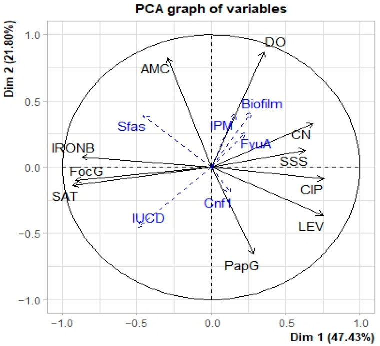
Correlation analyses between the two components and the initial variables (Table 6 and Figure 6) shows at the level of axis 1, a strong positive correlation with the variables “IRON B”, “SAT” and “FocG” and a strong negative correlation with the variables “LEV”, “CN”, “CIP” and “SSS” are positively correlated with axis 1. Thus, axis 1 expresses that the resistance of the isolated strains to the antibiotics “LEV”, “CN “, “CIP” and “SSS” is linked to the absence of the virulence genes “IRON B”, “SAT” and “FocG”. As for axis 2, it shows a positive correlation with the “AMC” and “DO” variables and a negative correlation with the “Pap G” variable. This axis indicates that the presence of the “pap G” virulence gene leads to the sensitivity of the “AMC” and “DO” strains to antibiotics.
Table 6.
Correlation between the starting variables and the principal components.
| Dim,1 | Dim,2 | |
|---|---|---|
| IRON.B | −0.870 | 0.076 |
| SAT | −0.928 | −0.136 |
| Foc.G | −0.911 | −0.100 |
| AMC | −0.292 | 0.828 |
| DO | 0.358 | 0.873 |
| LEV | 0.751 | −0.365 |
| CN | 0.685 | 0.331 |
| CIP | 0.755 | −0.090 |
| SSS | 0.632 | 0.128 |
| Pap.G | 0.285 | −0.657 |
Foc G: F1C fimbrial adhesin, SAT: secreted autotransporter toxin, Pap G: adhesin P-fimbria, IronB: salmochelin outer membrane receptor, AMC: amoxicillin/clavulanic acid, DO: doxycycline, LEV: levofloxacin, CN: gentamicin, CIP: ciprofloxacin, SSS: sulfonamide.
Figure 6.
Circle of correlation of variables with the two axes.
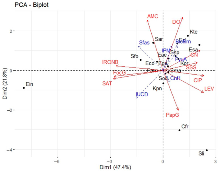
The projection of the different observations at the level of axes 1 and 2 (Figure 7) indicates that the species E. cloacae, E. sakazakii, K. ornithinolytica, and K. terrigena are mainly located in the positive parts of the two axes. These species are resistant to the antibiotics “DO”, “CN” and “SSS”. C. freundii and S. liquefaciens are mainly located in the positive part of axis 1 and the negative part of axis 2. S. liquefaciens better expresses the virulence gene Pap G. E. intermidius is located in the negative part of the two axes. This species expresses sat and Forc G virulence genes, which were not found in E. cloacae, E. sakazakii, K. ornithinolytica and K. terrigena. The species S. arizonae and S. fonticola are located in the negative part of axis 1 and the positive part of axis 2. It was noticeable that these species express both the IronB virulence gene and the AMC resistance gene, in contrast to the species C. freundii and S. liquefaciens.
Figure 7.
Scatterplot relating to the position of species with respect to axes 1 and 2. Legend: Cfr = C. freundii; Eco = E. coli; Eae = E. aerogenes; Eam = E. amnigenus; Ecl = E. cloacae; Ege = E. gergoviae; Ein = E. intermidius; Esa = E. sakazakii; Kor = K. ornithinolytica; Kpn = K. pneumoniae; Kte = K. terrigena; Sar = S. arizonae; Ssp = S. spp; Sfo = S. fonticola; Sli = S. liquefaciens; Sma = S. marcescens; Sod = S. odolifera.
3.8. The Relationship between Biofilm Production and Virulence Genes
Correlation analyses between biofilm production, virulence genes and resistance showed a positive correlation between biofilm production and the virulence gene FyuA and resistance to gentamicin. The more a species express FyuA (virulence gene and resistant to gentamicin), the more biofilm it produces. A positive correlation was also found between biofilm production and sex. Species isolated from male UTIs produced more biofilm than species isolated from female UTIs. The virulence genes PapG, iucD and IronB are negatively correlated with biofilm production. Therefore, the presence of the virulence genes PapG, iucD and IronB decreases the production of biofilm.
4. Discussion
To better understand the pathogenicity of strains causing infections and develop new vaccines and therapeutic targets, it is necessary to identify the susceptibility to antibiotics, the factors associated with the formation of biofilms and the virulence factors of these strains [7,30]. These potential predictors help clinicians manage patients and anticipate the evolution of the infection in the host organization [31]. In this study, we sought to determine the prevalence of Enterobacteriaceae strains, their antibiotic resistance profiles, their ability to form biofilms and the presence of some urinary tract infection virulence genes in Benin.
During our study, Enterobacteriaceae were isolated from patients mainly between 21 and 30 years old (22.23%). This age bias can be explained by the fact professional, cultural and sporting activities may contribute to the infection. The results of our study indicate the presence of Enterobacteriaceae strains in a proportion of 26.41% in urine samples. Our results are lower than those obtained in Mali [32], which found a prevalence of 76.7% of Enterobacteriaceae in urine. Enterobacteriaceae have also been shown to cause 84–87% of UTIs [33]. This difference may be due to sample size, laboratory strain detection techniques, social demographics, climatic conditions, people’s levels of personal hygiene and healthcare-seeking habits. Enterobacteriaceae are found in the urine because they can easily contaminate the urinary tract, especially in women, since they are normal flora of the large intestine [7].
In our study, 18 different species of Enterobacteriaceae were isolated, with a predominance of Escherichia coli (30.97%), followed by Serratia marcescens (11.65%) and Klebsiella ornithinolytica (8.73%). On the other hand, the weakly represented species were Serratia liquefaciens, Citrobacter freundii and Enterobacter intermidius (0.97%). As expected, E. coli was the major Enterobacterales species among the urinary tract samples, which is similar to previous works in other countries [34,35]. However, the rate of 30.97% in this study is lower than the 72% recorded in France [36]. Traditionally, E. coli has been the dominant uropathogen due to its expression of toxins, adhesins, pili, and fimbriae that allow it to adhere to the uroepithelium [37]. These protect bacteria from urinary elimination and allow for bacterial multiplication and invasion of uroepithelial tissues. The presence of S. marcescens as a second isolated species can be explained by the fact that it has a great affinity for the urinary tract [38].
The resistance of Enterobacteriaceae isolates varied according to the antibiotics. We observed variable resistance rates depending on the families of antibiotics and the species isolated. High resistance rates (≥80%) were obtained with molecules of the macrolide, cephalosporin and penicillin families. Resistance rates of more than 50% were obtained with molecules from the fluoroquinolone, aminoglycoside and tetracycline families. In a recent study conducted in Benin on surgical patients, a high resistance rate to many of the antibiotics tested was shown [39]. However, our results are superior to those found in Senegal [40] and close to those obtained in Algeria [41]. Antibiotic resistance and the rapid spread of aminoglycosides and lactams such as cephalosporins and fluoroquinolones against uropathogenic bacteria compromise the clinical management of the infection and lead to a poor prognosis [42]. The multiple-drug resistance (MDR) of pathogenic bacteria may be associated with severe morbidity in urinary tract infections, leading to a major global health problem [43,44,45]. The non-regulation of the use of antibiotics in patients with access to over-the-counter prescriptions, the misuse of certain classes of antimicrobials, frequent self-medication with often random and inappropriate dosages, the premature discontinuation of treatment, the use of antibiotics as growth promoters in agriculture, the use of contraband molecules that are often less dosed or devoid of active ingredients, and unfavorable economic and social conditions are the main drivers that promote the emergence of bacteria that are multi-resistant to antibiotics [46,47]. In order to control the spread of this antibiotic resistance, since it represents a serious health problem in Benin, actions to raise awareness of the proper use of antibiotics coupled with monitoring of the acquisition of antibiotics must be implemented. Weak resistance to imipenem was detected. This trend was also found by Hashemi et al. [48], with an Enterobacteriaceae resistance rate to imipenem of around 19%. Carbapenems, therefore, remain, to this day, the most active molecules against uropathogenic Enterobacteriaceae [49].
The overall frequency of ESBL-producing Enterobacteriaceae among uropathogenic strains in this study was 6%. A similar prevalence (6.05%) was estimated in Morocco in 2014 [50]. Recently, the highest rate (56.2%) was reported in Benin among Enterobacteriaceae samples collected at the Cotonou National Center Hubert Koutoukou Maga university Hospital, Benin [51]. The differences observed from the study conducted in Benin may be due to the fact that their samples included various infections, whereas our sampling targeted only UTIs. In Europe, the resistance of Enterobacteriaceae to third-generation cephalosporins ranged from 6.2 to 30.8% among bacterial isolates in 2019 [52]. Muriuki et al. [53] reports a similar finding for uropathogenic E. coli in Kenya between 2015 and 2018. The production of ESBL is probably due to the often-empirical prescriptions of ß-lactams, particularly in ambulatory medicine, while awaiting the results of ECBU. The development of resistance to third-generation cephalosporins is a major cause of prolonged hospitalization of infected patients and limits treatment options agaimst the bacteria [54]. A 25% prevalence of ESBL production, therefore, creates significant therapeutic problems and will limit or reduce treatment options [55]. Our results on the production of ESBL by our strains of uropathogenic Enterobacteriaceae are, therefore, reassuring, but should be monitored.
Of the 103 Enterobacteriaceae isolates tested for biofilm production, 40% formed a biofilm on the microplate. These results are similar to other previous studies [56]. Biofilms provide a survival strategy for bacteria by positioning them to efficiently use available nutrients and prevent access to antimicrobial agents, antibodies and white blood cells [57]. It has also been found that biofilms harbor a large number of enzymes that inactivate antibiotics, such as beta-lactamases, and thus, create an island of antimicrobial resistance [58]. Biofilms, therefore, make our strains more virulent and multi-resistant. Recent studies have revealed that a reduction in oxygen tension in the bladder, combined with the presence of terminal electron receptors in the urine, facilitates the preferential expression of E. coli [59]. The expression of other factors, such as cytochrome bd quinol oxidase, promotes biofilm complexity and resistance to extracellular stressors by altering the abundance of extracellular matrix components [59]. This biofilm formation may be due to curli (functional amyloid) fibers, which constitute the main protein component of many biofilms of Gram-negative bacteria [60]. The presence of curli fibers in these biofilms provides a competitive advantage in mouse models of urinary tract infection by promoting adhesion to bladder epithelial cells [61]. This adhesion is further increased by the presence of phospho-ethanolamine cellulose produced simultaneously by UPECs [61].
Regarding genes encoding for virulence factors, in this study, the distribution of fimbriae was observed. Enterobacteriaceae harbor the sfa gene (25.15%). The papG gene is present in 9.71% and the focG gene in 8.74% of the Enterobacteriaceae strains. These results are lower than those obtained in Romania [62], in Mongolia [63] and in Egypt [64]. Fimbriae are required by the bacterium to promote the colonization of surfaces, which helps prevent urinary outflow and allows for infection by the bacterium, which may indicate their critical role in the production and progression of urinary infections. They also play an important role in the formation of biofilms [65].
In our study, uropathogenic Enterobacteriaceae secreted toxins such as α-hemolysin (hly) and cytotoxic necrosis factor 1 (cnf1) with respective presence percentages of 2.91% and 15.53%. These results are similar to studies conducted in Romania (13%) [61] and Mexico (15.4%) [66]. These toxins promote the exfoliation of bladder cells and cell lysis, which makes available the iron and nutrients necessary for bacterial growth [29]. Alpha-hemolysin has been associated with clinical severity in patients with UI and CNF1 with bladder inflammation [67].
Genes encoding siderophores were detected in our study. Enterobacteriaceae possessed genes coding for salmochelins IronB (22.33%), followed by genes coding for aerobactin iucD (21.36%) and, finally, those coding for yersiniabactin fyuA (5.82%). Siderophores such as toxins allow the bacterium to mobilize iron. They are essential virulence factors in most pathogenic Gram-negative bacteria [68].
In the present study, we also identified the presence of an autotransporter (sat gene) in 6.79% of the isolated strains. This rate is much lower than the 31.1% found in Guadeloupe [29]. Autotransporters can be self-secreted through the membrane of Gram-negative bacteria [69] and modify the structure of the host cell. Thanks to these capacities, they make the strains more virulent.
The sfa gene was the most detected among the virulence genes in the present study. This high prevalence could lead us to consider sfa as a candidate for a potential vaccine. The difference in the prevalence of virulence genes between our studies and different studies abroad may be due to differences in sample size and methodology. Virulence factors are the product of different genes, which can be detected using the PCR method [70,71]. However, due to a possible mutation in the corresponding gene, PCR may not detect the presence of the gene [31]. Therefore, although this phenomenon is rare, a negative PCR result does not necessarily equate to the absence of the corresponding gene [31].
In our study, biofilm production is associated with the virulence gene fyuA and gentamicin resistance. This observation has been made in various other studies. Thus, Stephenson and Brown [72] reported that biofilm production was significantly associated with resistance to fluoroquinolones [72]. In another study by Zamani et al. [73], it was found that biofilm production in UPEC was significantly associated with the Fim gene [73]. Our study also found that the presence of PapG, iucD and IronB virulence genes decreases biofilm production. On the other hand, in a survey carried out in Uganda [17], biofilm production was not associated with any virulence gene or resistance to a particular antibiotic. Thus, further studies are required to better understand the relationship between virulence factors, antibiotic resistance and biofilm formation.
5. Conclusions
The bacteriological analyses that we carried out on urinary tract infections made it possible to identify microbial diversity in this study. Our results showed noticeable diversity in the distribution of Enterobacteriaceae species, namely strains of Escherichia coli, Serratia marcescens, Klebsiella ornithinolytica, Serratia liquefaciens, Citrobacter freundii, Enterobacter intermidius and Klebsiella pneumoniae. The highlighting of their resistance to antibiotics revealed the importance of the frequency of multi-resistant strains to various antibiotics that are specifically used for treatment. In addition, the majority of strains were biofilm-forming, and a small proportion were ESBL-producing. To measure the danger that the strains can represent, we noted the presence of genes encoding for virulence factors. Therefore, the habituation of physicians to requesting a cytobacteriological examination of urine with an antibiogram is essential for any patient presenting suspicious signs of urinary tract infection. It is also necessary to plan a good strategy for the supply and dispensing of antibiotics to avoid self-medication. Empirical treatment of urinary tract infections in our country should also be revised. Further research on the main uropathogenic genes detected that can be used for the manufacture of potential vaccines must be carried out in this area.
Acknowledgments
The authors would like to express their gratitude to the staff of the nine visited hospitals, namely the Natitingou Area Hospital, Djougou Area Hospital, Ménontin Area Hospital, Departmental Hospital and University Centers of Borgou/Alibori, Departmental Hospital Centers of Zou/Colline, Departmental Hospital Centers of Mono/Couffo, Departmental Hospital and University Centers of Ouémé/Plateau, Clinique bon Samaritan of Porto-Novo and Clinique Senalia. The authors also thank Raphaela Adjobimey of the Städtisches Gymnasium Sedanstraße, Wuppertal, Germany, for providing thorough language assistance.
Author Contributions
Conceptualization, J.F.M.; data curation, F.F.A., H.S., T.A., A.D.P.N., A.S., B.B., A.D.D., L.A. and B.B.S.K.; formal analysis, F.F.A., A.D.P.N., A.D.D. and B.B.S.K.; funding acquisition, F.F.A., H.S., A.A. and L.B.-M.; investigation, F.F.A., T.A., A.S., B.B. and L.A.; methodology, F.F.A., H.S., A.D.P.N., A.S., B.B., L.A. and B.B.S.K.; resources, H.S. and L.B.-M.; supervision, H.S., A.D.D., J.F.M., A.A. and L.B.-M.; validation, T.A., J.F.M., A.A. and L.B.-M.; writing—original draft, F.F.A. and A.D.P.N.; writing—review and editing, H.S., T.A., J.F.M., A.A. and L.B.-M. All authors have read and agreed to the published version of the manuscript.
Data Availability Statement
The data used to support the findings of this work are available from the corresponding author upon request.
Conflicts of Interest
The authors declare that they have no conflict of interest.
Funding Statement
This research received no external funding.
Footnotes
Disclaimer/Publisher’s Note: The statements, opinions and data contained in all publications are solely those of the individual author(s) and contributor(s) and not of MDPI and/or the editor(s). MDPI and/or the editor(s) disclaim responsibility for any injury to people or property resulting from any ideas, methods, instructions or products referred to in the content.
References
- 1.Griebling T.L. Urologic diseases in American project: Trends in resource use for urinary tract infections in women. J Urol. 2005;173:1281–1287. doi: 10.1097/01.ju.0000155596.98780.82. [DOI] [PubMed] [Google Scholar]
- 2.Tan C.W., Chlebicki M.P. Urinary tract infections in adults. Singap. Med. J. 2016;57:485–490. doi: 10.11622/smedj.2016153. [DOI] [PMC free article] [PubMed] [Google Scholar]
- 3.Agarwa J., Srivastava S., Singh M. Pathogenomics of uropathogenic Escherichia coli. Indian J. Med. Microbiol. 2012;30:141–149. doi: 10.4103/0255-0857.96657. [DOI] [PubMed] [Google Scholar]
- 4.Launay E., Bingen E., Cohen R. Stratégies thérapeutiques dans les infections urinaires du nourrisson et de l’enfant. Arch. Pediatr. 2012;19:S109–S116. doi: 10.1016/S0929-693X(12)71283-6. [DOI] [PubMed] [Google Scholar]
- 5.Toner L., Papa N., Aliyu S.H., Dev H., Lawrentschuk N., Al-Hayek S. Extended spectrum beta-lactamase-producing Enterobacteriaceae in hospital urinary tract infections: Incidence and antibiotic susceptibility profile over 9 years. World J. Urol. 2016;34:1031–1037. doi: 10.1007/s00345-015-1718-x. [DOI] [PubMed] [Google Scholar]
- 6.Spencer J.D., Schwaderer A.L., Becknell B., Watson J., Hains D.S. The innate immune response during urinary tract infection and pyelonephritis. Pediatr. Nephrol. 2013;29:1139–1149. doi: 10.1007/s00467-013-2513-9. [DOI] [PMC free article] [PubMed] [Google Scholar]
- 7.Flores-Mireles A.L., Walker J.N., Caparon M., Hultgren S.J. Urinary tract infections: Epidemiology, mechanisms of infection and treatment options. Nat. Rev. Microbiol. 2015;13:269–284. doi: 10.1038/nrmicro3432. [DOI] [PMC free article] [PubMed] [Google Scholar]
- 8.Ny S., Edquist P., Dumpis U., Gröndahl-Yli-Hannuksela K., Hermes J. Antimicrobial resistance of Escherichia coli isolates from outpatient urinary tract infections in women in six European countries including Russia. J. Glob. Antimicrob. Resist. 2018;17:25–34. doi: 10.1016/j.jgar.2018.11.004. [DOI] [PubMed] [Google Scholar]
- 9.Bader M.S., Loeb M., Brooks A.A. An update on the management of urinary tract infections in the era of antimicrobial resistance. Postgrad. Med. 2016;129:242–258. doi: 10.1080/00325481.2017.1246055. [DOI] [PubMed] [Google Scholar]
- 10.Ali I., Rafaque Z., Ahmed S., Malik S., Dasti J.I. Prevalence of multi-drug resistant uropathogenic Escherichia coli in Potohar region of Pakistan. Asian Pac. J. Trop. Biomed. 2016;6:60–66. doi: 10.1016/j.apjtb.2015.09.022. [DOI] [Google Scholar]
- 11.Neupane S., Pant N.D., Khatiwada S., Chaudhary R., Banjara M.R. Correlation between biofilm formation and resistance toward different commonly used antibiotics along with extended spectrum beta lactamase production in uropathogenic Escherichia coli isolated from the patients suspected of urinary tract infections visit. Antimicrob. Resist. Infect. Control. 2016;5:5. doi: 10.1186/s13756-016-0104-9. [DOI] [PMC free article] [PubMed] [Google Scholar]
- 12.Hammami S., Saidani M., Ferjeni S., Aissa I., Slim A., Boutiba-Ben Boubaker I. Characterization of extended spectrum beta-lactamase-producing Escherichia coli in community-acquired urinary tract infections in Tunisia. Microb. Drug Resist. 2013;19:231–236. doi: 10.1089/mdr.2012.0172. [DOI] [PubMed] [Google Scholar]
- 13.Barguigua A., El Otmani F., Talmi M., Zerouali K., Timinouni M. Prevalence and types of extended spectrum beta-lactamases among urinary Escherichia coli isolates in Moroccan community. Microb. Pathog. 2013;61–62:16–22. doi: 10.1016/j.micpath.2013.04.010. [DOI] [PubMed] [Google Scholar]
- 14.Paterson D.L., Bonomo R.A. Extended-spectrum beta-lactamases: A clinical update. Clin. Microbiol. Rev. 2005;18:657–686. doi: 10.1128/CMR.18.4.657-686.2005. [DOI] [PMC free article] [PubMed] [Google Scholar]
- 15.Naboka Y.L., Mavzyutov A.R., Kogan M.I., Gudima I.A., Dzhalagoniya K.T., Ivanov S.N., Naber K.G. The gene profile of Enterobacteriaceae virulence factors in relation to bacteriuria levels between the acute episodes of recurrent uncomplicated lower urinary tract infection. Expert Rev. Anti-Infect. Ther. 2021;19:1061–1066. doi: 10.1080/14787210.2021.1866986. [DOI] [PubMed] [Google Scholar]
- 16.Sarowska J., Futoma-Koloch B., Jama-Kmiecik A., Frej-Madrzak M., Ksiazczyk M., Bugla-PLoskonska G. Virulence factors, prevalence and potential transmission of extraintestinal pathogenic Escherichia coli isolated from different sources: Recent reports. Gut Pathog. 2019;11:10. doi: 10.1186/s13099-019-0290-0. [DOI] [PMC free article] [PubMed] [Google Scholar]
- 17.Katongole P., Nalubega F., Florence N.C., Asiimwe B., Andia I. Biofilm formation, antimicrobial susceptibility and virulence genes of Uropathogenic Escherichia coli isolated from clinical isolates in Uganda. BMC Infect. Dis. 2020;20:453. doi: 10.1186/s12879-020-05186-1. [DOI] [PMC free article] [PubMed] [Google Scholar]
- 18.Yazdanpour Z., Tadjrobehkar O., Shahkhah M. Significant association between genes encoding virulence factors with antibiotic resistance and phylogenetic groups in community acquired uropathogenic Escherichia coli isolates. MC Microbiol. 2020;20:241. doi: 10.1186/s12866-020-01933-1. [DOI] [PMC free article] [PubMed] [Google Scholar]
- 19.Khairy R.M., Mohamed E.S., Abdel Ghany H.M., Abdelrahim S.S. Phylogenic classification and virulence genes profiles of uropathogenic E. coli and diarrhegenic E. coli strains isolated from community acquired infections. PLoS ONE. 2019;14:e0222441. doi: 10.1371/journal.pone.0222441. [DOI] [PMC free article] [PubMed] [Google Scholar]
- 20.Donelli G., Vuotto C. Biofilm-based infections in long-term care facilities. Future Microbiol. 2014;9:175–188. doi: 10.2217/fmb.13.149. [DOI] [PubMed] [Google Scholar]
- 21.Karam M.R.A., Habibi M., Bouzari S. Relationships between virulence factors and antimicrobial resistance among Escherichia coli isolated from urinary tract infections and commensal isolates in Tehran, Iran. Osong Public Health Res. Perspect. 2018;9:217–224. doi: 10.24171/j.phrp.2018.9.5.02. [DOI] [PMC free article] [PubMed] [Google Scholar]
- 22.Schwartz D. La méthode statistique en médecine: Les enquêtes éthiologiques. Rev. Stat. Appliquée. 1960;8:5–27. [Google Scholar]
- 23.Riegel P., Archambaud M., Clavé D., Vergnaud M. Bactérie de Culture et D’identification Difficiles. Biomérieux; Nancy l’Etoile, France: 2006. pp. 93–112. [Google Scholar]
- 24.CASFM “Comité de L’antibiogramme de la Société Française de Microbi Logie: Recommandations. 2021. [(accessed on 25 September 2022)]. Available online: https://www.sfmmicrobiologie.org/2021/05/06/casfm-eucast-2021-v2/
- 25.Allouch P.Y., Labia R., Pina P., Morin E. Observatoires hospitaliers de la sensibilité de E. coli et de Klebsiella à l’association amoxicilline-acide clavulanique en 1994. Med. Mal. Infect. 1995;25:934–939. doi: 10.1016/S0399-077X(05)81150-3. [DOI] [Google Scholar]
- 26.Christensen G.D., Simpson W.A., Bisno A.L., Beachy E.H. Adherence of biofilm producing strains of Staphylococci epidermidis to smooth surfaces. Infect. Imm. 1982;37:318–326. doi: 10.1128/iai.37.1.318-326.1982. [DOI] [PMC free article] [PubMed] [Google Scholar]
- 27.Stepanović S., Vuković D., Dakić I., Savić B., Švabić-Vlahović M. A modified microtiter-plate test for quantification of staphylococcal biofilm formation. J. Microbiol. Methods. 2000;40:175–179. doi: 10.1016/S0167-7012(00)00122-6. [DOI] [PubMed] [Google Scholar]
- 28.Rasmussen R.S., Morrissey M.T. DNA-based methods for the identification of commercial fish and seafood species. Compr. Rev. Food Sci. Food Saf. 2008;7:280–295. doi: 10.1111/j.1541-4337.2008.00046.x. [DOI] [PubMed] [Google Scholar]
- 29.Maris S. Ph.D. Dissertation. Université du Québec, Institut National de la Recherche Scientifique; Quebec City, QC, Canada: 2016. [(accessed on 25 December 2021)]. Caractérisation de Souches d’Escherichia coli Pathogènes Urinaires Provenant de Guadeloupe: Portrait de la Diversité des Facteurs de Virulence Présentes. Available online: https://espace.inrs.ca/id/eprint/4854/1/Maris-S-M-Aout2016.pdf. [Google Scholar]
- 30.Römling U., Balsalobre C. Biofilm infections, their resilience to therapy and innovative treatment strategies. J. Intern. Med. 2012;272:541–561. doi: 10.1111/joim.12004. [DOI] [PubMed] [Google Scholar]
- 31.Tarchouna M., Ferjani A., Ben-Selma W., Boukadida J. Distribution of uropathogenic virulence genes in Escherichia coli isolated from patients with urinary tract infection. Int. J. Infect. Dis. 2013;17:e450–e453. doi: 10.1016/j.ijid.2013.01.025. [DOI] [PubMed] [Google Scholar]
- 32.Kalambry A.C. Profil de résistance aux bêtalactamines des entérobactéries isolées des prélèvements urinaires à l’Hôpital du Mali. Rev. Afr. Médecine Interne. 2019;14:1–10. [Google Scholar]
- 33.Thaden J.T., Pogue J.M., Kaye K.S. Role of newer and re-emerging older agents in the treatment of infections caused by carbapenem-resistant Enterobacteriaceae. Virulence. 2016;8:403–416. doi: 10.1080/21505594.2016.1207834. [DOI] [PMC free article] [PubMed] [Google Scholar]
- 34.Maraki S., Mantadakis E., Michailidis L., Samonis G. Changing antibiotic susceptibilities of community-acquired uropathogens in Greece, 2005–2010. J. Microbiol. Immunol. Infect. 2013;46:202–209. doi: 10.1016/j.jmii.2012.05.012. [DOI] [PubMed] [Google Scholar]
- 35.Soubra L., Kabbani S., Anwar M.F., Dbouk R. Spectrum and patterns of antimicrobial resistance of uropathogens isolated from a sample of hospitalised Lebanese patients with urinary tract infections. J. Glob. Antimicrob. Resist. 2014;2:173–178. doi: 10.1016/j.jgar.2014.01.007. [DOI] [PubMed] [Google Scholar]
- 36.Farfour E., Dortet L., Guillard T., Chatelain N., Poisson A., Mizrahi A., Fournier D., Bonnin R.A., Degand N., Morand P., et al. Antimicrobial Resistance in Enterobacterales Recovered from Urinary Tract Infections in France. Pathogens. 2022;11:356. doi: 10.3390/pathogens11030356. [DOI] [PMC free article] [PubMed] [Google Scholar]
- 37.Johnson B., Stephen B.M., Joseph N., Asiphas O., Musa K., Taseera K. Prevalence and bacteriology of culture-positive urinary tract infection among pregnant women with suspected urinary tract infection at Mbarara regional referral hospital, South-Western Uganda. BMC Pregnancy Childbirth. 2021;21:159. doi: 10.1186/s12884-021-03641-8. [DOI] [PMC free article] [PubMed] [Google Scholar]
- 38.Bush L.M., Vazquez-Pertejo M.T. Infections par Klebsiella, Enterobacter, et Serratia. Manuels Professional Mal Infect. 2022. [(accessed on 3 December 2022)]. Available online: https://www.msdmanuals.com/fr/professional/maladies-infectieuses/bacilles-gram-n%C3%A9gatifs/introduction-aux-bacilles-gram-n%C3%A9gatifs.
- 39.Yehouenou C.L., Kpangon A.A., Affolabi D., Rodriguez-Villalobos H., Van Bambeke F., Dalleur O., Simon A. Antimicrobial resistance in hospitalized surgical patients: A silently emerging public health concern in Benin. Ann. Clin. Microbiol. Antimicrob. 2020;19:54. doi: 10.1186/s12941-020-00398-4. [DOI] [PMC free article] [PubMed] [Google Scholar]
- 40.Sabor H. Phénotypes de résistance des entérobactéries isolées au CHUNU de fann de Dakar de 2014 à 2016. Mémoire DES de biologie clinique, Université Cheikh Anta Diop de Dakar, Faculté de Médecine de Pharmacie et d’Odontologie, Dakar, Senegal. 2017. [(accessed on 25 September 2022)]. Available online: http://196.1.97.20/greenstone/collect/mmoires/import/memm_2017_0201.pdf.
- 41.Djahida S., Imane S., Mourad D. Résistance aux antibiotiques des entérobactéries au niveau du CHU de Sidi Bel Abbes (Algerie) MHA. 2011;23:37–41. [Google Scholar]
- 42.Huemer M., Mairpady Shambat S., Brugger S.D., Zinkernagel A.S. Antibiotic resistance and persistence—Implications for human health and treatment perspectives. EMBO Rep. 2020;21:e51034. doi: 10.15252/embr.202051034. [DOI] [PMC free article] [PubMed] [Google Scholar]
- 43.Haque M., Sartelli M., McKimm J., Bakar M.A. Health care-associated infections–an overview. Infect Drug Resist. 2018;11:2321. doi: 10.2147/IDR.S177247. [DOI] [PMC free article] [PubMed] [Google Scholar]
- 44.Shafiq M., Zeng M., Permana B., Bilal H., Huang J., Yao F., Algammal A.M., Li X., Yuan Y., Jiao X. Coexistence of blaNDM−5 and tet(X4) in international high-risk Escherichia coli clone ST648 of human origin in China. Front. Microbiol. 2022;13:1031688. doi: 10.3389/fmicb.2022.1031688. [DOI] [PMC free article] [PubMed] [Google Scholar]
- 45.Bilal H., Zhang G., Rehman T., Han J., Khan S., Shafiq M., Yang X., Yan Z., Yang X. First Report of blaNDM-1 Bearing IncX3 Plasmid in Clinically Isolated ST11 Klebsiella pneumoniae from Pakistan. Microorganisms. 2021;9:951. doi: 10.3390/microorganisms9050951. [DOI] [PMC free article] [PubMed] [Google Scholar]
- 46.Namuwenge J.M., Mwesige O.G., Mukanga N. Over-the-counter suboptimal dispensing of antibiotics in Uganda. J. Multidiscip. Health. 2013;6:303–310. doi: 10.2147/JMDH.S49075. [DOI] [PMC free article] [PubMed] [Google Scholar]
- 47.Alonso C.A., Zarazaga M., Ben Sallem R., Jouini A., Ben Slama K., Torres C. Antibiotic resistance in Escherichia coli in husbandry animals: The African perspective. Lett. Appl. Microbiol. 2017;64:318–334. doi: 10.1111/lam.12724. [DOI] [PubMed] [Google Scholar]
- 48.Hashemi S.H., Esna-Ashari F., Tavakoli S., Mamani M. The prevalence of antibiotic resistance of Enterobacteriaceae strains isolated in community-and hospital-acquired infections in teaching hospitals of Hamadan, west of Iran. J. Res. Health Sci. 2013;13:75–80. [PubMed] [Google Scholar]
- 49.Hameed M.F., Chen Y., Wang Y., Shafiq M., Bilal H., Liu L., Ma J., Gu P., Ge H. Epidemiological Characterization of Colistin and Carbapenem Resistant Enterobacteriaceae in a Tertiary: A Hospital from Anhui Province. Infect. Drug Resist. 2021;14:1325–1333. doi: 10.2147/IDR.S303739. [DOI] [PMC free article] [PubMed] [Google Scholar]
- 50.Akel Z. Doctoral Dissertation. Mohammed V University of Rabat Faculty of Medicine and Pharmacy; Rabat, Morocco: 2014. [(accessed on 23 July 2022)]. Profil épidémiologique des Entérobactéries Productrices de Carbapénémases Isolées au CHU Ibn Sina-Rabat. Available online: http://ao.um5.ac.ma/xmlui/bitstream/handle/123456789/14692/P075%202014.pdf?sequence=2&isAllowed=y. [Google Scholar]
- 51.Affolabi D., Sogbo F., Haag U., Orekan J., Anagonou S. Bacteriological profile of Enterobacteriaceae producing broad-spectrum beta-lactamases at the Cotonou National Center Hubert Koutoukou Maga university Hospital, Benin. [(accessed on 3 January 2023)];Ann. Clin. Microbiol. Antimicrob. 2016 10 Available online: https://anafrimed.net/download/3319/?tmstv=1672855058. [Google Scholar]
- 52.European Centre for Disease Prevention and Control Surveillance Atlas of Infectious Diseases. 2019. [(accessed on 27 October 2022)]. Available online: http://atlas.ecdc.europa.eu/public/index.aspx.
- 53.Muriuki C.W., Ogonda L.A., Kyanya C., Matano D., Masakhwe C., Odoyo E. Phenotypic and genotypic characteristics of uropathogenic Escherichia coli isolates from Kenya. Microb. Drug Resist. 2022;28:31–38. doi: 10.1089/mdr.2020.0432. [DOI] [PMC free article] [PubMed] [Google Scholar]
- 54.Vazouras K., Velali K., Tassiou I., Anastasiou-Katsiardani A., Athanasopoulou K., Barbouni A. Antibiotic treatment and antimicrobial resistance in children with urinary tract infections. J. Glob. Antimicrob. Resist. 2019;20:4–10. doi: 10.1016/j.jgar.2019.06.016. [DOI] [PubMed] [Google Scholar]
- 55.Shakya P., Shrestha D., Maharjan E., Sharma V.K., Paudyal R. ESBL production among E. coli and Klebsiella spp. causing urinary tract infection: A hospital-based study. Open Microbiol. J. 2017;11:23–30. doi: 10.2174/1874285801711010023. [DOI] [PMC free article] [PubMed] [Google Scholar]
- 56.Sudheendra K.R., Basavaraj P.V. Analysis of antibiotic sensitivity profile of biofilm-forming uropathogenic Escherichia coli. J. Nat. Sci. Biol. Med. 2018;9:175. doi: 10.4103/jnsbm.JNSBM_209_17. [DOI] [Google Scholar]
- 57.Nandakumar V., Chittaranjan S., Kurian V.M., Doble M. Characteristics of bacterial biofilm associated with implant material in clinical practice. Polym. J. 2013;45:137. doi: 10.1038/pj.2012.130. [DOI] [Google Scholar]
- 58.Davies J., Davies D. Origins and evolution of antibiotic resistance. Microbiol. Mol. Biol. Rev. 2010;74:417–433. doi: 10.1128/MMBR.00016-10. [DOI] [PMC free article] [PubMed] [Google Scholar]
- 59.Klein R.D., Hultgren S.J. Urinary tract infections: Microbial pathogenesis, host-pathogen interactions and new treatment strategies. Nat. Rev. Microbiol. 2020;18:211–226. doi: 10.1038/s41579-020-0324-0. [DOI] [PMC free article] [PubMed] [Google Scholar]
- 60.Biesecker S.G., Nicastro L.K., Wilson R.P., Tukel C. The Functional Amyloid Curli Protects Escherichia coli against Complement-Mediated Bactericidal Activity. Biomolecules. 2018;8:5. doi: 10.3390/biom8010005. [DOI] [PMC free article] [PubMed] [Google Scholar]
- 61.Hollenbeck E.C., Antonoplis A., Chai C., Thongsomboon W., Fuller G.G., Cegelski L. Phosphoethanolamine cellulose enhances curli-mediated adhesion of uropathogenic Escherichia coli to bladder epithelial cells. Proc. Natl. Acad. Sci. USA. 2018;115:10106–10111. doi: 10.1073/pnas.1801564115. [DOI] [PMC free article] [PubMed] [Google Scholar]
- 62.Usein C.R., Damian M., Tatu-Chitoiu D., Capusa C., Fagaras R., Tudorache D. Prevalence of virulence genes in Escherichia coli strains isolated from Romanian adult urinary tract infection cases. J. Cell. Mol. Med. 2001;5:303–310. doi: 10.1111/j.1582-4934.2001.tb00164.x. [DOI] [PMC free article] [PubMed] [Google Scholar]
- 63.Munkhdelger Y., Gunregjav N., Dorjpurev A., Juniichiro N., Sarantuya J. Detection of virulence genes, phylogenetic group and antibiotic resistance of uropathogenic Escherichia coli in Mongolia. J. Infect. Dev. Ctries. 2017;11:51–57. doi: 10.3855/jidc.7903. [DOI] [PubMed] [Google Scholar]
- 64.Alabsi M.S., Ghazal A., Sabry S.A., Alasaly M.M. Association of some virulence genes with antibiotic resistance among uropathogenic Escherichia coli isolated from urinary tract infection patients in Alexandria, Egypt: A hospital-based study. J. Glob. Antimicrob. Resist. 2014;2:83–86. doi: 10.1016/j.jgar.2014.01.003. [DOI] [PubMed] [Google Scholar]
- 65.Lasaro M.A., Salinger N., Zhang J., Wang Y., Zhong Z., Goulian M., Zhu J. F1C fimbriae play an important role in biofilm formation and intestinal colonization by the Escherichia coli commensal strain Nissle 1917. Appl. Environ. Microbiol. 2009;75:246–251. doi: 10.1128/AEM.01144-08. [DOI] [PMC free article] [PubMed] [Google Scholar]
- 66.Paniagua-Contreras G.L., Monroy-Pérez E., Rodrı’guez-Moctezuma J.R., Domı’nguez-Trejo P., Vaca-Paniagua F., Vaca S. Virulence factors, antibiotic resistance phenotypes and O-serogroups of Escherichia coli strains isolated from community-acquired urinary tract infection patients in Mexico. J. Microbiol. Immunol. Infect. 2017;50:478–485. doi: 10.1016/j.jmii.2015.08.005. [DOI] [PubMed] [Google Scholar]
- 67.Yun K.W., Kim H.Y., Park H.K., Kim W., Lim I.S. Virulence factors of uropathogenic Escherichia coli of urinary tract infections and asymptomatic bacteriuria in children. J. Microbiol. Immunol. Infect. 2014;47:455–461. doi: 10.1016/j.jmii.2013.07.010. [DOI] [PubMed] [Google Scholar]
- 68.Lawlor M.S., O’connor C., Miller V.L. Yersiniabactin is a virulence factor for Klebsiella pneumoniae during pulmonary infection. Infect. Immun. 2007;75:1463–1472. doi: 10.1128/IAI.00372-06. [DOI] [PMC free article] [PubMed] [Google Scholar]
- 69.Bernstein H.D. Type V Secretion in Gram-Negative Bacteria. EcoSal Plus. 2019;8 doi: 10.1128/ecosalplus.ESP-0031-2018. [DOI] [PMC free article] [PubMed] [Google Scholar]
- 70.Kot B., Wicha J., Gruzewska A., Piechota M., Wolska K., Obrebska M. Virulence factors, biofilm-forming ability, and antimicrobial resistance of urinary Escherichia coli strains isolated from hospitalized patients. Turk. J. Med. Sci. 2016;46:1908–1914. doi: 10.3906/sag-1508-105. [DOI] [PubMed] [Google Scholar]
- 71.Tabasi M., Karam M.R.A., Habibi M., Mostafavi E., Bouzari S. Genotypic characterization of virulence factors in Escherichia coli isolated from patients with acute cystitis, pyelonephritis and asymptomatic Bacteriuria. J. Clin. Diagn. Res. 2016;10:DC01–DC07. doi: 10.7860/JCDR/2016/21379.9009. [DOI] [PMC free article] [PubMed] [Google Scholar]
- 72.Stephenson S.A.M., Brown P.D. Distribution of virulence determinants among antimicrobial-resistant and antimicrobial-susceptible Escherichia coli implicated in urinary tract infections. Indian J. Med. Microbiol. 2016;34:448–456. doi: 10.4103/0255-0857.195354. [DOI] [PubMed] [Google Scholar]
- 73.Zamani H., Salehzadeh A. Biofilm formation in uropathogenic Escherichia coli: Association with adhesion factor genes. Turk. J. Med Sci. 2018;48:162–167. doi: 10.3906/sag-1707-3. [DOI] [PubMed] [Google Scholar]
Associated Data
This section collects any data citations, data availability statements, or supplementary materials included in this article.
Data Availability Statement
The data used to support the findings of this work are available from the corresponding author upon request.


