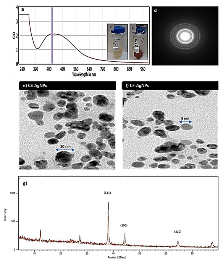
Figure 3.
(a) Surface plasmon absorption bands (SPR) of AgNPs formed in the presence of Streptomyces sp. ASM23 cell filtrate (CF-AgNPs). (b) The color change due to the reduction of metal ions and (c) the formation of AgNPs represented by (a) Control+ AgNO3 and (b) Streptomyces sp. ASM23 cell filtrate+ AgNO3. (d) Selected area (electron) diffraction (SAED) confirms the crystalization of nanoparticles. (e) Transmission electron microscope micrographs for (f) AgNPs biosynthesized by Streptomyces sp. ASM23 cell filtrate (CF-AgNPs) and cell supernatant (CS-AgNPs). (g) X-ray diffraction (XRD) pattern of silver nanoparticles.

