Abstract
Successful management of mandibular incisors with pulp canal obliteration using guided endodontics is described, for the first time in Iran. A 58-year-old man was referred for root canal treatment of teeth #24, #25 and #26. Upon radiographic examination, partial obliteration of the root canal system was detected. Cone-beam computed tomography (CBCT) was requested to enhance the diagnosis and detection of root canals. Next, a 3-dimensional (3D) guide was designed and printed to aid in localization and access to the root canal system with minimal destruction of the tooth structure. With the use of a targeted 3D guide, a conservative access cavity was prepared to avoid unnecessary removal of tooth structure. The teeth were successfully treated endodontically. Obtained results revealed that the technique can be effective and predictable for the management of calcified canals.
Key Words: Calcification, Cone-beam Computed Tomography, Guided Endodontics, Intra Oral Scanning, Minimally Invasive Access Cavity
Introduction
Chemo-mechanical preparation of the root canal system is a fundamental step in the success of root canal therapy [1]. It aims to disinfect the root canal space and remove pathogenic microorganisms that are the potential causes of apical periodontitis [2]. Root canal treatments performed under controlled conditions according to the standard protocol can bring about long-term successful outcomes [3]. Comprehensive debridement and disinfection of the root canal space are imperative for long-term success of endodontic treatment. However, some debris, pulpal residues, and bacteria may remain due to root canal irregularities even after precise chemo-mechanical preparation [3].
Calcific metamorphosis (CM) is a challenge encountered in root canal treatment of some teeth. According to the American Association of Endodontics, CM is defined as a pulpal response to trauma, which is characterized by rapid hard tissue deposition in the root canal space. The entire root canal space may appear obliterated on the radiographs as the result of extensive deposition; however, histological assessment may reveal some empty portions of the pulp space [4]. The affected teeth often have a darker hue compared with the adjacent teeth, showing a yellow discoloration. The clinical crown often loses its translucency due to increased thickness [5]. The discoloration and extent of calcification often become worse over time [6]. CM commonly occurs due to trauma and often affects the anterior teeth in young adults [7]. Some other etiologies have also been proposed in the literature such as pulpal response to caries, occlusal interferences, vital pulp therapy, and orthodontic treatment [8]. Partial or complete root canal obliteration may prevent complete instrumentation and disinfection of the root canal system, and may also increase the risk of iatrogenic procedural errors such as perforation and weakening of the remaining tooth structure [7]. In patients with CM, adequate access cavity preparation and negotiation of root canal orifices may be challenging, and require massive removal of tooth structure, which increases the risk of fracture [9]. Therefore, preoperative planning by using a 3-dimensional (3D) imaging modality is highly recommended [10].
Cone-beam computed tomography (CBCT) and dental operating microscope have been proposed for enhanced detection of some cases [11]. Guided endodontics recently gained much popularity for management of CM [12]. In guided endodontics, a digital impression is made and superimposed on CBCT scans. A drill path is created, and a guide is fabricated by using computer-aided design software to access the root canal. Finally, a 3D printer is used to print the guide [10].
The purpose of this case report is to describe the use of guided endodontics for mandibular central and lateral incisors with partial pulp canal obliteration.
Case Presentation
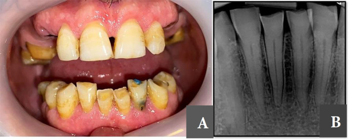
A 58-year-old Iranian male patient with no medical history was referred to the Department of Endodontics, Dental School, Mashad University of Medical Sciences, Iran, for root canal treatment of teeth #24, #25 and #26 due to prosthodontic reasons. On clinical examination, the patient was asymptomatic with no pain on percussion or sinus tract (Figure 1A). Radiographic examination revealed partial canal obliteration of anterior teeth (Figure 1B). Based on clinical and radiographic findings, a diagnosis of pulp necrosis was made for the respective teeth. A cone beam computed tomography (CBCT) was requested to further assess the pulp canal obliteration and root canal anatomy. CBCT (ProMax 3D MAX; Planmeca OY, Helsinki, Finland) revealed a calcified root canal in the coronal third of the root. To avoid iatrogenic damage during treatment, such as perforation or deviation from the main root canal path, a 3D- printed guide was designed to help navigate the procedure. Written informed consent was obtained from the patient prior to the procedure.
Figure 1.
A) Clinical view; B) Periapical radiograph, anterior teeth with attrition and partial calcifications in the root canal system
An intra-oral scan was performed (Trios 3 Shape, Warren, NJ, USA) to carefully plan and design the guide; and, both image data sets [CBCT digital imaging and communications in medicine (DICOM) files and scanner standard tessellation language (STL) files] were imported to a software program (Dental System v2017; 3Shape, Copenhagen, Denmark), which is originally used to design implant guides. During the planning phase, a path was specifically designed for size 1 Munce Discovery bur (CJM Engineering, Santa Barbara, CA, USA) by means of incorporating the size of the bur into the template (Figure 2). Finally, a 3D guide was printed to navigate the drill path by using a 3D printer (Sonic 4K 3D Printer; Phrozen Technology; Taiwan).
Figure 2.
Designing the template
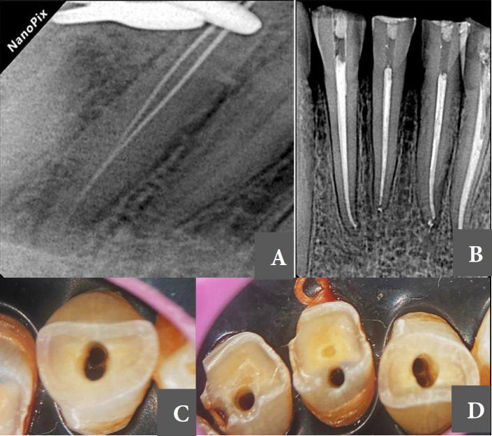
To check the insertion and stability of the guide, an alginate impression was made, and the plaster model was used to primarily check the printed guide. In the clinical session prior to the treatment onset, the guide was checked, and a pilot drill hole was created in the teeth using a diamond bur to further accommodate the drill placement through the guide. The access cavities were prepared by means of Munce Discovery bur (CJM Engineering, Santa Barbara, CA, USA). The template was periodically removed, and the access cavity was checked by a c pilot #10 file (VDW Endodontic Synergy, Munich, Germany). Finally, the canal was reached and patency was achieved. Since two canals were detected on CBCT image of tooth #26, after negotiation of the main canal, the guide was removed and the access cavity was manually expanded bucco-lingually to find the lingual canal. The working length in all canals was determined by an electronic apex locator (Propex IQ, Dentsply Sirona, Ballaigues, Switzerland) and confirmed radiographically (Figure 3A). During the procedure, the canals were irrigated with 25 mL of 5% NaOCl (Cerkamed, Stalowa Wola, Poland), and patency was checked in-between instrumentation. The canals were prepared up to size 25/0.04 in apical region with rotary files. (Hero 642, Micro Mega, France) and a final rinse with 3 mL of 17% EDTA (Cerkamed, Stalowa Wola, Poland) was performed. The canals were then dried with paper points (Meta Dental Co., Cheougja City, Korea) and obturated with warm vertical condensation technique using AH-Plus sealer (Dentsply Maillefer, Ballaigues, Switzerland). The access cavities were sealed with a temporary restorative material (3M ESPE, St. Paul, MN, USA), and the patient was referred to the prosthodontics department to further complete the restorative treatment (Figure 3B). At 6-month follow-up the teeth were asymptomatic with normal function.
Figure 3.
A) Presence of 2 canals in tooth #26; B) Post-obturation radiograph; C, D) Magnified view of the drill point (note the difference between the site of guided entry and the presumed site of root canal orifice)
Discussion
The use of 3D printed guides is becoming an accepted modality to manage complex endodontic cases [13]. Compared with the conventional techniques, guided endodontics significantly decreases the chair time and can be safely used in cases with calcified canals [12], anatomical malformations such as dens invagination [14] and dense evagination [15], and also periapical microsurgeries [16]. Furthermore, this technique is easy to use, and does not require complex expert training [17]. This case report presented the application of a guided endodontic procedure for nonsurgical endodontic treatment of three mandibular incisors of an elderly patient with pulp canal obliteration. To our knowledge, this is the first reported application of such targeted guides in endodontics in Iran, which was carried out in Mashhad University of Medical Sciences.
Pulp canal obliteration has always been a challenge for clinicians, and is associated with many iatrogenic mishaps including perforation and file fracture [9]. Although CBCT and dental operating microscope have been quite helpful in management of such cases [18], there is still need for further technological advancements to achieve better outcomes.
Since the first case report of guided endodontics by Krastl et al. [19] many studies used this protocol [12]. The 3D guides were originally developed for implant placement. For the present case, due to lack of a specific software for endodontics, an implant software program was used for our purpose. 3D printed guides have made it possible to have a direct access to the root canal [10] and this point of entry could be further away from the point the clinician would normally choose to start the access (Figures 3C and 3D).
CBCT is currently an essential imaging modality in endodontics. It enhances 3D visualization of teeth and greatly aids in accurate diagnosis and treatment planning [20]. In the present case, CBCT enabled the detection of second lingual canal of tooth #26, which had remained undetected on the periapical radiograph.
This case report indicated successful application of guided endodontics for mandibular incisors with pulp canal obliteration. Clinical studies are required on a larger scale (number of patients) with adequate follow-up period to further scrutinize the efficacy of this technique.
Conclusion
The guided endodontic technique was proven to be highly efficient in management of mandibular incisors with pulp canal obliteration. A pre-designed guide was used to design a minimally invasive access cavity, and the root canals were carefully negotiated. If performed correctly, this technique can significantly decrease the chair time and minimize the occurrence of iatrogenic errors in root canal treatment.
Conflict of Interest:
‘None declared’.
References
- 1.Carvalho MC, Zuolo ML, Arruda-Vasconcelos R, Marinho ACS, Louzada LM, Francisco PA, Pecorari VGA, Gomes B. Effectiveness of XP-Endo Finisher in the reduction of bacterial load in oval-shaped root canals. Braz Oral Res. 2019;33:e021. doi: 10.1590/1807-3107bor-2019.vol33.0021. [DOI] [PubMed] [Google Scholar]
- 2.Siqueira JF, Rôças I, Lopes H. Treatment of endodontic infections. Quintessence London; 2011. [Google Scholar]
- 3.Friedman S. Expected outcomes in the prevention and treatment of apical periodontitis. Essential endodontology: prevention and treatment of apical periodontitis. 2008. pp. 408–69. [Google Scholar]
- 4.Eleazer P, Glickman G, McClanahan S. AAE Glossary of endodontic terms. new york: American Association of Endodontists: 2020. [Google Scholar]
- 5.Amir FA, Gutmann JL, Witherspoon DE. Calcific metamorphosis: a challenge in endodontic diagnosis and treatment. Quintessence Int. 2001;32(6):447–55. [PubMed] [Google Scholar]
- 6.Oginni AO, Adekoya-Sofowora CA. Pulpal sequelae after trauma to anterior teeth among adult Nigerian dental patients. BMC Oral Health. 2007;7:11. doi: 10.1186/1472-6831-7-11. [DOI] [PMC free article] [PubMed] [Google Scholar]
- 7.McCabe PS, Dummer PM. Pulp canal obliteration: an endodontic diagnosis and treatment challenge. Int Endod J. 2012;45(2):177–97. doi: 10.1111/j.1365-2591.2011.01963.x. [DOI] [PubMed] [Google Scholar]
- 8.de Toubes KMS, de Oliveira PAD, Machado SN, Pelosi V, Nunes E, Silveira FF. Clinical approach to pulp canal obliteration: a case series. Iran Endod J. 2017;12(4):527–33. doi: 10.22037/iej.v12i4.18006. [DOI] [PMC free article] [PubMed] [Google Scholar]
- 9.Cvek M, Granath L, Lundberg M. Failures and healing in endodontically treated non-vital anterior teeth with posttraumatically reduced pulpal lumen. Acta Odontol Scand. 1982;40(4):223–8. doi: 10.3109/00016358209019816. [DOI] [PubMed] [Google Scholar]
- 10.Zehnder MS, Connert T, Weiger R, Krastl G, Kuhl S. Guided endodontics: accuracy of a novel method for guided access cavity preparation and root canal location. Int Endod J. 2016;49(10):966–72. doi: 10.1111/iej.12544. [DOI] [PubMed] [Google Scholar]
- 11.Quaresma SA, Costa RPd, Petean IBF, Silva-Sousa AC, Mazzi-Chaves JF, Ginjeira A, Sousa-Neto MD. Root canal treatment of severely calcified teeth with use of cone-beam computed tomography as an intraoperative resource: new strategies for root canal treatment of severely calcified teeth with use of intraoperative CBCT. Iran Endod J. 2022;17(1):39–47. doi: 10.22037/iej.v17i1.36153. [DOI] [PMC free article] [PubMed] [Google Scholar]
- 12.Diniz JMB, Oliveira HFD, Coelho RCP, Manzi F, Silva FE, Sobrinho APR, Machado VC, Tavares WLF. Guided endodontic approach in teeth with pulp canal obliteration and previous iatrogenic deviation: a case series. Iran Endod J. 17(2):78–84. doi: 10.22037/iej.v17i2.36830. [DOI] [PMC free article] [PubMed] [Google Scholar]
- 13.Garcia-Sanchez A, Bakhsh K, Sanchez SE, Tadinada A, Chen I-P. The use of three-dimensional (3D)-printed guide for identifying root canals in endodontic treatment. J Dent Treat Oral Care. 2020;3(1):104. [Google Scholar]
- 14.Ali A, Arslan H. Guided endodontics: a case report of maxillary lateral incisors with multiple dens invaginatus. Restor Dent Endod. 2019;44(4):e38. doi: 10.5395/rde.2019.44.e38. [DOI] [PMC free article] [PubMed] [Google Scholar]
- 15.Mena-Alvarez J, Rico-Romano C, Lobo-Galindo AB, Zubizarreta-Macho A. Endodontic treatment of dens evaginatus by performing a splint guided access cavity. J Esthet Restor Dent. 2017;29(6):396–402. doi: 10.1111/jerd.12314. [DOI] [PubMed] [Google Scholar]
- 16.Smith BG, Pratt AM, Anderson JA, Ray JJ. Targeted Endodontic Microsurgery: Implications of the Greater Palatine Artery. J Endod. 2021;47(1):19–27. doi: 10.1016/j.joen.2020.10.005. [DOI] [PubMed] [Google Scholar]
- 17.Freire BB, Vianna S, Nascimento EHL, Freire M, Chilvarquer I. Guided endodontic access in a calcified central incisor: a conservative alternative for endodontic therapy. Iran Endod J. 2021;16(1):56–9. doi: 10.22037/iej.v16i1.27427. [DOI] [PMC free article] [PubMed] [Google Scholar]
- 18.Perrin P, Neuhaus KW, Lussi A. The impact of loupes and microscopes on vision in endodontics. Int Endod J. 2014;47(5):425–9. doi: 10.1111/iej.12165. [DOI] [PubMed] [Google Scholar]
- 19.Krastl G, Zehnder MS, Connert T, Weiger R, Kühl S. Guided Endodontics: a novel treatment approach for teeth with pulp canal calcification and apical pathology. Dent Traumatol. 2016;32(3):240–6. doi: 10.1111/edt.12235. [DOI] [PubMed] [Google Scholar]
- 20.Tavares WLF, Machado VdC, Fonseca FO, Vasconcellos BC, Magalhães LC, Viana ACD, Henriques LCF. Guided endodontics in complex scenarios of calcified molars. Iran Endod J. 2020;15(1):50–6. doi: 10.22037/iej.v15i1.26709. [DOI] [PMC free article] [PubMed] [Google Scholar]


