Abstract
The current case study evaluated the effect of vital pulp therapy on a human dental pulp after a long-term period using micro-computed tomography (MCT) for the first time. In the presented report, the successful outcomes of full pulpotomy using calcium-enriched mixture (CEM) cement on an irreversible pulpitis case were documented clinically/radiographically over 5 years. Due to an unrestorable crown fracture at the 5-year recall, the tooth was extracted and evaluated by MCT; the images showed that CEM pulpotomy allowed the dental pulp to create complete dentinal bridges without pulp canal obliteration (PCO). These MCT results showed that CEM pulpotomy, as a bio-regenerative treatment, caused no negative consequence of PCO or calcific metamorphosis on dental pulp over the long term.
Key Words: Calcific metamorphosis, Calcium compounds, Calcium-enriched mixture, Tricalcium silicate, Micro-CT, Pulp canal obliteration, Pulpotomy, Vital pulp therapy
Introduction
Full pulpotomy, as a simple vital pulp therapy (VPT) modality, is highly recommended for the management of carious pulp exposure in mature and/or immature permanent teeth. This therapeutic procedure is considered a more accessible, affordable, available, easier, safer and less invasive treatment modality compared to conventional root canal treatment (RCT), and has demonstrated favorable clinical success as well as acceptable effectiveness [1, 2]. It has been claimed that VPT does not compromise the subsequent use of RCT should it fail; except for cases in which hard tissue is deposited along the internal walls of root canal(s) and fills most of the pulpal system leaving it narrowed and restricted; i.e. pulp canal obliteration (PCO).
Seemingly, PCO after VPT is a challenging task for endodontists [3]. Nonetheless, there is no supporting evidence on the nature of human dental pulp tissue after long-term and successful VPT treatments. Additionally, most of the current evidence obtained from trials has evaluated animal or human dental pulp responses in short-term intervals [4, 5]. In this case study, a micro-computed tomography (MCT) assessment of pulpotomy is reported, using calcium-enriched mixture (CEM) cement on a human dental pulp after 5 years for the first time.
Case Report
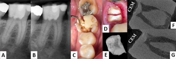
A 33-year-old female, with a history of symptomatic irreversible pulpitis related to her mandibular right first molar, was treated with full pulpotomy using CEM cement (BioniqueDent, Tehran, Iran) and permanent restoration using resin-based dental composite restorative materials (Z250/Z350, 3M, ESPE, USA) (Figure 1A). The treated tooth was asymptomatic and fully functional in routine follow-ups, with radiographic evaluations clearly showing hard tissue formation at root canal orifices at 5-year recall (Figure 1B). However, at the end of year 5, she reported persistent pain after accidentally biting into a stone in her food. Clinical examination revealed a vertical coronal fracture that separated the buccal cusps from the main structure of the tooth, making it unrestorable (Figure 1C). After obtaining informed consent and during the tooth extraction, the remaining crown broke, and to protect the alveolar bone from further traumatic damage, the roots were carefully separated and then, cautiously extracted (Figure 1D).
Figure 1.
Periapical radiographs (PR), clinical photographs, and micro-CT images of the case. A) Immediate post-treatment PR shows a mandibular right first molar treated with a CEM pulpotomy; B) PR at 5-year recall shows dentinal bridge formation at canal orifices and normal PDL, indicating successful results; C) Clinical photograph shows a vertical fracture which made the tooth unrestorable; D) Separation of the mesial and distal roots during surgical tooth extraction; E) MCT image from the apical aspect of the root specimen, showing two open root canals connected with an isthmus; F-G) MCT images from the coronal plan, illustrating dentinal bridge formation at buccal and lingual canal orifices
Subsequently, the mesial root was scanned using a micro-computed tomography (micro-CT) system. The micro-CT images proved the unblocking of root canals at the apical end of the mesial root and displayed the deposition of formed hard tissue over the two canal orifices capped with CEM cement (Figures 1E and 1F). Moreover, the mineralized bridge showed identical density with its surrounding normal dentin without any tunnel defects, and entirely re-closed the remaining radicular pulp similar to normal conditions (Figure 1G).
Discussion
Generally, there is little chance for long-term micro-CT evaluations of a human dental pulp that has been treated with VPT; i.e. full pulpotomy. In the unprecedented presented case, micro-CT evaluations revealed that there was no PCO or calcific metamorphosis in root canals after 5 years of CEM pulpotomy; however, the canal orifices had been closed due to effective dentinogenesis [6]. In the above-mentioned process, using an endodontic biomaterial, the remaining radicular pulp as well as periradicular tissues stayed protected from further excessive irritation [7, 8]. If the stated phenomenon had not occurred, it could have led to progressive reactionary responses; e.g. PCO or calcific metamorphosis after calcium hydroxide pulpotomy, specifically following traumatic injuries.
Furthermore, using CEM biomaterial, the full pulpotomy promoted of dentinal bridge formation at canal orifices [9] and protected the remaining radicular pulp from further pathological consequences; i.e. PCO or calcific metamorphosis.
Acknowledgment
The author would like to thank Dr. Hadi Assadian for his help in the preparation of micro-computed tomography images.
Conflict of Interest:
‘None declared’.
References
- 1.Yazdani S, Jadidfard MP, Tahani B, Kazemian A, Dianat O, Alim Marvasti L. Health technology assessment of CEM pulpotomy in permanent molars with irreversible pulpitis. Iran Endod J. 2014;9(1):23–9. [PMC free article] [PubMed] [Google Scholar]
- 2.Asgary S, Fazlyab M, Sabbagh S, Eghbal MJ. Outcomes of different vital pulp therapy techniques on symptomatic permanent teeth: a case series. Iran Endod J. 2014;9(4):295–300. [PMC free article] [PubMed] [Google Scholar]
- 3.Kumar D, Antony S. Calcified canal and negotiation-A review. J Pharm and Tech. 2018;11(8):3727–30. [Google Scholar]
- 4.Haghgoo R, Asgary S, Mashhadi Abbas F, Montazeri Hedeshi R. Nano-hydroxyapatite and calcium-enriched mixture for pulp capping of sound primary teeth: a randomized clinical trial. Iran Endod J. 2015;10(2):107–11. [PMC free article] [PubMed] [Google Scholar]
- 5.Asgary S, Parirokh M, Eghbal MJ, Ghoddusi J. SEM evaluation of pulp reaction to different pulp capping materials in dog's teeth. Iran Endod J. 2006;1(4):117–23. [PMC free article] [PubMed] [Google Scholar]
- 6.Mehrdad L, Malekafzali B, Shekarchi F, Safi Y, Asgary S. Histological and CBCT evaluation of a pulpotomised primary molar using calcium enriched mixture cement. Eur Arch Paediatr Dent. 2013;14(3):191–4. doi: 10.1007/s40368-013-0038-3. [DOI] [PubMed] [Google Scholar]
- 7.Asgary S. A successful pulpotomy-treated permanent molar withstood recurrent decay after 10 years of treatment. J Dent Sci. 2022;17:1827–8. doi: 10.1016/j.jds.2022.06.024. [DOI] [PMC free article] [PubMed] [Google Scholar]
- 8.Asgary S, Parhizkar A. The role of vital pulp therapy in the management of periapical lesions - Letter to the Editor. Eur Endod J. 2021;6(1):130–1. doi: 10.14744/eej.2020.04706. [DOI] [PMC free article] [PubMed] [Google Scholar]
- 9.Kabbinale P, Chethena K, Kuttappa M. Role of calcium-enriched mixture in endodontics. Arch Med Health Sci. 2015;3(1):80. [Google Scholar]


