

Abstract
Rare case of Ewing’s Sarcoma of maxilla managed with newly proposed multimodalityapproach in which First neo-adjuvant chemotherapy was done to shrink the size oftumor mass and control possible occult distant metastasis and in the second phase surgical management that is followed by adjuvant chemotherapy was done. During Surgical phase, instead of doing a complete surgical resection, we have decided intraoperatively and performed only curettage of the tumor mass left after shrinkage of the tumor mass post chemotherapy phase. This concludes that there is the scope of trying newer ideas for management and, thus, more contemporary approaches for this rare entity. Like in our case, curettage of the tumor rather than radical resection as a part of multimodality approach also gave optimal outcome.
Introduction
Ewing’s sarcoma is a small round cell malignant neoplasm which categorized under Ewing’s sarcoma family of tumors that includes Ewing’s sarcoma, atypical Ewing’s sarcoma, peripheral primitive neuroectodermal tumors (pPNET). Involvement of the osseous or soft tissue structures of the head and neck is rare, that is 1–4% of the cases, and it is mostly in the mandible and its primary maxilla involvement is rare. This rare case report highlights the critical need for a multidisciplinary approach to manage head and neck sarcomas in children and how a newly proposed technique becomes a favorable approach for the patient where Ewing’s sarcoma of the maxilla treated with neoadjuvant chemotherapy that was followed by surgical management and the surgical approach was chosen to be curettage of the tumor that is different from what literature shows in most of the case reports/case series jaw resection was done.
Case Report
A 13-year-old male patient reported with complains of swelling and pain over right middle one-third of face since 7 weeks for which he underwent extraction of his mobile deciduous teeth. He has noticed swelling and pain starts increasing in a more rapid pace 6 days post-extraction. On examination diffuse swelling of approx 5 × 3 cm size seen in right middle one-third of face and skin over swelling appears to be normal, and surface appears smooth (Fig. 1a), No lymph node involvement and intra-orally expansion of buccal cortical plate present from 11 to 16, obliteration of buccal vestibule, mobile and extruded 11, 12, 13, deep bite, occlusion deranged, mouth opening adequate (Fig. 1b).
Fig. 1.

a, b Diffuse swelling involving right maxillary region causing obliteration of nasolabial fold and elevation of ala of the nose and intraoral swelling obliterating right maxillary vestibule, causing extrusion of adjacent teeth’s and overlying mucosa appears normal in color with dilated capillaries visible over the surface
NCCT face and OPG findings suggestive of ill-defined lobulated soft tissue attenuated lesion noted in right maxilla causing surrounding bony destruction,extending into right maxillary sinus, nasal cavity and causing displacement of 12,13.
F-18- FDG PET suggestive of soft tissue density lesion in right anterior maxilla with extension to right nasal cavity and hard palate. No evidence of distant metastasis.
Histopathology report shows sheets of uniform small round cells separated by dense fibrous tissue. Differential diagnosis of small round tumors is lymphoma, neuroblastoma, rhabdomyosarcoma, primitive neuroectodermal tumors and Ewing’s sarcoma. Immunohistochemistry was done that shows diffuse and strong positivity for vimentin, MIC 2 and NKK 2.2 occasional tumor cells showing positivity for p63 while negative for LCA, CK, desmin and p53 that leads to confirm diagnosis of malignant small round cell tumor consistent with Ewing’s sarcoma.
Our patient being a very young boy, we decided to avoid extensive resection of anterior maxilla which could have led to extreme disfigurement of the face and also would have affected the functionality adversely. Also patient and his parents were very scared and reserved for such extensive surgery. So, in conjunction with pediatric oncology we adopted this new multimodality approach for managing cases of Ewing’s sarcoma.
Firstly, patient was referred to pediatric oncology and the chemotherapy protocol adopted was neoadjuvant chemotherapy in the form of 7 alternate cycle of VDC IE at 2-weekly interval in which dosage of Inj. Vincristine 2 mg /m2, Inj. Cyclophosphamide 1200/m2, Inj. Doxorubicin 37.5 mg/m2 and Inj. Ifosfamide 1800/ m2, Inj. Etoposide 100 mg/m2 according to body surface area. Complication of these chemotherapy drugs is nausea, vomiting, febrile neutropenia with high risk of infections and high risk of hemorrhagic cystitis with ifosfamide and cyclophosphamide and to counteract these effects antiemetic drugs, i.e., Tab Granisetron 1 mg PO OD, antibiotics, i.e., Inj. Amikacin, Inj. Ceftazidime, Tab. Fluconazole, Tab. Septran to lessen the chance of infection, Inj. Filgrastim 300 mcg SC to treat neutropenia and Inj. Mesna 20% of the dosage of cyclophosphamide and ifosfamide at 2 h, 6 h and 12 h of completion of infusion of these drugs was given to prevent complication of hemorrhagic cystitis, and it was observed that size of tumor reduced greatly at the end of 7th cycle where extra-orally nasolabial fold is recovered and ala of the nose comes to near-normal position and intraoral right maxillary vestibular depth is achieved.
FDG PET CT done post-neo-adjuvant chemotherapy suggests decreased FDG uptake and decreased metabolic activity; then, it was decided to proceed with the surgical phase (Fig. 2a, b).
Fig. 2.

a, b High FDG uptake suggesting increased metabolic activity in right maxillary region before chemotherapy and showing complete resolution of metabolic activity post-chemotherapy
During surgical phase intra-operatively, it is observed that only thin lining of tumor was left post-chemotherapy (Fig. 3) so it was decided to perform curettage of the lesion rather than going for what usually done in the literature for such type of cases that is aggressive jaw resection considering the patients age, and it is followed by 2 more cycle of VDC IE in the form of adjunct chemotherapy.
Fig. 3.
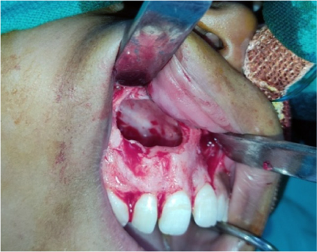
Only thin lining of tumor mass seen for which curettage of the lesion done intra-operatively
Follow-up
There is a specific protocol adopted by our institute for follow-up of these patients. During the chemotherapy phase, patient is called regularly at 3-weeks interval in which physical examination of the lesion, lymph nodes examination and blood counts, viz. CBC, RBC, WBC, platelets, MCH, MCHC, red cell width, LDH and other routine investigations, are done. After the completion of chemotherapy and surgery, the patient is followed up regularly at 1-month interval with all the blood investigations mentioned above for a period of 2 years. After that follow-up done at every 6-month interval for a period of next 2 years. Patient is also recalled annually for upto 5 years of completion of the treatment. This particular patient was followed up for upto 2 years after surgery with no fresh complaints and no evidence of disease recurrence (Fig. 4a, b).
Fig. 4.

a, b Swelling completely reduced and nasolabial fold depth regains completely and showing that maxillary vestibule depth completely regains post-surgery with maxilla preserved
Discussion
Ewing’s sarcoma of maxilla and zygoma is a biggest diagnostic challenge due to its rare involvement and its absence of pathognomonic radiography and clinical features. CT and MRI show an expanding and destructive lesion of the involved bone, and literature shows whole-body 18F-FDG PET images showed no abnormal activity except for the involved bone [1] Literature suggests it is difficult to differentiate Ewing sarcoma from other similar tumors; hence, it requires a histopathological examination, immunohistochemistry and a cytogenetic analysis to make the diagnosis [2]. In Ewing family tumor (EFT), the most common translocation that is diagnostic for Ewing’s sarcoma is t(11,22)(q24;q12) translocation, accounting more than 85% of cases. [3, 4] However, in our case, we did not performed any cytogenetic test due to a cost-related issues. The diagnosis of Ewing sarcoma was mainly based on the histological features, its confirmatory immunohistochemical staining, and FDG PET was done to rule out distant metastasis.
For this tumor various combination of management is been tried, unlike other sites definitive surgical resection of primary head and neck sarcomas is undesirable and constrained by a proximity to nonexpendable structures and concerns over cosmetic outcome [5, 6]. Ewing’s sarcoma is treated in a multidisciplinary manner including chemotherapy, surgery and radiotherapy [7]. Approximately only 10% of the patients with Ewing's sarcoma survived before the introduction of chemotherapy as a management modality, which has now drastically improved with 75% survival rates in patients with localized tumors [8]. Less than 20% of the cases will have distant metastasis to lungs. Very rarely, it can metastasize to other bones and lymph nodes [9].
In our case, we have used the multimodality approach in which first neo-adjuvant chemotherapy was done to shrink the size of tumor mass and control possible occult distant metastasis and in the second phase surgical management that is followed by adjuvant chemotherapy was done. During surgical phase, instead of doing a complete surgical resection, we have decided intra-operatively and performed only curettage of the tumor mass left after shrinkage of the tumor mass post-chemotherapy phase. We have performed this new approach considering the age of the patient, and as currently no gold standard treatment exists for these types of tumor and that helped us to avoid the requirement of reconstruction surgery and that gives an idea that these tumors can be managed by more conservative approach rather than doing radical resection.
Conclusion
Considering the rare occurrence of head and neck Ewing’s sarcoma and gold standard management not yet developed for this rare entity. There is the scope of trying newer ideas for management. Like in our case, curettage of the tumor rather than radical resection as a part of multimodality approach also gives optimal outcome. And most importantly, whatever will be the approach long-term follow-up is warranted considering the aggressive nature of the disease.
Funding
This study received no specific grant from any funding agency in the public, commercial, or not-for-profit sectors.
Footnotes
Publisher's Note
Springer Nature remains neutral with regard to jurisdictional claims in published maps and institutional affiliations.
References
- 1.Takami Y, Aga F, Mitamura K, Norikane T, Okuda H, Yamamoto Y, Miyake M, Nishiyama Y. A Case of Ewing sarcoma of the mandible on 18F-FDG PET/CT. Asia Ocean J Nuclear Med Biol. 2020;8(1):84–87. doi: 10.22038/aojnmb.2019.13876. [DOI] [PMC free article] [PubMed] [Google Scholar]
- 2.Satish D, Nanadakumar R, Balasubramanya AM, Mathew N. A rare case of Ewing’s sarcoma in the sinonasal tract. Int J Otorhinolaryngol Head Neck Surg. 2018;4(1):304–307. doi: 10.18203/issn.2454-5929.ijohns20175649. [DOI] [Google Scholar]
- 3.Iwamoto Y. Diagnosis and treatment of Ewing’s sarcoma. Jpn J Clin Oncol. 2007;37(2):79–89. doi: 10.1093/jjco/hyl142. [DOI] [PubMed] [Google Scholar]
- 4.Unni K, FMCDMF Pathology and genetics of tumours of soft tissue and bone. Surg Oncol. 2004;13(1):297. [Google Scholar]
- 5.Wexler LH, Kacker A, Piro JD, Haddad J, Jr, Close LG. Combined modality treatment of Ewing's sarcoma of the maxilla. Head Neck. 2003;25(2):168–172. doi: 10.1002/hed.10156. [DOI] [PubMed] [Google Scholar]
- 6.Infante-Cossio P, Gutierrez-Perez JL, Garcia-Perla A, Noguer-Mediavilla M, Gavilan-Carrasco F. Primary Ewing's sarcoma of the maxilla and zygoma: report of a case. J Oral Maxillofac Surg. 2005;63(10):1539–1542. doi: 10.1016/j.joms.2005.06.011. [DOI] [PubMed] [Google Scholar]
- 7.Krishna KBB, Thomas V, et al. A radiological review of Ewing's sarcoma of mandible: a case report with one year follow-up. Int J Clin Pediatr Dent. 2013;6(2):109–114. doi: 10.5005/jp-journals-10005-1200. [DOI] [PMC free article] [PubMed] [Google Scholar]
- 8.Balamuth NJ, Womer RB. Ewing's sarcoma. Lancet Oncol. 2010;11(2):184–192. doi: 10.1016/S1470-2045(09)70286-4. [DOI] [PubMed] [Google Scholar]
- 9.Mohiyuddin SMA, Deo RP, Jyothi DN, Kumar HML, Sagayaraj A. Ewing's sarcoma of Maxilla: a rare presentation. Int J Head Neck Surg. 2015;6(2):96–98. doi: 10.5005/jp-journals-10001-1232. [DOI] [Google Scholar]