

Abstract
Surgical removal of an impacted tooth is considered to be one of the most frequently performed minor oral surgical procedures due to the plethora of indications associated with it. Like any other surgical intervention, the surgical removal of impacted third molars is also associated with numerous complications. Lot of emphasis has been laid to prevent the complications associated with the surgical removal of impacted third molars. However, at times in spite of delivering the utmost caution, complications occur as a consequence of surgical removal of impacted third molars. We report a rare case in which iatrogenic mandibular ramus fracture occurred in an attempt to surgically removal of an impacted third molar.
Keywords: Third molar, Mandibular fracture, Iatrogenic injury
Introduction
Owing to the surplus indications for the surgical removal of an impacted tooth, it is considered to be the most frequently performed minor oral surgical procedure [1]. Surgical removal of deeply impacted third molar poses a significant surgical challenge to even an experienced operator. Numerous complications can occur as a consequence of surgical removal of deeply seated impacted third molars. They may manifest in the form of alveolar osteitis, secondary infection, dysesthesia, pain, hemorrhage, etc. The incidence of such complications ranges from 0.2 to 6% [2, 3].
The incidence of iatrogenic mandibular fracture associated with the surgical removal of an impacted third molar range from 0.0034 to 0.0075% [4]. Literature reveals that the presence of an unerupted lower third molar significantly weakened the mandibular angle region [5]. Hence, iatrogenic mandibular fracture usually occurs at the mandibular angle region. We report a case of a rare mandibular ramus fracture that occurred as a consequence of surgical removal of an impacted third molar.
Case Report
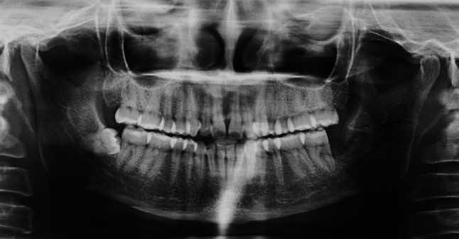
A 40-year-old female reported to our unit complaining of pain and difficulty in opening her mouth. She gives a history of pain in the left lower back tooth region since 2 days for which she visited a local dental clinic. Following a clinical & radiological examination, she was diagnosed with an infection in relation to her deeply impacted lower left third molar and she was advised to undergo surgical removal of her impacted lower left molar (Fig. 1). She gives a history of undergoing a surgical intervention for the same which was traumatic and is of long duration under local anesthesia by a general dentist. With great difficulty the impacted tooth was removed in multiple pieces and the patient was discharged from the OPD. In spite of the usage of medication, her pain did not subside and the mouth opening started reducing gradually.
Fig. 1.

Preoperative radiograph showing impacted left lower third molar
A thorough clinical examination by us revealed that the sutures were intact at the surgical site and the healing of the soft tissue was progressing normally. There was tenderness at the retromolar region and anterior border of the ramus. A postoperative OPG was done which revealed a left mandibular ramus fracture as a consequence of surgical removal of impacted third molar (Fig. 2). Patient was advised open reduction and internal fixation for the management of her mandibular ramus fracture. However, considering the physical & psychological trauma the patient underwent during the course of time in addition to the fact that the fracture was a simple and an undisplaced fracture, a conservative treatment option in the form of intermaxillary fixation (IMF) with ties and arch bars for 6 weeks was done instead of the surgical option. Patient was followed up at regular intervals and evaluated for pain and occlusal stability. IMF was removed after 6 weeks. Following the removal of IMF, an aggressive mouth opening exercises was performed and 6 months follow-up of the patient revealed a pain free adequate mouth opening with good occlusal stability.
Fig. 2.
Postoperative radiograph showing the removal of impacted tooth that lead to iatrogenic mandibular ramus fracture
Discussion
Literature reveals that iatrogenic fractures may occur during a surgical procedure or within 4 weeks after the intervention [6]. Occurrence of an iatrogenic mandibular fracture during the surgical removal of the impacted third molar is very rare [7, 8]. Review of literature indicating the site of iatrogenic mandibular fracture and the type of management employed is shown in Table 1. Studies have revealed that there is a risk of fracture of the mandibular angle because the presence of an unerupted third molar. [9, 10] Deeply impacted third molars occupy a large osseous space and thereby weaken the mandibular angle by decreasing the cross-sectional area of bone and causing the loss of support. Studies have shown that iatrogenic mandibular fractures occur mostly during the surgical removal of an erupted vertically impacted third molar and unerupted mesio-angular third molar [11, 12]. Iatrogenic mandibular fracture occurring at the ramus in an attempt to remove a horizontally impacted tooth is very rare.
Table 1.
Review of literature indicating the site of iatrogenic mandibular fracture following surgical removal of third molars and the type of management employed
| S. no. | References | Intraoperative/late | Iatrogenic mandibular fracture following surgical removal of impacted third molars | Type of management |
|---|---|---|---|---|
| 1. | Krimmel and Reinert [6] | Late postoperative | Mandibular angle fracture | ORIF |
| 2. | Wagner [14] | Late postoperative | Mandibular angle fracture | ORIF + autologous iliac crest graft |
| 3. | Woldenberg et al. [7] | Intraoperative | Mandibular angle fracture | Closed reeduction |
| 4. | Cankaya et al. [15] | Intraoperative | Mandibular angle fracture | Closed reduction |
| 5. | Tomruk and Arslan [13] | Late postoperative | Mandibular angle fracture | ORIF |
| 6. | Mihmanli et al. [8] | Intraoperative | Mandibular ramus fracture | Closed reduction |
| 7. | Silva et al. [19] | Intraoperative | Mandibular angle fracture | ORIF |
Iatrogenic mandibular fractures occur as a result of improper instrumentation and application of excessive force to the bone during surgical removal of third molars. Contrary to that, mandibular fractures can occur in the late postoperative phase probably due to the application of high level of biting forces during mastication or a failure in not having soft diet in the postoperative phase [13]. A study reveals that iatrogenic mandibular fractures occur predominantly on the left side compared to the right side since visualization of the right side is better and control of the forces is easier compared to the left side [14].On the contrary, another study found no difference in the occurrence of fractures on the right and left sides [11].
The extent of tooth impaction, the volume of the impacted tooth, and the relative portion of mandibular volume are also important contributing factor for iatrogenic mandibular fractures following surgical removal of impacted third molars [11, 15]. Preexisting pathological findings at the surgical site such as pericoronitis, periodontal pockets, and cysts may also weaken the mandible [15]. In addition to this, increased age leading to reduction in the bony elasticity and patients suffering from osteoporosis are at high risk for iatrogenic mandibular fractures following surgical removal of impacted third molars. [11, 15, 16].
The operators experience is an essential element in iatrogenic injury. A recent study conducted to evaluate the perception among the medical fraternity and general public pertaining to the scope of specialty of Oral and maxillofacial surgery revealed that 80.6% of the general public and 95% of medical professionals would seek the help of a general dentist to undergo surgical removal of their impacted third molars [17]. Iatrogenic mandibular fracture occurring at the ramus in an attempt to remove a horizontally impacted tooth by a general dentist supports the above study. However, there is enough evidence in the literature to support that iatrogenic mandibular fractures can occur even in the hands of the most experienced Oral and maxillofacial surgeons [18]. This necessitates the need to plan each individual case of third molar extraction case accurately by both clinical examination radiographic evaluation to lessen the risk of complications [19].
In developing countries like India, this is the most common reason for most of the complications seen following surgical removal of third molars particularly the iatrogenic mandibular fractures even in healthy patients where the tooth may or may not be deeply impacted. It is a well-known fact that a duly qualified and trained operator can deliver flawless treatment with their trained and skillful hands and this awareness needs to be increased keeping the best interests of the patient which is the main purpose of this article.
Conclusion
The ability to predict the surgical difficulty of surgical removal of impacted third molars is very critical in designing a treatment plan. An operator needs to evolve with the aid of the available current literature, sophisticated equipment’s and personal experience to understand the reasons for iatrogenic mandibular fractures and prevent this complication that can occur as a consequence of surgical removal of impacted third molars.
Compliance with Ethical Standards
Conflict of interest
All the authors declare that they have no conflict of interest.
Footnotes
Publisher's Note
Springer Nature remains neutral with regard to jurisdictional claims in published maps and institutional affiliations.
References
- 1.Uppada UK. A modification of ward’s incision for third molar surgery. J Dent Res Rev. 2019;6:77–78. doi: 10.4103/jdrr.jdrr_66_19. [DOI] [Google Scholar]
- 2.Goldberg MH, Nemarich AN, Marco WP., II Complications after mandibular third molar surgery: a statistical analysis of 500 consecutive procedures. J Am Dent Assoc. 1985;111:277–279. doi: 10.14219/jada.archive.1985.0098. [DOI] [PubMed] [Google Scholar]
- 3.Osborn TP, Frederickson G, Small IA, Torgerson TS. A prospective study of complications related to mandibular third molar surgery. J Oral Maxillofac Surg. 1985;43:767–769. doi: 10.1016/0278-2391(85)90331-3. [DOI] [PubMed] [Google Scholar]
- 4.Libersa P, Roze D, Cachart T, Libersa JC. Immediate and late mandibular fractures after third molar removal. J Oral Maxillofac Surg. 2002;60:163–166. doi: 10.1053/joms.2002.29811. [DOI] [PubMed] [Google Scholar]
- 5.Reitzik M, Lownie JF, Cleaton-Jones P, Austin J. Experimental fractures of monkey mandibles. Int J Oral Surg. 1978;7:100. doi: 10.1016/S0300-9785(78)80054-4. [DOI] [PubMed] [Google Scholar]
- 6.Krimmel M, Reinert S. Mandibular fracture after third molar removal. J Oral Maxillofac Surg. 2000;58:1110–1112. doi: 10.1053/joms.2000.9566. [DOI] [PubMed] [Google Scholar]
- 7.Woldenberg Y, Gatot I, Bodner L. Iatrogenic mandibular fracture associated with third molar removal: Can it be prevented? Med Oral Patol Oral Cir Bucal. 2007;12:E70–E72. [PubMed] [Google Scholar]
- 8.Mihmanli, et al. Fracture of the mandibular ramus during third molar removal: case report. Bezmialem science. 2014;2:9–100. [Google Scholar]
- 9.Rajkumar K, Sinha R, Chowdhury R, Chattopadhyay PK. Mandibular third molars as a risk factor for angle fractures: a retrospective study. J Maxillofac Oral Surg. 2009;8:237–240. doi: 10.1007/s12663-009-0058-z. [DOI] [PMC free article] [PubMed] [Google Scholar]
- 10.Revanth Kumar S, Sinha R, Uppada UK, Ramakrishna Reddy BV, Paul D. Mandibular third molar position influencing the condylar and angular fracture patterns. J Maxillofac Oral Surg. 2015;14:956–961. doi: 10.1007/s12663-015-0777-2. [DOI] [PMC free article] [PubMed] [Google Scholar]
- 11.Bodner K, Brennan PA, McLeod NM. Characteristics of iatrogenic mandibular fractures associated with tooth removal: review and analysis of 189 cases. Br J Oral Maxillofac Surg. 2011;49(7):567–572. doi: 10.1016/j.bjoms.2010.09.007. [DOI] [PubMed] [Google Scholar]
- 12.Wolujewic MA. Fractures of the mandible involving the impacted third molar tooth: an analysis of 47 cases. Br J Oral Surg. 1980;18:125–131. doi: 10.1016/0007-117X(80)90028-1. [DOI] [PubMed] [Google Scholar]
- 13.Tomruk CO, Arslan A. Mandibular angle fractures during third molar removal: a report of two cases. Aust Dent J. 2012;57:231–235. doi: 10.1111/j.1834-7819.2012.01674.x. [DOI] [PubMed] [Google Scholar]
- 14.Wagner KW, Schoen R, Wongchuensoontorn C, Schmelzeisen R. Complicated late mandibular fracture following third molar removal. Quintessence Int. 2007;38:63–65. [PubMed] [Google Scholar]
- 15.Cankaya AB, Erdem MA, Cakarer S, Cifter M, Korhan C. Iatrogenic mandibular fracture associated with third molar removal. Int J Med Sci. 2011;8(7):547–553. doi: 10.7150/ijms.8.547. [DOI] [PMC free article] [PubMed] [Google Scholar]
- 16.Chrcanovic BR, Custodio ALN. Considerations of mandibular angle fractures during and after surgery for removal of third molars: a review of the literature. Oral Maxillofac Surg. 2010;14:71–80. doi: 10.1007/s10006-009-0201-5. [DOI] [PubMed] [Google Scholar]
- 17.Vadepally AK, Sinha R, Uppada UK, Rama Krishna Reddy BV, Agarwal A. Oral and maxillofacial surgery: perception of its scope among the medical fraternity and general public. J Cranio Max Dis. 2015;4:21–27. doi: 10.4103/2278-9588.151898. [DOI] [Google Scholar]
- 18.Pires WR, et al. Late mandibular fracture occurring in the postoperative period after third molar removal: systematic review and analysis of 124 cases. Int J Oral Maxillofac Surg. 2017;46(1):46–53. doi: 10.1016/j.ijom.2016.09.003. [DOI] [PubMed] [Google Scholar]
- 19.Silva TCG, et al. Mandibular fracture after third molar removal: a case report. Gen Dent. 2019;67(4):e7–e10. [PubMed] [Google Scholar]