

Abstract
Effective use of underutilised fish processing by-products could open avenues for new industries if they are used to extract high-valued bioactive compounds. Therefore, discarded skin offcuts of three main commercial fish species of Sri Lanka were used to extract collagen with acetic acid and the extracted collagen was evaluated for industrial suitability. The yields of acid-soluble collagens from Yellowfin tuna (Thunnus albacares), Seer fish (Scomberomorus commerson) and Asian sea bass (Lates calcarifer) were 61.26%, 58.21% and 59.31%, respectively on a dry-weight basis. Fourier Transform Infra-Red spectra and X-ray Diffraction spectra confirmed that all collagens were in type I and preserved the native triple-helical structure during extraction. The UV absorption spectra confirmed a high collagen purity in all species. These results confirm that the extracted collagen consists of the characteristics required for collagen-based industries such as food, pharmaceutical, and biomedical. The high availability of skin offcuts from the processing industry and the higher collagen yields revealed in this study confirm the possibility of using discarded skin offcuts of the three fish species as a potential source of type I collagen for industrial purposes.
Keywords: Fish skin, Collagen, SEM, FTIR, XRD, UV spectra
Introduction
Collagen is the most abundant protein, which consists of around 30% of the total body protein, in vertebrates (Li et al. 2013; Singh et al. 2011) and invertebrates (Gobeaux et al. 2008). It is a structural (Chen et al. 2016; Li et al. 2013), extracellular matrix protein (Sun et al. 2017) found in the connective tissues such as skin, bones, tendons and cartilage (Zhang et al. 2016). Although there are 28 genetically distinct collagen types in the collagen family (Ricard-Blum 2011), which differ in their amino acid composition and sequence; molecular structure and function (Li et al. 2013), the type-l collagen, which is the fibril-forming collagen type (Ricard-Blum 2011), is the most abundant in vertebrates and also as the major structural protein in invertebrates (Gobeaux et al. 2008). Type-I collagen has a wide variety of applications in the biomedical; pharmaceutical; food; cosmetic; leather; and film industries (Sionkowska et al. 2015; Zeng et al. 2009).
Mammalian sources are the most common and widely used form of type-I collagen, but its usage has been limited due to the risk of human infections with zoonotic diseases like bovine spongiform encephalopathy or mad cow disease; foot-and-mouth disease and transmissible spongiform encephalopathy (Muralidharan et al. 2013). But fish collagen has not been reported to be linked with such diseases (Li et al. 2013), therefore it has drawn greater attention as a safer alternative to mammalian collagen. Moreover, some religious and cultural barriers prevent the use of bovine and porcine origin collagen (Sun et al. 2017). Further, fish collagen has a higher biological value and a higher essential amino acid content than mammalian collagen (Tanaka et al. 2018).
If the fish collagen can be extracted from the fish processing byproducts, which are generally discarded, it could be a great potential for new collagen-based industries because generally, seafood processing recovers only 20–50% as edible portions depending on the processes used and types of products. Simultaneously, 50–80% of remaining offcuts are discarded as by-products after filleting (Bae et al. 2008), with a global annual average of 20 million Mt (Pal and Suresh 2016). These fish processing by-products generally include damaged muscle, skin, bones, fins and scales (Muralidharan et al. 2013) and they consist of valuable compounds such as collagen, peptides, oils (Lakmini et al., 2022), chitin (Liyanage et al., 2022), vitamins, minerals, enzymes, and pigments which could be used in high-end industries (Pal and Suresh 2016).
In Sri Lanka, most of these fish processing by-products are used for low levels of value addition (Xu et al. 2017) such as fishmeal, fish oil, silage, animal feed, compost and direct fertiliser production while the rest is dumped without getting any use but creating environmental issues. Anyhow, during the last decade, more than 75 medium and large-scale processing plants were established as the export-oriented Sri Lankan fish processing industry was expanding (EDB (Export Development Board (EDB) 2019).
The backbone of the Sri Lankan fish exports industry is Yellowfin tuna (Thunnus albacares), and Seer fish (Scomberomorus commerson) (Ministry of Fisheries, 2020). Recently, the Asian sea bass (Lates calcarifer) or Sri Lankan Barramundi which is coming from aquaculture is also becoming a competitive species contributing to the country's economy. Even though very high quantities of offcuts of these three fish species are generated at fish processing plants, inadequate knowledge hinders its potential usage for extracting bioactive compounds for potential high-end industries. Therefore, the present study was conducted to extract type-I collagen from the discarded skin offcuts of three main commercial fish species, Yellowfin tuna, Seer fish and Asian sea bass to contrast the yields and evaluate the characteristics of extracted collagen to understand the potential of using such discards as a source of type-I collagen.
Material and methods
Raw material
Skin offcuts of Yellowfin tuna (Thunnus albacares), Seer fish (Scomberomorus commerson) and Asian sea bass (Lates calcarifer), which were produced as by-products during the fish filleting process for export were obtained from a fish processing plants located in Ja-Ela, Western Province of Sri Lanka. Samples were brought to the laboratory under frozen conditions. Samples were free of dust and sand as they were already washed at the processing plant, however, they have washed again with distilled water to remove any extraneous matter. They were stored at − 20 °C in a freezer until used for the experiment.
Moisture, protein and fat contents
The moisture content of the skins was determined according to the oven drying method (at 105 °C for 24 h). The protein content of the skins and extracted collagen was determined according to the Kjeldahl method. The fat content of the skins was determined according to the method described by Bligh and Dyer (1959).
Pre-treatment of fish skin
From each of the three fish species, a sample of 30 g of the skin was cut separately into 0.3 cm × 0.3 cm pieces (Fig. 1a, c, e) and treated with 0.8 mol/L sodium chloride (NaCl) at a ratio of 1:6 (w/v) for 10 min to remove impurities and pigments. This process was repeated three times and then washed with cold distilled water as described by Muralidharan et al (2013) with modifications as described below. NaCl-treated skins were soaked in cold distilled water at a ratio of 1:50 (w/v) for 5 min with stirring. This washing process was repeated four times. This modification was done to reduce the amount of wastewater. The sodium hydroxide (NaOH) pre-treatment to remove non-collagenous proteins was carried out as described by Sato et al (1986) as follows. The washed samples were soaked in 0.1 mol/L NaOH at a ratio of 1:10 (w/v) for 3 days with gentle stirring. NaOH solution was changed every day. Treated skins were then washed with cold distilled water until neutral pH by soaking in cold distilled water at a ratio of 1:50 (w/v) for 5 min with stirring and repeated four times. The entire process was carried out at 4 °C using cold room conditions.
Fig. 1.

Raw skin offcuts of Yellowfin tuna (a), Seer fish (c) and Asian sea bass (e) on the left column. The external morphology of lyophilised collagen of Yellowfin tuna (b), Seer fish (d), and Asian sea bass (f) on the right column
Extraction of collagen
Acid soluble collagen (ASC) from pre-treated fish skins was extracted following the protocol described by Nagai and Suzuki (2000), with modifications as described below. Samples were treated with 0.5 mol/L acetic acid at a ratio of 1:30 (w/v) for 3 days and then the solution was centrifuged at low centrifugation speed (1,896 × g) for 30 min at 4 °C to cut off the unnecessary production cost. The supernatant was collected, and the residue was re-extracted using the same procedure. The supernatant which was collected from both extractions was salted out by adding NaCl to a final concentration of 0.9 mol/L. The precipitated collagen was centrifuged at 1,896 × g for 30 min at 4 °C. The resultant precipitate was re-dissolved in 0.5 mol/L acetic acid and dialysed first against 0.1 M acetic acid and for 24 h with a change of solution at 12 h, and then against distilled water for 24 h with a change of solution at 12 h, to remove impurities of chemicals used in the extraction process. The resultant solution was then lyophilised and stored at − 20 °C until further experimentation.
Determination of collagen yield
The yield of extracted collagen was calculated according to the method described by Chen et al. (2016), based on the dry weight of starting material:
Characterisation of collagen
Scanning electron microscopy (SEM) images
The method described by Zhang et al. (2016) was slightly modified to observe the morphology of collagen. A collagen film of 2 mm was mounted on to aluminium stub using double-sided tape to ensure high electrical conductivity between the specimen and the stub. Then it was sputter-coated with 1 angstrom of gold using a sputter coating machine and viewed in the Carl ZEISS EVO 18 Scanning Electron Microscope using secondary electron mode.
Fourier transform infrared (FTIR) spectra
FTIR spectra of lyophilised collagen samples were obtained to analyse the type of extracted collagen based on the functional groups and the interactions between the bonds. ATR-FTIR spectra of all acid soluble collagen (ASC) were obtained according to the method described by Singh et al. (2011) with slight modifications, using an FTIR spectrophotometer (Bruker Model ALPHA). A collagen film of 5 mm was placed on the crystal cell and the transmittance signals were collected for 40 scans over the range of 4000 to 400 cm−1 at a resolution of 2 cm−1. Baseline correction was done using a background spectrum collected from the clean empty cell. Analysis was done at 25 °C. Analysis of spectral data was carried out using the OPUS data collection software program.
X-Ray Diffraction (XRD) spectra
The crystal structures of lyophilised collagen samples were analysed to determine the organisation structure of collagen as described by Alves et al. (2017). The XRD pattern was determined using a CuKα (λ = 1.5405Å) radiation from Regaku Ultima IV X-ray diffractometer (XRD). A collagen film of 5 cm × 5 cm was placed on general purposes sample holder. Then scan was performed in a range of 2θ = 5 to 30° and at a scanning speed of 2°/min. The crystal phases were identified by using the PDF2 database from International Center for Diffraction Data (ICDD).
UV absorption spectra
UV–Visible spectrophotometric analysis was performed to determine collagen’s purity based on the absorption at a specific wavelength. The method described by Zeng et al. (2009) was slightly modified for UV analysis. A sample of 0.2 mg of freeze-dried fish collagen was dissolved in 500 mL of 0.02 M sodium acetate buffer, pH 4.8 containing 2 M urea. The solution was placed into a quartz cell with a path length of 1 mm. The UV spectrum was measured at wavelength 190–400 nm at a scan speed of 2 nm/s with an interval of 1 nm using a Thermo GENESYS 10 UV–Vis Spectrophotometer.
Statistical analysis
Collagen extraction was triplicated for each of the three fish species. For the collagen yield calculations, the mean values were calculated from three determinants and expressed as mean ± standard deviation. The results were statistically interpreted using a one-way analysis of variance (ANOVA) at a 0.05 significance level. Minitab 18 statistical software package was used for the data analysis.
Results and discussion
Moisture, protein and fat content
The moisture content of the fish skin samples and the protein content of both fish skin and extracted collagen are given in Table 1. The percentage moisture contents of the Yellowfin tuna, Seer fish and Asian sea bass were 57.47 ± 0.31, 61.27 ± 0.31 and 61.07 ± 1.94, respectively. The percentage protein contents of the skin of Yellowfin tuna, Seer fish and Asian sea bass were 69.28 ± 0.46, 65.62 ± 0.33 and 65.86 ± 0.11, respectively on dry weight basis while the percentage protein contents of the extracted collagen from the Yellowfin tuna, Seer fish and Asian sea bass were 92.72 ± 0.96, 90.34 ± 1.09 and 92.07 ± 1.39, respectively on dry weight basis. Even though the extracted collagen revealed 90–92% of protein, probably the collagen, the remaining balance could probably be the partially removed lipids and minerals. The slight difference in percentage protein contents of the extracted collagen could probably be due to the different lipid and collagen protein contents in the skin offcuts of the three species. The fat contents of Yellowfin tuna, Seer fish and Asian sea bass were 2.7 ± 0.06%, 0.77 ± 0.05% and 0.81 ± 0.05% respectively on a wet weight basis. The quality of the extracted collagen is based on the lipid content of the used raw materials since the remaining lipid content directly affects the extracted collagen’s odour (Sadowska et al. 2003). If the skin fat is not properly removed during the pre-treatment and remains in the extracted collagen, it develops a rancid fish oil odour (Sadowska et al. 2003). However, the extracted collagen in this study had only a barely detectable fishy odour may be due to the very small amount of fat content in skin offcuts and proper removal of fat during the pre-treatment. Besides, extraction of collagen immediately after receiving the raw materials and storing the extracted collagen in vacuum conditions at − 20 °C could have prevented the development of rancid off-odour for upto eight months in this study.
Table 1.
Yield of collagen (in dry weight basis), moisture and protein content (in dry weight basis) of the skin and collagen
| Fish species | Skin | Collagen | |||
|---|---|---|---|---|---|
| Moisture % | Protein % | Protein % | Protein recovery % | Yield % | |
| Yellowfin tuna | 57.47 ± 0.31 | 71.92 ± 0.43 | 92.72 ± 0.96 | 78.98 ± 1.07 | 61.26 ± 0.63 |
| Seer fish | 61.27 ± 0.31 | 67.66 ± 0.23 | 90.34 ± 1.09 | 77.73 ± 1.14 | 58.21 ± 0.27 |
| Asian sea bass | 61.07 ± 1.94 | 68.49 ± 0.14 | 92.07 ± 1.39 | 79.73 ± 1.32 | 59.31 ± 2.87 |
Yield of collagen
Average yields of collagen obtained from skin offcuts of the three fish species were almost up to 60% (Table 1) on a dry weight basis. The remaining wastage could consist of non-collagenous components such as non-collagenous proteins, fat, pigments (Li et al. 2013), and non-extracted enzyme soluble collagen (Bhuimbar et al. 2019). Moreover, no significant differences were obtained among the three species (P = 0.29). Different collagen yields have been recorded in previous studies from the skin of different fish species. Anand et al. (2013) reported collagen yields of 8% (Asian Sea bass (Lates calcarifer)) and 7.5% (Australasian Snapper (Pagrus auratus)) on a dry weight basis, using the acetic acid extraction method. Nagai and Suzuki (2000) reported collagen yields of 51.4% (Japanese seabass (Lateolabrax japonicus), 49.8% (chub mackerel (Scomber japonicus)), and 50.1% (bullhead shark (Heterodontus japonicus)) on a lyophilised dry weight basis, using acetic acid extraction method. The collagen yield obtained by Li et al (2013) from Spanish mackerel (Scomberomorous niphonius) using the acetic acid extraction method was 58.62% on a dry weight basis. A collagen yield of 5.4% on a dry weight basis was obtained from bluefin tuna (Thunnus orientalis) by Tanaka et al., (2018) using the combined treatment of acetic acid and pepsin enzyme. Bhuimbar et al (2019) recorded a collagen yield of 25% from black ruff (Centrolophus niger) on a wet weight basis. The yield of collagen from the skin may vary due to several factors: fish species; biological conditions of fish such as age; method of extraction i.e. acetic acid extraction, pepsin extraction; parameters of extraction i.e. acid concentration, the ratio of raw materials to acid volume, extraction temperature, time (Pal and Suresh, 2016). Yield data of the present study depict that skin offcuts of these three species are a viable and promising source of collagen to gain economic benefits rather than discarded as waste.
Characterisation of collagen
Morphology
The morphology of the lyophilised collagen was observed under the naked eye and SEM. Collagen film appeared as a white sponge with a porous structure on its surface under the naked eye (Fig. 1b, d, f) for all three fish species. The SEM images of the lyophilised collagen from skin offcuts of three fishes showed the characteristic porous, fibrous and sheet-like morphology which should be preserved for further value-addition of collagen (Fig. 2). Similar morphological results were reported in previous studies using fish as the source of collagen: skin of Centrolophus niger (Bhuimbar et al. 2019); skin and bones of Scomberomorous niphonius (Li et al. 2013).
Fig. 2.

SEM images of collagen extracted from skin offcuts of Yellowfin tuna (a) × 500; (b) × 1,560, Seer fish (c): × 500; (d): × 1,560 and Asian sea bass (e): × 500; (f): (× 1,560)
FTIR spectra
The FTIR spectra of Yellowfin tuna (Fig. 3a), Seer fish (Fig. 3c) and Asian sea bass (Fig. 3e) exhibited peaks of amide A, amide B, amide I, amide II, and amide III which are characteristics of type I collagen (Alves et al. 2017). These characteristic peaks were similar to the FTIR spectra of other fish species reported in previous studies; tilapia (Oreochromis niloticus) (Chen et al. 2016); striped catfish (Pangasianodon hypophthalmus) (Singh et al. 2011) and channel catfish (Ictalurus punctaus) (Liu et al. 2007). The amide A bands of Yellowfin tuna, Seer fish and Asian sea bass were found at wavenumbers of 3298.81 cm−1, 3281.37 cm−1 and 3379.29 cm−1, respectively. The characteristic infrared absorption of the amide A band is relative to the N–H stretching vibrations (Xu et al. 2017; Li et al. 2013) and is commonly observed in the range between 3,400 and 3,440 cm−1 (Singh et al. 2011). The position shifts to a lower frequency, usually around 3300 cm−1, when the N–H group of a peptide bond is involved in hydrogen bonding probably with a carbonyl group (Singh et al. 2011). The amide B bands of Yellowfin tuna, Seer fish and Asian seabass were found at wavenumbers of 2921.57 cm−1, 2921.90 cm−1 and 2926.01 cm−1, respectively. Amide B band is associated with the asymmetrical stretching of CH2 groups (Anand et al. 2013; Li et al. 2013) and is commonly observed near 2920 cm−1. The amide I band of Yellowfin tuna, Seer fish and Asian sea bass were found at wavenumbers of 1653.74 cm−1, 1635.30 cm−1 and 1641.42 cm−1, respectively. The amide I band is associated with the stretching vibrations of the carbonyl group (C = O bond) along the polypeptide backbone and is commonly observed in the range from 1,600 to 1,700 cm−1 (Singh et al. 2011). Amide I band is responsible for the degree of molecular order found in collagen (Muyonga et al. 2004), and is a sensitive marker of the peptide secondary structure (Singh et al. 2011). The amide II band is commonly observed in the range between 1,550 and 1,600 cm−1 (Duan et al. 2009). In this study, amide II bands of Yellowfin tuna, Seer fish and Asian sea bass were found at wavenumbers of 1540.65 cm−1, 1539.74 cm−1 and 1539.19 cm−1, respectively. The shifting of amide II band positions to lower frequencies was due to more H bonds indicating a good activity of collagen and triple-helical structure (Riaz et al. 2018). The amide III bands of Yellowfin tuna, Seer fish and Asian sea bass were found at wavenumbers of 1234.56 cm−1, 1234.83 cm−1 and 1247.94 cm−1, respectively. The position of the amide III band around 1240 cm−1 is an indication of the existence of a triple-helical structure (Liu et al. 2007). The amide II and amide III bands represent N–H bending vibration coupled with C–N stretching vibration and C–H stretching (Liu et al. 2007; Singh et al. 2011; Riaz et al. 2018).
Fig. 3.

FTIR spectra of collagen extracted from Yellowfin tuna (a), Seer fish (c) and Asian sea bass (e); XRD spectra of collagen extracted from skin offcuts of Yellowfin tuna (b), Seer fish (d) and Asian sea bass (f)
Additionally, a peak occurred at 1455.37 cm−1, 1451.87 cm−1 and 1450.47 cm−1, respectively for Yellowfin tuna, Seer fish and Asian sea bass. An absorption ratio of approximately 1, between the amide III and the 1454–1450 cm −1 peak is an indication of preserving the triple helical structure of collagen molecule during the extraction procedure (Li et al. 2013). In this study, absorption ratio values of 0.98, 1.01 and 0.98 were obtained for Yellowfin tuna, Seer fish and Asian sea bass respectively, indicating that the native triple helical structures were maintained. Native triple helical structure determines the functions of type I collagen, therefore, it is important for further practical applications (Liu et al. 2007). These spectral results confirm that the collagen extracted from the skin offcuts of Yellowfin tuna, Seer fish and Asian sea bass belonging to type I and collagen extraction conditions had no pronounced negative effect on the triple-helical structure. Therefore, the method and conditions provided in this study are suitable to extract collagen from the skin offcuts of these three selected fish species, in non-denatured form.
XRD spectra
As evident in Fig. 3b, d, f, the XRD diagrams of lyophilised collagen extracted from the skin offcuts of Yellowfin tuna, Seer fish and Asian sea bass exhibited two peaks characteristic of collagen (Sun et al. 2017). The Bragg equation: 2dsinθ = λ (λ = 0.15405), was used to calculate the minimum of the repeated interval (d), where λ is the X-ray wavelength (its value is 1.5405Å), and θ is the Bragg diffraction angle (Wang et al. 2009). The calculated d values are given in Table 2. The first diffraction peak of a collagen XRD spectrum which is relatively sharp is related to the distance between molecular chains of collagen fibres (Alves et al. 2017; Sun et al. 2017). The corresponding diffraction angles (2θ) at the first peak for Yellowfin tuna, Seer fish and Asian sea bass were found at 8.2°, 7.8° and 7.5°, respectively. Accordingly, the calculated distances between molecular chains were 10.77 Å, 11.32 Å and 11.78 Å, for Yellowfin tuna, Seer fish and Asian sea bass, respectively. The distance between molecular chains is important depending on the application of extracted collagen, for example, collagen with a higher distance between molecular chains is more suitable as a drug delivery agent (Chen et al. 2016). The second diffraction peak of a collagen XRD diagram reflects the diffused scatter caused by many structural layers of collagen fibres (Chen et al. 2016) or in other words the distance between collagen skeletons (Alves et al. 2017; Sun et al. 2017). The corresponding diffraction angles (2θ) at the second peak were found at 20.8°, 24.9° and 22° for Yellowfin tuna, Seer fish and Asian sea bass, respectively. Accordingly, the calculated distance between collagen skeletons was 4.27 Å, 3.57 Å and 4.04 Å, for Yellowfin tuna, Seer fish and Asian sea bass, respectively. These results are similar to the findings of previous studies including fish skin collagen extracted from Tilapia (Oreochromis niloticus) by Chen et al. (2016) and Atlantic salmon (Salmo salar) by Alves et al. (2017). Calculated distance values were consistent with the diameter of a single left-handed helix chain and a triple-helical collagen molecule, thus confirming the collagen extracted from the skin offcuts of Yellowfin tuna, Seer fish and Asian sea bass were non-denatured with native triple helical conformation (Sun et al. 2017).
Table 2.
Distance values of XRD peaks of collagen extracted from skin offcuts of Yellowfin tuna, Seer fish and Asian sea bass
| Sample | Peak 1 | Peak 2 | ||||
|---|---|---|---|---|---|---|
| Peak position | Distance between collagen molecules | Peak position | Distance between collagen skeletons | |||
| 2θ | Å | nm | 2θ | Å | nm | |
| Yellowfin tuna | 8.2 | 10.77 | 1.08 | 20.8 | 4.27 | 0.43 |
| Seer fish | 7.8 | 11.32 | 1.13 | 24.9 | 3.57 | 0.36 |
| Asian sea bass | 7.5 | 11.78 | 1.18 | 22.0 | 4.04 | 0.40 |
UV absorption spectra
Pure type I collagen gives the maximum absorption peak in the wavelength range from 220 to 240 nm. This peak is associated with the –C = O, –COOH, –CONH2 in polypeptide chains of collagen molecules (Bhuimbar et al. 2019; Zeng et al. 2009). If the extracted collagen is associated with minor amounts of residual amino acids from non-collagenous proteins, a small peak is given at 280 nm corresponding to tyrosine and tryptophan amino acids (Xu et al. 2017; Zeng et al. 2009). Hence, the absence of a peak around 280 nm confirms that pre-treatment has been effective to remove all non-collagenous proteins, and the collagen is pure. In this study, UV absorption spectra were recorded between the 190 nm and 400 nm wavelength range for the three selected fish species (Fig. 4), and the maximum absorption wavelength of the three selected fish species occurred at 217 nm, which was similar to the acid-soluble collagen extracted from several fish species in previous studies including Pacific cod (Gadus macrocephalus) skin (Sun et al. 2017); southern catfish (Silurus meridionalis) skin (Xu et al. 2017) and Nile tilapia (Oreochromis niloticus) skin (Zeng et al. 2009). The absence of the peaks at 280 nm of the three species indicates the high purity of collagen.
Fig. 4.
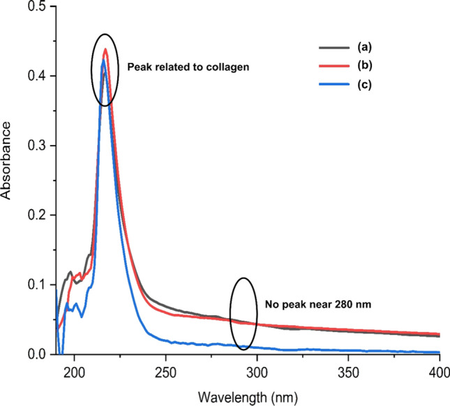
UV absorption spectra of collagen extracted from skin offcuts of Yellowfin tuna (a), Seer fish (b) and Asian sea bass (c)
Conclusion
Collagens were successfully extracted from the skin offcuts of three main commercial fish species in Sri Lanka: Yellowfin tuna (Thunnus albacares), Seer fish (Scomberomorus commerson) and Asian sea bass (Lates calcarifer) using the acetic acid extraction method. Extracted collagen yields were approximately similar from all the three species and were between 55 and 63% on a dry weight basis. All collagens were characterised as type I collagen and maintained their native triple-helical structure which is essential for further value addition. Therefore, the skin offcuts of these three fish species which are discarded in a considerable quantity from fish processing factories can potentially be used to extract collagen for industrial purposes.
Author contribution
A.G.D.M.A. did experiments, curated and analysed data, validated data, and wrote the original draft of the manuscript; S.T.G. conceptualised the research idea, curated and analysed data, validated data, engaged in funding acquisition, administered and supervised the project, and reviewed and edited drafts; C.A.N.F. analysed data, engaged in funding acquisition, administered and supervised the project, provided laboratory resources, validated data, and reviewed and edited drafts; M.D.S.T.D.C. conceptualised the research idea, curated and analysed data, engaged in funding acquisition, administered and supervised the project, provided laboratory resources, validated data, and reviewed and edited drafts.
Funding
This work was supported by the development-oriented research grant (AHEAD/DOR-80/AQF/WUSL) from World Bank under the grant scheme of Accelerating Higher Education Expansion and Development (AHEAD) through the government of Sri Lanka.
Data availability
The datasets used and/or analysed during the current study are available from the corresponding author on reasonable request.
Declarations
Conflicts of interest
The authors declare that they have no competing interests.
Footnotes
Publisher's Note
Springer Nature remains neutral with regard to jurisdictional claims in published maps and institutional affiliations.
Contributor Information
A. G. D. M. Ampitiya, Email: ampitiyaduleeka@gmail.com
S. T. Gonapinuwala, Email: suchimatg@wyb.ac.lk
C. A. N. Fernando, Email: canfernando9@gmail.com
M. D. S. T. de Croos, Email: dileepa_dc@yahoo.com, Email: dileepad@wyb.ac.lk
References
- Alves AL, Marques AL, Martins E, Silva TH, Reis RL. Cosmetic potential of marine fish skin collagen. Cosmetics. 2017;4(4):39. doi: 10.3390/cosmetics4040039. [DOI] [Google Scholar]
- Anand S, Kamath S, Chuang L, Kasapis S, Lopata AL. Biochemical and thermo-mechanical analysis of collagen from the skin of Asian Sea bass (Lates calcarifer) and Australasian Snapper (Pagrus auratus), an alternative for mammalian collagen. Eur Food Res Technol. 2013;236(5):873–882. doi: 10.1007/s00217-013-1950-9. [DOI] [Google Scholar]
- Bae I, Osatomi K, Yoshida A, Osako K, Yamaguchi A, Hara K. Biochemical properties of acid-soluble collagens extracted from the skins of underutilised fishes. Food Chem. 2008;108(1):49–54. doi: 10.1016/j.foodchem.2007.10.039. [DOI] [Google Scholar]
- Bhuimbar MV, Bhagwat PK, Dandge PB. Extraction and characterization of acid soluble collagen from fish waste: Development of collagen-chitosan blend as food packaging film. J Environ Chem Eng. 2019;7(2):102983. doi: 10.1016/j.jece.2019.102983. [DOI] [Google Scholar]
- Bligh EG, Dyer WJ. A rapid method of total lipid extraction and purification. Can J Biochem Physiol. 1959;37(8):911–917. doi: 10.1139/o59-099. [DOI] [PubMed] [Google Scholar]
- Chen J, Li L, Yi R, Xu N, Gao R, Hong B. Extraction and characterization of acid-soluble collagen from scales and skin of tilapia (Oreochromis niloticus) LWT-Food Sci Technol. 2016;66:453–459. doi: 10.1016/j.lwt.2015.10.070. [DOI] [Google Scholar]
- Duan R, Zhang J, Du X, Yao X, Konno K. Properties of collagen from skin, scale and bone of carp (Cyprinus carpio) Food Chem. 2009;112(3):702–706. doi: 10.1016/j.foodchem.2008.06.020. [DOI] [Google Scholar]
- EDB (Export Development Board (EDB) (2019) Industrial Capability Report—Fisheries Sector – 2019. https://www.srilankabusiness.com/ebooks/fish---fishery-products---industry-capability-report---december-2019.pdf. Accessed 13 April 2022.
- Gobeaux F, Mosser G, Anglo A, Panine P, Davidson P, Giraud-Guille MM, Belamie E. Fibrillogenesis in dense collagen solutions: a physicochemical study. J Mol Biol. 2008;376(5):1509–1522. doi: 10.1016/j.jmb.2007.12.047. [DOI] [PubMed] [Google Scholar]
- Lakmini KPC, Gonapinuwala ST, Senarath HPS, Fernando CAN, Wijesekara I, De Croos MDST. Effect of autoclaving as a pre-treatment in the wet reduction process for extracting fish oil from yellowfin tuna heads. Sri Lanka J Aquat Sci. 2022;27(1):43–61. doi: 10.4038/sljas.v27i1.7596. [DOI] [Google Scholar]
- Li ZR, Wang B, Chi CF, Zhang QH, Gong YD, Tang JJ, Luo HY, Ding GF. Isolation and characterization of acid soluble collagens and pepsin soluble collagens from the skin and bone of Spanish mackerel (Scomberomorous niphonius) Food Hydrocoll. 2013;31(1):103–113. doi: 10.1016/j.foodhyd.2012.10.001. [DOI] [Google Scholar]
- Liu H, Li D, Guo S. Studies on collagen from the skin of channel catfish (Ictalurus punctaus) Food Chem. 2007;101(2):621–625. doi: 10.1016/j.foodchem.2006.01.059. [DOI] [Google Scholar]
- Liyanage CS, Gonapinuwala ST, Fernando CAN, De Croos MDST. Physico-chemical properties of chitosan extracted from Whiteleg shrimp (Litopenaeus vannamei) processing shell waste in Sri Lanka. Sri Lanka J Aquat Sci. 2022;27(2):23–36. [Google Scholar]
- Ministry of Fisheries. (2020). Fisheries statistics - 2020. https://www.fisheriesdept.gov.lk/web/-images/Statistics/FISHERIES-STATISTICS- -2020-.pdf. Accessed 11 April 2022.
- Muralidharan N, Shakila RJ, Sukumar D, Jeyasekaran G. Skin, bone and muscle collagen extraction from the trash fish, leather jacket (Odonus niger) and their characterization. J Food Sci Technol. 2013;50(6):1106–1113. doi: 10.1007/s13197-011-0440-y. [DOI] [PMC free article] [PubMed] [Google Scholar]
- Muyonga JH, Cole CGB, Duodu KG. Characterisation of acid soluble collagen from skins of young and adult Nile perch (Lates niloticus) Food Chem. 2004;85(1):81–89. doi: 10.1016/j.foodchem.2003.06.006. [DOI] [Google Scholar]
- Nagai T, Suzuki N. Isolation of collagen from fish waste material—skin, bone and fins. Food Chem. 2000;68(3):277–281. doi: 10.1016/S0308-8146(99)00188-0. [DOI] [Google Scholar]
- Pal GK, Suresh PV. Sustainable valorisation of seafood by-products: recovery of collagen and development of collagen-based novel functional food ingredients. Innov Food Sci Emerg Technol. 2016;37:201–215. doi: 10.1016/j.ifset.2016.03.015. [DOI] [Google Scholar]
- Riaz T, Zeeshan R, Zarif F, Ilyas K, Muhammad N, Safi SZ, Rahim A, Rizvi SAA, Rehman IU. FTIR analysis of natural and synthetic collagen. Appl Spectrosc Rev. 2018;53(9):703–746. doi: 10.1080/05704928.2018.1426595. [DOI] [Google Scholar]
- Ricard-Blum S. The collagen family. Cold Spring Harb Perspect Biol. 2011;3(1):a004978. doi: 10.1101/cshperspect.a004978. [DOI] [PMC free article] [PubMed] [Google Scholar]
- Sadowska M, Kołodziejska I, Niecikowska C. Isolation of collagen from the skins of Baltic cod (Gadus morhua) Food Chem. 2003;81(2):257–262. doi: 10.1016/S0308-8146(02)00420-X. [DOI] [Google Scholar]
- Sato K, Yoshinaka R, Sato M, Ikeda S. A simplified method for determining collagen in fish muscle. Bull Jpn Soc Sci Fish. 1986;52(5):889–893. doi: 10.2331/suisan.52.889. [DOI] [Google Scholar]
- Singh P, Benjakul S, Maqsood S, Kishimura H. Isolation and characterisation of collagen extracted from the skin of striped catfish (Pangasianodon hypophthalmus) Food Chem. 2011;124(1):97–105. doi: 10.1016/j.foodchem.2010.05.111. [DOI] [Google Scholar]
- Sionkowska A, Kozłowska J, Skorupska M, Michalska M. Isolation and characterization of collagen from the skin of Brama australis. Int J Biol Macromol. 2015;80:605–609. doi: 10.1016/j.ijbiomac.2015.07.032. [DOI] [PubMed] [Google Scholar]
- Sun L, Li B, Song W, Si L, Hou H. Characterization of Pacific cod (Gadus macrocephalus) skin collagen and fabrication of collagen sponge as a good biocompatible biomedical material. Process Biochem. 2017;63:229–235. doi: 10.1016/j.procbio.2017.08.003. [DOI] [Google Scholar]
- Tanaka T, Takahashi K, Tsubaki K, Hirata M, Yamamoto K, Biswas A, Moriyama T, Kawamura Y. Isolation and characterization of acid-soluble bluefin tuna (Thunnus orientalis) skin collagen. Fish Aquatic Sci. 2018;21(1):7. doi: 10.1186/s41240-018-0084-1. [DOI] [Google Scholar]
- Wang C, Chen Z, He Y, Li L, Zhang D. Structure, morphology and properties of Fe-doped ZnO films prepared by facing-target magnetron sputtering system. Appl Surf Sci. 2009;255(15):6881–6887. doi: 10.1016/j.apsusc.2009.03.008. [DOI] [Google Scholar]
- Xu S, Yang H, Shen L, Li G. Purity and yield of collagen extracted from southern catfish (Silurus meridionalis Chen) skin through improved pretreatment methods. Int J Food Prop. 2017;20(sup1):S141–S153. doi: 10.1080/10942912.2017.1291677. [DOI] [Google Scholar]
- Zeng SK, Zhang CH, Lin H, Yang P, Hong PZ, Jiang Z. Isolation and characterisation of acid-solubilised collagen from the skin of Nile tilapia (Oreochromis niloticus) Food Chem. 2009;116(4):879–883. doi: 10.1016/j.foodchem.2009.03.038. [DOI] [Google Scholar]
- Zhang Q, Wang Q, Lv S, Lu J, Jiang S, Regenstein JM, Lin L. Comparison of collagen and gelatin extracted from the skins of Nile tilapia (Oreochromis niloticus) and channel catfish (Ictalurus punctatus) Food Biosci. 2016;13:41–48. doi: 10.1016/j.fbio.2015.12.005. [DOI] [Google Scholar]
Associated Data
This section collects any data citations, data availability statements, or supplementary materials included in this article.
Data Availability Statement
The datasets used and/or analysed during the current study are available from the corresponding author on reasonable request.