Abstract
G protein-coupled receptors (GPCRs) are embedded in phospholipids that strongly influence drug-stimulated signaling. Anionic lipids are particularly important for GPCR signaling complex formation, but a mechanism for this role is not understood. Using NMR spectroscopy, we visualized the impact of anionic lipids on the function-related conformational equilibria of the human A2A adenosine receptor (A2AAR) in bilayers containing defined mixtures of zwitterionic and anionic phospholipids. Anionic lipids primed the receptor to form complexes with G proteins through a conformational selection process. Without anionic lipids, signaling complex formation proceeded through a less favorable induced fit mechanism. In computational models, anionic lipids mimicked interactions between a G protein and positively charged residues in A2AAR at the receptor intracellular surface, stabilizing a pre-activated receptor conformation. Replacing these residues strikingly altered the receptor response to anionic lipids in experiments. High sequence conservation of the same residues among all GPCRs supports a general role for lipid-receptor charge complementarity in signaling.
Introduction
The essential roles of lipids as key regulators of the structures and functions of membrane proteins are well established1–7. For the 826 human G protein-coupled receptors (GPCRs), mounting evidence documented in the literature has increasingly highlighted the critical roles of lipids in regulating ligand-induced cellular signaling, both through their bulk physical properties, such as membrane curvature8, and as specific chemical partners acting as orthosteric ligands9,10 or as allosteric modulators11–13.
Recent structural and biophysical studies point to special roles of anionic lipids in regulating GPCR activity. Biochemical data indicated anionic lipids strongly influenced selectivity of the β2-adrenergic receptor (β2AR) for different G proteins through lipid-protein charge complementarity14, and anionic lipids impacted the affinity and efficacy of β2AR ligands11. Anionic lipids, including POPS, were found to be critical for activation of the CB2 cannabinoid receptor15. Mass spectrometry data supported the role of the anionic phospholipid PtdIns(4,5)P2 (PIP2) stabilizing complexes of human GPCRs with their partner G proteins16. Anionic phospholipids such as PIP2 have also been observed bound to the human serotonin receptor 5-HT1A in cryo-electron microscopy structures17, supporting the idea that direct lipid-protein interactions modulate receptor activity.
Knowledge of GPCR structural plasticity is critical to developing an understanding of signaling mechanisms. Nuclear magnetic resonance (NMR) spectroscopy is well-suited to experimentally investigate and measure GPCR structural plasticity, as it provides the unique capability to detect multiple, simultaneously populated receptor conformations and link observed conformational equilibria to efficacies of bound drugs18,19. We leveraged this capability to investigate the impact of anionic lipids on the structural plasticity of the human A2A adenosine receptor (A2AAR), a representative class A GPCR and exemplary GPCR for investigating receptor-lipid interactions. Our investigation builds on accumulated data from NMR spectroscopic studies of A2AAR interactions with small molecule ligands12,20–26, providing a firm foundation for evaluating the impact of anionic lipids. In both mass spectrometry experiments16 and molecular dynamics simulations27, anionic lipids were observed to strongly influence the formation of A2AAR signaling complexes. However, a mechanistic basis for these observations has not been determined.
Using nanodiscs, we precisely controlled phospholipid composition to investigate the response of A2AAR over a wide range of defined binary lipid mixtures. In 19F-NMR data, we observed the impact of anionic lipids on the conformational equilibria of A2AAR to be of a similar magnitude as bound drugs. Synergy observed between the presence of anionic lipids and efficacy of bound drugs indicated that sensitivity to lipid composition depended on the receptor conformation. In both NMR experiments and computational models, positively charged residues on the A2AAR intracellular surface near regions that interact with partner signaling proteins appeared to facilitate the influence of anionic lipids. Integrating these data with correlative signaling assays and NMR experiments of judiciously selected A2AAR variants provided a mechanistic view on the role of lipids in A2AAR signaling. Key residues identified in these experiments are conserved among not only class A but all receptor classes, indicating this mechanism may be shared across the GPCR superfamily.
Results
Molecular recognition of drug compounds by A2AAR investigated across a wide range of phospholipid compositions
We expressed human A2AAR containing a single extrinsic cysteine introduced into position 289, A2AAR[A289C] (Supplementary Fig. 1). The extrinsic cysteine, located near the intracellular-facing surface of transmembrane (TM) helix VII, was introduced for 19F-NMR experiments, and this A2AAR variant was previously shown to retain pharmacological activity of the native receptor22,28. Purified A2AAR[A289C] was reconstituted into lipid nanodiscs formed with the membrane scaffold protein MSP1D129 containing defined binary mixtures of different molar ratios of zwitterionic phospholipids, POPC (1-palmitoyl-2-oleoyl-glycero-3-phosphocholine) or POPE (1-palmitoyl-2-oleoyl-sn-glycero-3-phosphoethanolamine), and anionic lipids, POPS (1-palmitoyl-2-oleoyl-sn-glycero-3-phospho-L-serine), POPA (1-palmitoyl-2-oleoyl-sn-glycero-3-phosphate), POPG (1-palmitoyl-2-oleoyl-sn-glycero-3-phospho-(1’-rac-glycerol)) or PI(4,5)P2 (1,2-dioleoyl-sn-glycero-3-phospho-(1’-myo-inositol-4’,5’-biphosphate)). Analytical characterization of purified lipid nanodiscs containing A2AAR[A289C] showed highly monodispersed and homogenous samples (Supplementary Fig. 1). 31P-NMR in aqueous solutions was used to verify the lipid composition within the nanodisc samples studied in subsequent biophysical and NMR spectroscopy experiments (Supplementary Fig. 2). As the lipid headgroups showed resolved signals with unique chemical shifts, the intensities of the observed 31P signals were used to quantify the relative amounts of each lipid species present. For all samples and for all employed lipids, the relative amounts of each lipid species in the nanodisc samples determined by 31P-NMR closely agreed with the amounts we intended to incorporate in the nanodiscs (Supplementary Fig. 2).
To confirm that A2AAR[A289C] was folded in nanodiscs across the range of studied lipid compositions, we recorded fluorescence thermal shift assays for A2AAR[A289C] in complex with the antagonist ZM241385 and the agonist NECA (5’-N-ethylcarboxamidoadenosine) in nanodiscs composed of a wide range of different binary mixtures of zwitterionic and anionic lipids. The assay employed a thiol-reactive dye, N-[4-(7-diethylamino-4-methyl-3-coumarinyl)phenyl]maleimide (CPM), which reacts with cysteines that become solvent accessible upon thermal denaturation30. As the nanodisc scaffold protein MSP1D1 contains no cysteines, the assay thus observed a direct response of the thermal unfolding of the receptor. In nanodiscs containing mixtures of different relative amounts of POPC and POPS, we observed thermal unfolding curves consistent with well-folded receptors for both complexes with the antagonist ZM241385 (Supplementary Fig. 3a) and complexes with the agonist NECA (Supplementary Fig. 3b). Interestingly, as the molar fraction of POPS increased, the observed thermal melting temperature of A2AAR[A289C] also increased by ~10 °C from nanodiscs containing only POPC to nanodiscs containing only POPS (Supplementary Fig. 3a). This overall trend was also observed for A2AAR[A289C] in nanodiscs composed of different ratios of POPE and POPS, and POPC with POPA or POPG (Supplementary Fig. 3b), though the trend was most pronounced for nanodiscs containing mixtures of POPC or POPE mixed with POPS. Further, the increase in melting temperature associated with increasing amounts of anionic lipids was observed for both the complex with an antagonist and the complex with an agonist (Supplementary Fig. 3b). To confirm the fluorescence shift assay reported specifically on the thermal unfolding of the receptor within formed nanodiscs rather than disassembly of the nanodiscs, we recorded variable temperature circular dichroism (CD) data of nanodiscs without receptor (Supplementary Fig. 4). These data showed the thermal unfolding of the nanodiscs containing only lipids was higher than 90 °C, the upper limit of our instrument, confirming the fluorescence thermal shift data reported specifically on the unfolding of the receptor. These data appear in line with earlier observations from differential scanning calorimetry measurements, which reported nanodiscs containing binary mixtures of lipids remained assembled up to temperatures from 90 °C to over 105 °C 31.
The pharmacological activity of A2AAR[A289C] in lipid nanodiscs containing different binary lipid mixtures was measured in radioligand competition binding assays for both complexes with the antagonist ZM241385 and the agonist NECA. KD values were determined in nanodiscs containing only POPC lipids and mixtures of POPC with one of three different anionic lipids. Measured KD values for the antagonist ZM241385 varied only by a factor of ~2 among the different lipid compositions (Supplementary Fig. 5). Measured KI values for the agonist NECA varied only by a factor of ~3 among different lipid compositions (Supplementary Fig. 5). These relatively small differences indicated A2AAR[A289C] is pharmacologically active in the range of lipid compositions studied and, the presence of anionic lipids appeared to not impose a significant difference on the pharmacological activity of A2AAR.
The conformational ensemble of activated A2AAR strongly depends on the membrane environment
To prepare samples for NMR studies, a 19F-NMR reporter group was introduced by reacting the solvent-accessible cysteine at position 289 with 2,2,2-trifluoroethanethiol (TET) using an in-membrane chemical modification approach32, yielding A2AAR[A289CTET]. Earlier studies employing both the same stable-isotope labeling methodology and A2AAR variant demonstrated no other cysteines were available for 19F labeling22. The A289C 19F-NMR reporter was highly sensitive to function-related changes in the efficacies of bound drugs, providing ‘fingerprints’ for the corresponding functional states22.
19F-NMR spectra of A2AAR[A289CTET] in DDM (n-Dodecyl-β-D-Maltopyranoside)/CHS (cholesteryl hemisuccinate) mixed micelles with receptor prepared from Pichia pastoris (Fig. 1a) were in overall good agreement with 19F-NMR spectra reported for the same A2AAR variant expressed in insect cells and solubilized with the same detergent22. The present spectrum of an A2AAR[A289CTET] complex with the antagonist ZM241385 contained two signals at δ ≈ 11.3 ppm (P3) and δ ≈ 9.5 ppm (P1) with the signal P3 being the dominant signal in the spectrum (Fig. 1a). 19F spectra of the A2AAR[A289CTET] complex with the agonist NECA contained a signal at the chemical shift of P1 and two new signals, P2 and P4, at δ ≈ 10.7 ppm and δ ≈ 13.1 ppm, respectively, and no signal intensity at the chemical shift for P3 (Fig. 1a).
Fig. 1. NMR-observed conformational states of A2AAR-ligand complexes compared in two different membrane mimetics.
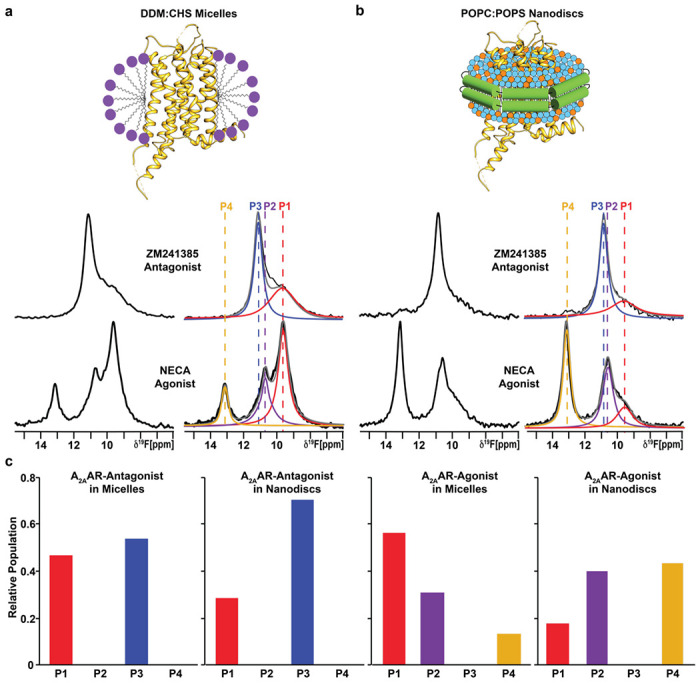
a The 1-dimensional 19F-NMR spectra of A2AAR[A289CTET] reconstituted into DDM/CHS mixed micelles and in complexes with the antagonist ZM241385 and agonist NECA. On the right, the NMR spectra shown on the left are interpreted by Lorenztian deconvolutions with the minimal number of components that provided a good fit, labeled P1 to P4. The chemical shifts of P1 to P4 are indicated by the colored dashed vertical lines. b The 1-dimensional 19F-NMR spectra of A2AAR[A289CTET] reconstituted into lipid nanodiscs containing the lipids POPC and POPS in a 70:30 molar ratio and in complexes with the same antagonist and agonist. Same presentation details as in a. c Relative populations of each state represented in a bar chart format.
We prepared samples of A2AAR[A289CTET] in lipid nanodiscs formed with the membrane scaffold protein MSP1D1 containing a mixture of the zwitterionic lipid POPC and anionic lipid POPS at a molar ratio of 70 to 30, respectively. 19F-NMR spectra of A2AAR[A289CTET] in nanodiscs with this ratio of lipid species showed the same number of signals with highly similar chemical shifts as observed in 19F-NMR spectra of A2AAR[A289CTET] in detergent micelles for both antagonist-bound and agonist-bound receptors (Fig. 1b). Only a small difference was observed for the chemical shift of state P3 for A2AAR in complex with the antagonist ZM241385, δ ≈ 10.9 ppm in lipid nanodiscs versus δ ≈ 11.3 ppm in detergent micelles (Fig. 1). Chemical shifts for states P1, P2, and P4 were practically indistinguishable between A2AAR[A289CTET] in DDM/CHS micelles and the POPC/POPS lipid nanodisc preparation. The highly similar chemical shifts observed for complexes in DDM/CHS mixed micelles and POPC/POPS lipid nanodiscs indicates that the introduced 19F-NMR probe is responsive to changes in receptor conformation rather than changes in the employed membrane mimetic.
The highly similar chemical shifts between A2AAR[A289CTET] in DDM/CHS micelles and the POPC/POPS lipid nanodisc preparation allowed us to quantitatively compare relative peak intensities between the two different membrane mimetics to investigate the impact of the membrane environment on the fingerprint of A2AAR functional states. For the complex with the antagonist ZM241385, the relative peak intensities for P1 and P3 were only marginally different between A2AAR in detergent and A2AAR in lipid nanodiscs (Fig. 1a). Ratios of peak intensities for apo A2AAR were also highly similar between detergent and lipid nanodisc environments (Supplementary Fig. 6a). In contrast, we observed striking differences in the relative intensities of peaks for the A2AAR complex with agonist. State P4, observed only for agonist-bound A2AAR, showed nearly a factor of 4 increase in intensity for POPC/POPS nanodiscs (Fig. 1b). State P1, which was the dominant peak observed in DDM/CHS, showed an intensity of nearly a factor of 4 smaller for POPC/POPS nanodiscs (Fig. 1b). The large differences in relative peak intensities were observed only for agonist-bound A2AAR, indicating a synergy between drug efficacy and the surrounding membrane environment. The ratio of populations of individual conformers for agonist-bound A2AAR is highly sensitive to changes in the surrounding membrane environment, but not for antagonist-bound A2AAR, as quantified in Fig. 1c. We also observed different rates of conformational exchange between A2AAR in DDM/CHS micelles and in lipid nanodiscs in 2-dimensional [19F,19F]-exchange spectroscopy (EXSY) (Supplementary Fig. 7). In the 2D-EXSY spectrum recorded with agonist-bound A2AAR in POPC/POPS lipid nanodiscs, we did not observe the presence of any crosspeaks. This is in contrast to previously reported EXSY spectra of agonist-bound A2AAR in DDM/CHS micelles that reported exchange crosspeaks between peaks P1 and P222, indicating the rate of exchange for populations P1 and P2 must be at least an order of magnitude slower in lipid nanodiscs.
Agonist-bound A2AAR adopts a predominantly inactive conformation in the absence of anionic lipids
To investigate the impact of anionic lipids on the observed conformational ensemble for A2AAR-agonist complexes, we recorded 19F-NMR data with A2AAR[A289CTET] bound to the agonist NECA in lipid nanodiscs containing only the zwitterionic phospholipid POPC (Fig. 2). Surprisingly, we observed a conformational ensemble that more closely resembled the fingerprint of inactive A2AAR even though a saturating amount of agonist was present, showing a dominant peak P3 and minor populations of peaks P1 and P2 (Fig. 2a). Fluorescence thermal shift assays of the A2AAR-agonist complex in POPC showed a well-folded protein (Supplementary Fig. 3), and radioligand binding experiments showed A2AAR in POPC nanodiscs bound the agonist NECA with a measured dissociation constant that was only a factor of ~2.5 times different from nanodiscs containing POPC and POPS (Supplementary Fig. 5). Thus, the 19F-NMR data show a conformational ensemble of functional, agonist-bound A2AAR largely in an inactive conformation. This indicates that even in the presence of saturating amounts of an activating drug, A2AAR adopts predominantly an inactive conformation in the absence of anionic lipids. 19F-NMR data of the A2AAR[A289CTET]-agonist complex in POPC nanodiscs measured at the higher temperature of 300 K were highly similar to spectra measured at 280 K with only a minor increase in the population of state P4 (Supplementary Fig. 8).
Fig. 2. NMR-observed conformation of an A2AAR-agonist complex and ternary complex in nanodiscs containing only zwitterionic lipids.
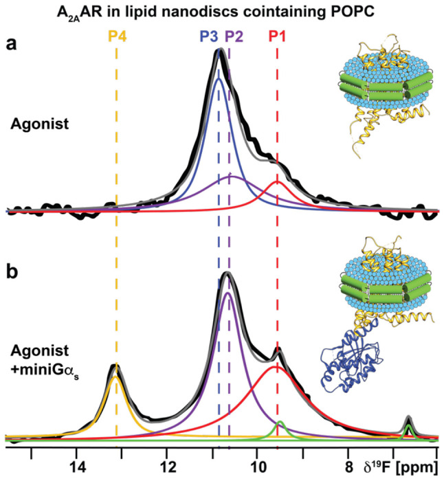
a A 1-dimensional 19F-NMR spectrum of an A2AAR[A289CTET] complex with the agonist NECA in lipid nanodiscs containing the zwitterionic lipid POPC. b The 1-dimensional 19F-NMR spectrum of an A2AAR ternary complex with the agonist NECA and an engineered Gαs protein in lipid nanodiscs containing POPC. Other figure presentation details the same as in Fig. 1, a and b.
To test whether A2AAR in POPC nanodiscs could still form signaling complexes, we added to the same sample the engineered Gαs protein, ‘mini-Gαs’, a partner protein designed to induce conformational changes in ternary complexes with A2AAR that mimic those of the native Gαs protein33,34. Analytical size exclusion chromatography and SDS-PAGE analysis confirmed the formation of a the A2AAR ternary complex with NECA and mini-Gαs (Supplementary Fig. 9). 19F-NMR data of A2AAR[A289CTET] in POPC nanodiscs showed a clear response to the addition of mini-Gαs (Fig. 2b). The NMR data appeared qualitatively similar to data measured for agonist-bound A2AAR in nanodiscs containing POPC and POPS, where the population of state P3 was significantly reduced and the populations of states P2 and P4, specific to the agonist complex, increased (Fig. 2b). This data allowed us to unambiguously assign the identity of state P4 to a conformation of activated A2AAR in the ternary complex. However, for the ternary complex, the relative population of state P4 was ~1/3 to 1/4 of the intensity of P4 observed for A2AAR-agonist complex in POPC/POPS nanodiscs. This indicated that while A2AAR could still populate a fully active conformation, the much lower P4 signal intensity indicated that the corresponding population was much lower than that observed for A2AAR-agonist in the presence of POPS even in the absence of a partner signaling protein (Fig. 1b).
A2AAR activation depends on lipid headgroup charge rather than chemical scaffold
To investigate whether observations in Figure 2 were specific to POPS or applied more generally to additional anionic lipids, we prepared a series of agonist-bound A2AAR samples in nanodiscs containing binary mixtures of POPC and a second, different anionic lipid. 19F-NMR data of agonist-bound A2AAR were all qualitatively highly similar for nanodiscs containing POPC mixed with one type of anionic lipid, including with POPS, POPA, POPG, or PI(4,5)P2 (Fig. 3b). In all mixtures of POPC with anionic lipids, we saw a significant population of the peak P4 corresponding to activated A2AAR (Fig. 3b). To also investigate the possibility that POPC played a special role in the binary mixtures, we recorded NMR data with agonist-bound A2AAR in nanodiscs containing POPE mixed with POPS. NMR data with these samples was highly similar to samples prepared with POPC and POPS, showing the presence of activated A2AAR with a population that increased proportionally with increasing amounts of anionic lipids (Supplementary Fig. 10). These data support that activation of agonist-bound A2AAR could be achieved in the presence of any anionic lipid studied regardless of the phospholipid headgroup chemical structure. We also determined the integrals of each peak from the deconvolutions of all the spectra and tabulated these values as a fraction of the total integrated signal intensities (Supplementary Table 1). For all spectra shown in Figure 3b we observed comparable integrated intensities for states, with the state P4 showing the largest fraction in the presence of PIP2 (Supplementary Table 1). We also observed larger line widths for states P4 and P2 for the A2AAR agonist complex in nanodiscs containing mixtures of POPC and POPA, suggesting the active state exhibits a larger degree of structural plasticity for this lipid composition.
Fig. 3. A2AAR conformational response to the presence of anionic lipids with varying chemical scaffolds.
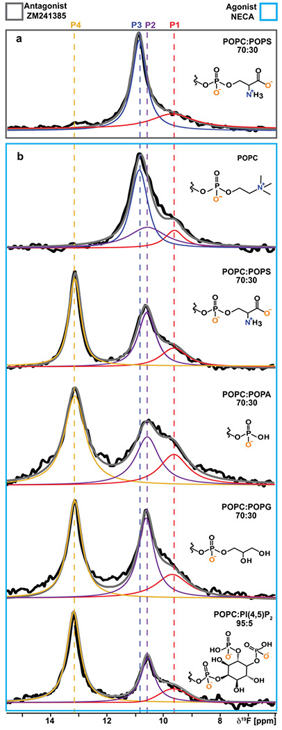
a A 1D 19F-NMR spectrum of the antagonist-A2AAR[A289CTET] complex in lipid nanodiscs containing a mixture of POPC and POPS lipids. b The 1D 19F NMR spectra of an agonist-A2AAR[A289CTET] complex in lipid nanodiscs containing either only POPC lipids or a mixture of POPC and anionic lipids, as indicated on the right of each spectrum. The chemical structures of the lipid headgroups are shown with charges expected at physiological pH.
Positively charged amino acids at the A2AAR intracellular surface of TM VI direct lipid-induced conformational changes
Our 19F data showed anionic lipids impacted the conformational ensemble of agonist-bound but not antagonist-bound A2AAR. We therefore hypothesized positively charged residues located near the intracellular surface that show significant differences in conformation between active and inactive states could play important roles mediating the impact of anionic lipids. We identified 4 positively charged residues predicted by the Orientation of Proteins in Membranes (OPM) database35 to be near the lipid-bilayer boundary: R1995.60, H2306.32, K2336.35 and R2917.56 (superscripts denote the Ballesteros-Weinstein nomenclature36) and in different conformations between agonist-bound and antagonist-bound A2AAR crystal structures (Fig. 4a). Though our experiments were carried out at neutral pH, H2306.32 has been proposed to be positively charged37 and was thus included in the current study. Positively charged amino acids are frequently found in position 6.32 in other GPCRs (see Discussion).
Fig. 4. Impact of positively charged amino acids near the intracellular signaling on NMR response to anionic lipids.
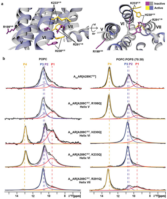
a Location of several charged amino acids near the lipid bilayer boundary. b 19F-NMR spectra of A2AAR[A289CTET] and several A2AAR variants in complex with the agonist NECA in lipid nanodiscs containing only POPC lipids (left column), and in complex with the agonist NECA and in lipid nanodiscs containing a mixture of POPC and POPS lipids at a 70:30 molar ratio (right column). Other figure presentation details the same as in Fig. 1, a and b.
We prepared A2AAR variants by individually replacing each of these four residues with the polar but neutral amino acid glutamine. Each of the resulting variants was folded and demonstrated closely similar pharmacological activity compared with native A2AAR (Supplementary Fig. 11). In thermal shift assays, the variants also showed an increase in melting temperature with increasing amounts of anionic lipids, similar to A2AAR[A289C] (Supplementary Fig. 11). For two of the variants, A2AAR[A289CTET,R199Q] and A2AAR[A289CTET,R291Q], 19F-NMR spectra for complexes with the agonist NECA were similar to the data of A2AAR[A298CTET] in both nanodiscs containing POPC or a mixture of 70/30 POPC/POPS (Fig. 4b). For A2AAR[A289CTET,R291Q], state P4 showed a higher relative intensity in nanodiscs containing 70/30 POPC/POPS but the same overall response to the presence of anionic lipids as A2AAR[A289CTET].
For the two variants with mutations in helix VI, A2AAR[A289CTET,H230Q] and A2AAR[A289CTET,K233Q], we observed a completely different response to the presence of anionic lipids and in striking contrast to A2AAR[A289CTET]. In the absence of anionic lipids, the 19F NMR spectra of these two variants in complex with the agonist NECA resembled the spectra of A2AAR[A289CTET] measured in the presence of anionic lipids (Fig. 4). 19F spectra of A2AAR[A289CTET,H230Q] and A2AAR[A289CTET,K233Q] in complex with NECA and in nanodiscs containing a mixture of POPC and POPS (70:30) appeared similar to spectra of inactive A2AAR[A298CTET] (Fig. 4). Intriguingly the individual mutations in Helix VI appeared to completely reverse the sensitivity of A2AAR to anionic lipids.
We tested signaling complex formation with A2AAR[A289CTET,K233Q] with mini Gas in nanodiscs containing POPC and POPS (70:30) . Upon addition of mini-Gαs, 19F-NMR spectra showed the presence of the state P4 (Supplementary Fig. 12), indicating complex formation can still occur for this variant. Cyclic AMP accumulation assays showed dose-dependent responses to agonists for the four A2AAR variants were similar to wild-type A2AAR (Supplementary Fig. 13), with A2AAR[A289CTET,R291Q] showing a measurably higher basal level of activity, consistent with the relatively higher signal intensity for peak P4 in 19F-NMR data (Fig. 4). Consideration of the 19F-NMR data and correlative functional experiments together indicates anionic lipids may not be strictly required for A2AAR activation but shift the conformational equilibria toward a fully active conformation even in the absence of a partner G protein.
Anionic lipids prepare A2AAR to recognize intracellular G proteins
To further investigate the molecular mechanism by which anionic lipids modulated A2AAR activation, a series of all-atom molecular dynamics (MD) simulations were implemented with several different A2AAR conformations and with different lipid compositions. We first noted H2306.32, K2336.35 and R2917.56 form a triad that coordinates engagement with mini-Gαs through interactions with glutamic acid in the end of the α5 helix of mini-Gαs at position 392 (Fig. 5). The difference between inactive and the fully-active G protein-coupled states is signaled by the distribution of distances between H2306.32 and R2917.56. For the A2AAR complex with mini-Gαs, the median of this distance distribution shifts toward longer distances and the distribution broadens (Fig. 5 and Supplementary Figs. 14 and 15). We hypothesized that negatively charged headgroups of anionic phospholipids mimic interactions of E392 in mini-Gαs, preorganizing the intracellular face for engaging with the G protein. To test this, we simulated the fully active receptor but without mini-Gαs. Simulations were initialized from the A2AAR ternary complex with the agonist NECA and mini-Gαs (PDB ID 5G53)33 and then deleting the mini-Gαs in-silico and allowing lipids to access the G protein-binding interface. Simulations were first performed with the backbone of the protein restrained to keep it close to the fully active receptor. In simulated membranes containing only POPC, the median H2306.32 and R2917.56 distance for agonist-bound A2AAR is similar to that observed for the A2AAR inactive state (Fig. 5) — a surprising result, given that the backbone of the protein is kept close to the fully active state by the restraints. In contrast, in the presence of POPS the median H2306.32 and R2917.56 distance shifts to longer distance, as a PS headgroup intercalates between H2306.32 and R2917.56 (Fig. 5 bottom right panel). If the restraints are then released and the simulation continued, the median H2306.32 and R2917.56 distance shifts to still longer distances (Fig. 5b, blue arrow), mimicking the Gαs protein-bound state. The interaction of the COO- group in PS headgroups with these residues mimics the interactions of the COO- group on E392, but only when the receptor is in the fully active state (Fig. 5).
Fig. 5. A2AAR-lipid interactions observed in molecular dynamics simulations.
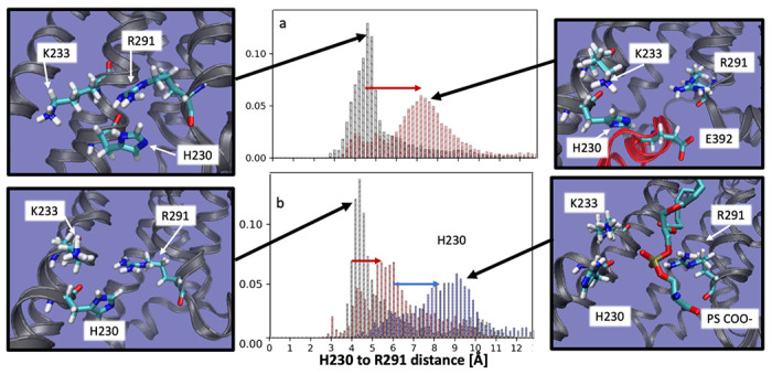
Distance distribution between H2306.32 and R2917.56 shifts to farther distance (red arrow, panel ‘a’) when the receptor switches from the inactive state (panel ‘a’, black) to the fully active, G protein-coupled state (panel ‘a’, red). When simulated in the fully active state but without the G-protein and no POPS (and with the backbone restrained to stay close to the fully active state), the H2306.32 to R2917.56 distance distribution (panel ‘b’, black) is similar to the inactive state, but the shift to longer distances is partially recapitulated (red arrow, panel ‘b’) when simulated in a membrane containing 85% phosphatidyl serine (panel ‘b’, red). Upon release of the backbone restraints the 85% PS simulation shifts to longer distances (blue arrow, panel ‘b’), with a distribution very similar to the fully active, G-protein coupled system. Inspection of the structures representative of the most likely distance for each case reveals that when the H2306.32 to R2917.56 distance is shorter, reflecting an inactive-like state, H2306.32, K2336.35 R2917.56 form a tight cluster (far left panels, top and bottom). When the distance is longer, reflecting an active-like state, this cluster is split, and coordinates a glutamic acid from the α5 helix of the G-protein (top right panel), or a PS headgroup in the absence of G-protein (lower right panel).
Discussion
Results presented in Figures 3 and 4 are intriguing in the context of literature data on the influence of phospholipids on GPCR activity. In earlier spectrophotometric studies of rhodopsin, anionic lipids shifted the equilibrium of intermediate activation states38. In studies of β2AR signaling, anionic lipids impacted the preference of the receptor to interact with Gαi over Gαs through lipid-G protein charge complementarity14. IC50 values for β2AR ligands varied among different lipid compositions in nanodiscs by a factor of ~3 for antagonists and ~7 for agonists11, though no clear relationship was observed between lipid headgroup type and measured IC50 values. In the present work, we observed a variation in KD values of the antagonist ZM241385 and agonist NECA among different lipid compositions by a factor of ~2 and ~3, respectively, also with no obvious correlation between lipid headgroup and determined KD value (Supplementary Fig. 5). Our data are more in line with earlier observations of the neurotensin 1 receptor, which reported similar binding affinities of the agonist neurotensin in nanodiscs containing either POPC or mixtures of POPC and POPG39. As all NMR samples employed a saturating amount of ligand, the relatively small differences among KD values do not explain the striking differences observed in 19F spectra observed between nanodisc preparations containing only zwitterionic lipids versus a mixture of zwitterionic and anionic lipids (Fig. 3). Thus, observed differences in our NMR data between different lipid compositions must be due to lipid-dependent changes in the A2AAR conformational equilibria.
Comparing spectra from Figures 1 and 2, anionic lipids appear to not be strictly required for A2AAR complex formation with mini-Gαs, rather they shift the conformational equilibria to favor a fully active A2AAR conformer. 19F-NMR data of agonist-bound A2AAR[A289C,K233Q] show this variant can still form signaling complexes even though an active state P4 is not observed in nanodiscs containing anionic lipids. These data are consistent with the cAMP functional data (Supplementary Fig. 13) and support a model whereby anionic lipids control the mechanism of A2AAR-G protein recognition (Fig. 6). In the absence of anionic lipids, A2AAR can form signaling complexes through an induced fit mechanism. In the presence of anionic lipids, a fully active receptor conformation is populated, leading to a conformational selection process directing complex formation (Fig. 6). Thus, anionic lipids appear to “prime” A2AAR for complex formation with partner proteins. This priming effect is reminiscent of earlier studies that proposed G proteins could synergistically prime a receptor for coupling with other G protein subtypes40.
Fig. 6. Visualization of the role of anionic lipids in complex formation of A2AAR with Gαs.
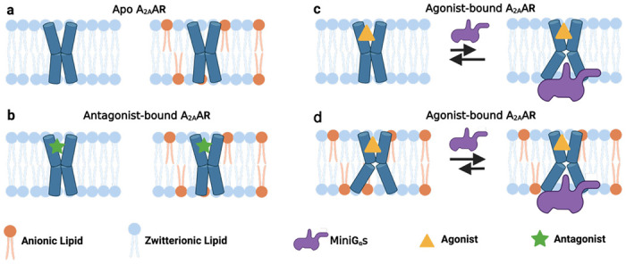
a and b Schematic side views of a apo and b antagonist-bound A2AAR in phospholipid membranes without and with anionic lipids, colored blue and orange, respectively. c and d Schematic view of agonist-bound A2AAR and mini-Gαs in membranes containing c only zwitterionic phospholipids and d zwitterionic and anionic phospholipids. The black arrows signify an equilibrium between mini-Gαs bound A2AAR and A2AAR bound to agonist alone, and the length of the arrows signifies how anionic lipids shift this equilibrium toward formation of the ternary complex. This figure was prepared with Biorender.com.
In earlier 19F-NMR studies22 and in the present study, the presence of state P4 is also observed for agonist-bound A2AAR in mixed micelles composed of DDM and CHS (Fig. 1). Since the pKA of the carboxylic acid group of CHS is ~5.8, at the pH value of 7 used for samples in the current study we anticipate the vast majority of CHS molecules should be negatively charged, consistent with earlier experiments with CHS41. We therefore hypothesize that the negative charge on CHS may mimic the impact observed on the conformational equilibria of A2AAR by the negative charges for anionic lipids.
Our data and proposed mechanism provide an opportunity to evaluate experimental results in the context of observations from computational modeling. MD simulations of A2AAR in complex with mini-Gαs indicated PIP2 strengthened interactions within the complex by “bridging” between basic residues of A2AAR and mini-Gαs27. 19F-NMR spectra of A2AAR in nanodiscs containing 5% PIP2 and in the absence of partner G protein show the presence of fully active A2AAR (Fig. 3), suggesting PIP2 may also enhance complex formation by populating a fully active receptor conformation prior to complex formation and play multiple roles in stabilizing the active complex. In MD simulations of β2AR38 and A2AAR37, active conformations of the receptors were proposed to be stabilized by increasing the receptor affinity for agonists and interactions with membrane-facing residues in TM6 and TM7. In contrast, experimentally we observed only relatively smaller differences in agonist binding among different lipid compositions, and we also propose that penetration of anionic lipids into the G protein binding site is important to stabilizing an active A2AAR conformation (Fig. 5).
Conservation of basic residues in helix VI among class A GPCRs and GPCRs from all classes suggest the present observations may extend to many additional receptors. Using structure-based alignment tools in GPCRdb42,43, we compared sequences of GPCRs among class A and among all classes. In class A receptors, one of these three residues is observed at position 6.32 or 6.35 with frequencies of 75.5% and 59.3%, respectively (Fig. 7 and Table 1). Expanding this search to include 1 position before or after these positions increases the frequency to 88.3% and 64.5%. In the opioid receptor family, position 6.32 is an arginine and has been noted to be critical for the activation of Gi signaling by the mu opioid receptor44. Conservation of a positively charged amino acid is also observed among the 402 non-olfactory GPCRs from all families. Notably, for class F GPCRs, either an arginine or lysine is always found in position 6.32 and mutations to a non-positively charged amino acid destroy G protein signaling45. In class B receptors, position 6.35 is typically a lysine and is noted to be critical for receptor activation46. Our study suggests that lipid-receptor interactions may play a critical role in regulating the drug response and activity of GPCRs across all receptor families.
Figure 7. Conservation of residues at the intracellular end of helix 6 among 290 class A GPCRs.

The size of each one-letter amino acid corresponds to the frequency of occurrence for that amino acid type. Numbers indicate the residue position in the Ballesteros-Weinstein nomenclature36. Sequences were obtained and aligned in the GPCRdb42,43. The figure was created using the online tool available at https://weblogo.berkeley.edu/logo.cgi.
Table 1.
Sequence conservation of positively charged amino acids on the intracellular ends of helices V, VI, and VII.
| Residue position | Amino acid type in A2AAR | Occurrence frequency of amino acid types K, R, or H (%)1 | |||
|---|---|---|---|---|---|
| 290 class A GPCRs | 290 class A GPCRs within ±1 residue2 | 402 non-olfactory GPCRs3 | 402 non-olfactory GPCRs ±1 residue 4 | ||
| 5.60 | R | 39.6% | 41.03% | 34.57% | 40.80% |
| 6.32 | H | 75.5% | 88.27% | 60.19% | 70.90% |
| 6.35 | K | 59.3% | 64.48% | 45.77% | 64.94% |
| 7.56 | R | 9.3% | 11.03% | 8.46% | 14.18% |
Frequency determined by sequence alignment and comparison in GPCRdb (see Methods). Numbers correspond to the frequency of finding a lysine, arginine, or histidine residue in the specified position.
Frequency of finding a lysine, arginine, or histidine residue in the specific position or within one residue before or after among non-olfactory class A GPCRs.
Frequency of finding a lysine, arginine, or histidine residue in the specific position among non-olfactory GPCRs from all classes.
Frequency of finding a lysine, arginine, or histidine residue in the specific position or within one residue before or after among non-olfactory GPCRs from all classes.
Methods
Molecular cloning.
The gene encoding human A2AAR (1-316) was cloned into a pPIC9K vector (Invitrogen) at the BamHI and NotI restriction sites. The gene contained a single amino acid replacement (N154Q) to remove the only glycosylation site in the receptor, an N-terminal FLAG tag, and a 10 X C-terminal His tag. We used PCR-based site-directed mutagenesis with the Accuprime Pfx SuperMix (ThermoFisher Scientific) to replace A2897.54 with cysteine, creating A2AAR[A289C]. This plasmid was used as a template for generating the additional A2AAR variants R2917.56Q, K2336.35Q, H2306.32Q and R1995.60Q via the same site directed mutagenesis approach using nucleic acid oligomers listed in Supplementary Table 2.
Small-scale protein expression optimization.
Plasmids containing A2AAR were introduced into a BG12 strain of Pichia pastoris (Biogrammatics) via electroporation. Clones exhibiting high protein expression were identified using small-scale protein production and screening approaches as previously reported21. Glycerol stocks of highly-expressing clones were stored at −80 °C for future use.
A2AAR expression, purification, and 19F-labeling via chemical modification.
All A2AAR variants were expressed in P. pastoris following previously reported protocols21. Briefly, 4 mL cultures in buffered minimal glycerol (BMGY) media were inoculated from glycerol stocks and allowed to grow at 30 °C for 48 h at 200 RPM. These cultures were used to inoculate 50 mL BMGY medium allowed to grow at 30 °C for 60 h with 200 RPM shaking. Each 50 mL culture was subsequently used to inoculate 500 mL BMGY medium and allowed to incubate at 30 °C for 48 h and 200 RPM. Cultures were then centrifuged at 3000 x g for 15 minutes, the supernatant discarded, and then resuspended in 500 mL of buffered minimal methanol (BMMY) medium without methanol. Cultures were allowed to grow for 6 h at 28 °C to remove any remaining glycerol. Protein expression was induced by the addition of methanol to a final concentration of 0.5% w/v. Two further aliquots of 0.5% w/v methanol were added to the cultures at 12 h intervals after induction for a total expression time of 36 h. Cells were harvested by centrifugation at 3000 x g for 15 minutes. Cell pellets were frozen in liquid nitrogen and stored at −80 °C until needed. Cell pellets were resuspended and lysed in lysis buffer (50 mM sodium phosphate pH 7.0, 100 mM NaCl, 5% glycerol (w/v), and in-house prepared protease inhibitor cocktail solution). Cell membranes containing A2AAR were isolated and collected by ultracentrifugation at 200,000 x g for 30 minutes, frozen in liquid nitrogen, and stored at − 80 °C for future use.
19F-TET was introduced to A2AAR variants using the in-membrane chemical modification (IMCM) method as previously reported32. In brief, isolated membrane pellets were resuspended in buffer (10 mM HEPES pH 7.0, 10 mM KCl, 20 mM MgCl2, 1 M NaCl, 4 mM theophylline) and incubated with 1 mM of 4,4’-dithiodipyridine (aldrithiol-4) and protease inhibitor cocktail solution (prepared in-house) for 1 h at 4 °C. The suspension was pelleted using ultracentrifugation, and excess aldrithiol was washed off using the same buffer. Membranes were resuspended and incubated with 1 mM of 2,2,2-trifluoroethaethiol (TET) for 1 h at 4°C. The suspension was pelleted using ultracentrifugation and the resulting pellet washed with buffer. Membranes were resuspended in the same buffer and incubated with 1 mM theophylline and protease inhibitor cocktail solution (prepared in-house) for 30 min at 4 °C. The membrane suspension was mixed 1:1 with a solubilizing buffer (50 mM HEPES pH 7.0, 500 mM NaCl, 0.5% (w/v) n-Dodecyl-β-D-Maltopyranoside (DDM), and 0.05% cholesteryl hemisuccinate (CHS)) for 6 h at 4 °C. Insolubilized material was separated by ultracentrifugation at 200,000 x g for 30 min, and the supernatant was incubated overnight with Co2+-charged affinity resin (Talon, Clontech) and 30 mM imidazole at 4 °C.
After overnight incubation, the Co2+-resin was washed with 20 column volumes (CV) of wash buffer 1 (50 mM HEPES pH 7.0, 500 mM NaCl, 10 mM MgCl2, 30 mM imidazole, 8 mM ATP, 0.05% DDM, and 0.005% CHS), and washed 2 subsequent times with 20 CV of wash buffer 2 (25 mM HEPES pH 7.0, 250 mM NaCl, 5% glycerol, 30 mM imidazole, 0.05% DDM, 0.005% CHS, and an excess of ligand). A2AAR was eluted with buffer 3 (50 mM HEPES pH 7.0, 250 mM NaCl, 5% glycerol, 300 mM imidazole, 0.05% DDM, 0.005% CHS, and ligand). The eluted protein was exchanged into buffer (25 mM HEPES pH 7.0, 75 mM NaCl, 0.05% DDM, 0.005% CHS, 100 μM trifluoroacetic acid (TFA), and ligand) using a PD-10 desalting column (Cytiva) and stored at 4 °C for nanodisc assembly. All buffers prepared with ligands used a saturating concentration of the required ligand. Apo A2AAR was prepared without ligand added in the purification buffers.
Nanodisc assembly.
Assembly of lipid nanodiscs containing A2AAR followed protocols from previous studies47–49 optimized to facilitate samples prepared with a wider range of lipid compositions. The scaffold protein MSP1D1 was expressed and purified similarly as described in previous studies47,48. 100 mM stock solutions of all lipids were prepared in a cholate buffer (25 mM Tris-HCl, pH 8.0, 150 mM NaCl, and 200 mM sodium cholate). To initiate nanodisc assembly, 27 μM of purified A2AAR in DDM/CHS micelles was mixed with purified MSP1D1 and detergent-solubilized lipids in a molar ratio of 1:5:250, respectively. The mixture was incubated for 1-2 h at 4 °C and incubated overnight with pre-washed bio-beads (Bio-Rad Laboratories) at 4 °C. Following the overnight incubation, the bio-beads were removed, and the resulting mixture was incubated with Ni-NTA resin (GoldBio) for 24 h at 4 °C. The Ni-resin was collected after 24 h and washed with 2 CV of a wash buffer (50 mM HEPES, pH 7.0, 150 mM NaCl, and 10 mM imidazole). Nanodiscs containing A2AAR were eluted with buffer (50 mM HEPES, pH 7.0, 150 mM NaCl, 300 mM imidazole and ligand) and exchanged into a final buffer used for all experiments (25 mM HEPES pH 7.0, 75 mM NaCl, 100 μM TFA and ligand) using a PD-10 desalting column (Cytiva). All ligand-containing buffers were prepared with a saturating concentration of ligand.
Mini-Gαs expression and purification.
The protocol for mini-Gαs expression and purification was adapted from previously described studies33,34. The purified protein was exchanged into storage buffer (10 mM HEPES pH 7.5, 100 mM NaCl, 10% v/v glycerol, 1 mM MgCl2, 1 μM GDP, 0.1 mM TCEP), concentrated to 1 mM, aliquoted, frozen in liquid nitrogen and stored at −80 °C for future use.
A2AAR-Mini-Gαs Complex Formation.
Assembly of A2AAR ternary complexes with agonist and mini-Gαs followed a protocol adapted from previous studies33,34. Nanodiscs containing A2AAR were mixed with a 2-fold molar excess of mini-Gαs, 1 mM MgCl2 and apyrase (0.1 U), and the mixture was incubated at 4 °C overnight. The samples were purified by SEC using a Superdex 200 Increase 10/300 GL column (Cytiva) pre-equilibrated with SEC buffer (25 mM HEPES pH 7.0, 75 mM NaCl, 5 mM MgCl2). Peak fractions, containing the A2AAR–mini-Gαs complex, were pooled and concentrated to 180-200 μM for NMR experiments.
NMR spectroscopy.
Nanodisc samples were concentrated to ~200 μM in 280 μL in a Vivaspin-6 concentrator with a 30 kDa MWCO (Sartorius). 20 μL D2O was added and gently mixed into the sample. 19F-NMR and 31P-NMR experiments were measured on a Bruker Avance III HD spectrometer operating at 600 MHz 1H nutation frequency using Topspin 3.6.2 and equipped with a Bruker 5-mm BBFO probe. To make direct comparisons with previously published 19F-NMR data50, 19F-NMR spectra were measured at 280 K. 31P-NMR experiments were measured at 300 K to obtain improved spectral resolution. Temperatures were calibrated from a standard sample of 4% methanol in D4-MeOH.
1-dimensional 19F data were recorded with a data size of 32k complex points, an acquisition period of 360 ms, 16k scans, 120 μs dwell time, and 0.3 s recycle delay for a total experimental time of about 3 hours per experiment. All 31P NMR experiments were acquired with an acquisition time of 900 ms, 2k scans, and 0.3 s recycle delay for a total experiment time of 42 min per experiment.
2-dimensional [19F,19F]-EXSY experiments were recorded with a data size of 120 and 8192 complex points in the indirect and direct dimensions, respectively. We recorded 256 scans for each experiment with 100 ms of mixing time.
Radioligand binding assays.
Competition binding assays with nanodiscs containing A2AAR were recorded as previously described28. Ligand binding was measured with 0.125-0.25 μg nanodiscs containing A2AAR per sample incubated in buffer containing 25 mM HEPES pH 7.0, 75 mM NaCl, [3H]ZM241385 (American radiolabeled chemicals) and increasing amounts of ZM241385 or NECA for 60 min at 25 °C. The binding reaction was terminated by filtration with a Microbeta filtermat-96 cell harvester (PerkinElmer). Radioactivity was counted using a MicroBeta2 microplate counter (PerkinElmer). ZM241385 and NECA binding affinities (KD or KI) were determined using competition binding experiments. Specific binding of A2AAR and A2AAR variants were determined as the difference in binding obtained in the absence and presence of 10 μM ZM241385. Radioligand experiments were conducted in triplicate and IC50 values determined using a nonlinear, least-square regression analysis (Prism 8; GraphPad Software, Inc.). The IC50 values were converted to KI values using the Cheng-Prusoff equation51. Error bars for each sample were calculated as the standard error of mean (s.e.m) for n=3 independent experiments.
Fluorescent thermal shift experiments.
Fluorescent thermal shift experiments followed a protocol adapted from earlier publications30,52. For each sample, 10 μg of nanodisc sample was incubated in buffer (50 mM HEPES pH 7.0, 150 mM NaCl) containing N-[4-(7-diethylamino-4-methyl-3-coumarinyl)phenyl]maleimide (CPM; Invitrogen) at a final concentration of 10 μM and incubated for 30 min in the dark on ice. Fluorescence thermal shift experiments were carried out with a Cary Eclipse spectrofluorometer using quartz cuvettes (Starna Cells, Inc.) over a linear temperature range from 20 °C to 90 °C at a constant heating rate of 2 °C/min. The excitation and emission wavelengths were 387 nm and 463 nm, respectively. Fluorescence thermal shift data were analyzed in Origin (OriginLab Corporation). The raw data were fit to a Boltzmann sigmoidal curve to determine the melting temperature (Tm). Error bars for each sample were calculated as the standard error of mean (s.e.m) for n≥3 independent experiments.
NMR data processing and analysis.
All NMR data were processed and analyzed in Topspin 4.0.8 (Bruker Biospin). All 1-dimensional 19F-NMR data were processed identically. Prior to Fourier transformation, the spectra were zero-filled to 64k points and multiplied by an exponential window function with 40 Hz line broadening. 19F spectra were referenced to the TFA signal at −75.8 ppm, which was set to 0 ppm. Deconvolution of the 19F-NMR data followed previously published procedures50 and was done with MestreNova version 14.1.1-24571 (MestreLab Research S.L). The 19F-NMR spectra were fit with a double- or triple-Lorentzian function. The quality of the fits was assessed from the residual difference between the experimental data and the sum of the computed components.
All 31P NMR data were processed identically. Prior to Fourier transformation, 31P spectra were zero-filled to 64k points and multiplied by an exponential window function with 50 Hz line broadening.
The 2-dimensional [19F, 19F] EXSY experiments were processed by zero-filling to 1k (t1) * 2k (t2) points and 100 Hz of exponential line broadening was applied prior to Fourier transformation.
Computational Simulations.
Three different A2AAR conformations were used as starting configurations: the complex with the antagonist ZM241385 (PDB ID: 3EML)53, the ternary complex with the full agonist NECA and an engineered mini-G protein (PDB ID: 5G53)33, and a conformation of the same ternary complex with the mini-G protein deleted. For the latter simulation, production simulations were run with the backbone heavy atoms softly restrained to stay close to the fully active state by harmonic restraints with a 0.5 kJ/mol/Å2 spring constant. The T4-lysozyme was removed from the inactive structure and initial coordinates for residues 209-219 were obtained from the structure of a thermostabilized A2AAR variant mutant (PDB: 3PWH)54 for which those residues are resolved. The termini were capped with methylamine (N-terminus) and acetyl (C-terminus) groups.
Each system was prepped using the CHARMMGUI membrane builder tools55. The protein was embedded in a mixture of POPS and POPC (see Supplementary Table 3 for simulated systems), solvated with TIP3P water56, neutralized with Na+ ions if needed, and NaCl added to bring the ionic strength to 150 mM. Lipids and protein were modeled with the CHARMM36 force field57,58; ligands were modeled with the CHARMM general force field59. Final system sizes were at least 15 nm in each dimension, yielding lipid-to-protein ratios of ~750:1.
Each system was prepared individually for production simulation through a series of 6 minimization and heating steps: (i) steepest descent to minimize the initial configuration; (ii) 125,000 steps of leapfrog dynamics with a 1 fsec timestep and velocities reassigned every 500 steps; (iii) 125,000 steps of leapfrog dynamics with a 1 fsec timestep, pressure controlled by the Parinello-Rahman barostat60 and velocities reassigned every 500 steps, then a total of 750,000 steps of leapfrog dynamics with a 2 fsec timestep and hydrogen positions constrained by LINCS61, pressure controlled by the Parinello-Rahman barostat, and velocities reassigned every 500 steps. During equilibration, double bonds were restrained in the cis configuration to prevent isomerization; these restraints are gradually reduced during the final three stages of the equilibration protocol. Production simulations (NPT ensemble) were integrated with leapfrog using the Parinello-Rahman barostat to control pressure (time constant 5 psec; compressibility 4.5e−5 bar−1; coupled anisotropically to allow independent fluctuation of the in-plane and normal directions) and temperature controlled using Nose-Hoover62 (time constant 1 psec) at a temperature of 25 °C. Hydrogens were constrained with LINCS (expansion order 4), a 2 fsec timestep was used, short range electrostatics were computed directly within 1.2 nm, and long-range electrostatics were computed every timestep using particle mesh Ewald63 with a grid spacing of 1 Å and cubic interpolation. Long range dispersion was smoothly truncated over 10-12 nm using a force-switch cutoff scheme. Residue-residue distances as reported in Fig. 5 were measured between the closest sidechain nitrogen atoms in H2306.32 and R2917.56.
cAMP Accumulation Assays.
Plasmids encoding wild-type or variant human A2A adenosine receptors were transfected into CHO cells using lipofectamine 2000. 24 hours after transfection, cells were detached and grown in 96-well plates in medium containing equal volume of DMEM and F12 supplemented with 10% fetal bovine serum, 100 Units/ml penicillin, 100 μg/ml streptomycin, and 2 μmol/ml glutamine. After growing for 24 hours, culture medium was removed and cells were washed twice with PBS. Cells were then treated with assay buffer containing rolipram (10 μM) and adenosine deaminase (3 units/ml) for 30 min followed by addition of agonist and incubated for 20 min. The reaction was terminated upon removal of the supernatant, and addition of 100 μl Tween-20 (0.3%). Intracellular cAMP levels were measured with an ALPHAScreen cAMP assay kit (PerkinElmer) following the manufacture’s protocol.
Supplementary Material
Acknowledgements
This work is supported by the National Institutes of Health, NIGMS MIRA grant R35GM138291 (M.T.E., N.T., A.R. and N.G.P) and by the University of Florida College of Liberal Arts and Sciences. A portion of this work was supported by the McKnight Brain Institute at the National High Magnetic Field Laboratory’s AMRIS Facility, which is funded by National Science Foundation Cooperative Agreement No. DMR-1644779 and the State of Florida. The authors also acknowledge support from the NIH/NIDDK Intramural Research Program (ZIA DK031117). LS and EL were supported by NIH award RO1GM120351. This work used the Extreme Science and Engineering Discovery Environment (XSEDE), which is supported by National Science Foundation grant number ACI-1548562.
Footnotes
Code Availability
Code used in this study is available from the corresponding author upon reasonable request.
Data Availability
Data presented in this paper are available from the corresponding author upon reasonable request.
References
- 1.Phillips R., Ursell T., Wiggins P. & Sens P. Emerging roles for lipids in shaping membrane-protein function. Nature 459, 379–385 (2009). [DOI] [PMC free article] [PubMed] [Google Scholar]
- 2.Sprong H., van der Sluijs P. & van Meer G. How proteins move lipids and lipids move proteins. Nat. Rev. Mol. Cell Biol. 2, 504–513 (2001). [DOI] [PubMed] [Google Scholar]
- 3.Palsdottir H. & Hunte C. Lipids in membrane protein structures. Biochim. Biophys. Acta Biomembr. 1666, 2–18 (2004). [DOI] [PubMed] [Google Scholar]
- 4.Agasid M.T. & Robinson C.V. Probing membrane protein–lipid interactions. Curr. Opin. Struct. Biol. 69, 78–85 (2021). [DOI] [PubMed] [Google Scholar]
- 5.Laganowsky A. et al. Membrane proteins bind lipids selectively to modulate their structure and function. Nature 510, 172–175 (2014). [DOI] [PMC free article] [PubMed] [Google Scholar]
- 6.Corradi V. et al. Emerging diversity in lipid–protein interactions. Chem. Rev. 119, 5775–5848 (2019). [DOI] [PMC free article] [PubMed] [Google Scholar]
- 7.Harayama T. & Riezman H. Understanding the diversity of membrane lipid composition. Nat. Rev. Mol. Cell Biol. 19, 281–296 (2018). [DOI] [PubMed] [Google Scholar]
- 8.Rosholm K.R. et al. Membrane curvature regulates ligand-specific membrane sorting of GPCRs in living cells. Nat. Chem. Biol. 13, 724–729 (2017). [DOI] [PubMed] [Google Scholar]
- 9.Byrne E.F.X. et al. Structural basis of Smoothened regulation by its extracellular domains. Nature 535, 517–522 (2016). [DOI] [PMC free article] [PubMed] [Google Scholar]
- 10.Luchetti G. et al. Cholesterol activates the G-protein coupled receptor Smoothened to promote Hedgehog signaling. eLife 5, e20304 (2016). [DOI] [PMC free article] [PubMed] [Google Scholar]
- 11.Dawaliby R. et al. Allosteric regulation of G protein–coupled receptor activity by phospholipids. Nat. Chem. Biol. 12, 35–39 (2015). [DOI] [PMC free article] [PubMed] [Google Scholar]
- 12.Huang S.K. et al. Allosteric modulation of the adenosine A2A receptor by cholesterol. eLife 11, e73901 (2022). [DOI] [PMC free article] [PubMed] [Google Scholar]
- 13.Manna M. et al. Mechanism of allosteric regulation of β2-adrenergic receptor by cholesterol. eLife 5, e18432 (2016). [DOI] [PMC free article] [PubMed] [Google Scholar]
- 14.Strohman M.J. et al. Local membrane charge regulates β2 adrenergic receptor coupling to Gi3. Nat. Comm. 10, 2234 (2019). [DOI] [PMC free article] [PubMed] [Google Scholar]
- 15.Kimura T. et al. Recombinant cannabinoid type 2 receptor in liposome model activates g protein in response to anionic lipid constituents. J. Biol. Chem. 287, 4076–4087 (2012). [DOI] [PMC free article] [PubMed] [Google Scholar]
- 16.Yen H.-Y. et al. PtdIns(4,5)P2 stabilizes active states of GPCRs and enhances selectivity of G-protein coupling. Nature 559, 423–427 (2018). [DOI] [PMC free article] [PubMed] [Google Scholar]
- 17.Xu P. et al. Structural insights into the lipid and ligand regulation of serotonin receptors. Nature 592, 469–473 (2021). [DOI] [PubMed] [Google Scholar]
- 18.Shimada I., Ueda T., Kofuku Y., Eddy M.T. & Wüthrich K. GPCR drug discovery: integrating solution NMR data with crystal and cryo-EM structures. Nat. Rev. Drug Disc. 18, 59–82 (2019). [DOI] [PMC free article] [PubMed] [Google Scholar]
- 19.Bostock M.J., Solt A.S. & Nietlispach D. The role of NMR spectroscopy in mapping the conformational landscape of GPCRs. Curr. Opin. Struct. Biol. 57, 145–156 (2019). [DOI] [PubMed] [Google Scholar]
- 20.Mizumura T. et al. Activation of adenosine A2A receptor by lipids from docosahexaenoic acid revealed by NMR. Science Advances 6, eaay8544 (2020). [DOI] [PMC free article] [PubMed] [Google Scholar]
- 21.Eddy M.T. et al. Allosteric coupling of drug binding and intracellular signaling in the A2A adenosine receptor. Cell 172, 68–80 (2018). [DOI] [PMC free article] [PubMed] [Google Scholar]
- 22.Sušac L., Eddy M.T., Didenko T., Stevens R.C. & Wüthrich K. A2A adenosine receptor functional states characterized by 19F-NMR. Proc. Natl. Acad. Sci. 115, 12733–12738 (2018). [DOI] [PMC free article] [PubMed] [Google Scholar]
- 23.Eddy M.T. et al. Extrinsic tryptophans as NMR probes of allosteric coupling in membrane proteins: application to the A2A adenosine receptor. J. Am. Chem. Soc. 140, 8228–8235 (2018). [DOI] [PMC free article] [PubMed] [Google Scholar]
- 24.Huang S.K. et al. Delineating the conformational landscape of the adenosine A2A receptor during G protein coupling. Cell 184, 1884–1894 (2021). [DOI] [PMC free article] [PubMed] [Google Scholar]
- 25.Ye L., Van Eps N., Zimmer M., Ernst O.P. & Scott Prosser R. Activation of the A2A adenosine G-protein-coupled receptor by conformational selection. Nature 533, 265–268 (2016). [DOI] [PubMed] [Google Scholar]
- 26.Eddy M.T., Martin B.T. & Wüthrich K. A2A adenosine receptor partial agonism related to structural rearrangements in an activation microswitch. Structure 29, 170–176 (2021). [DOI] [PMC free article] [PubMed] [Google Scholar]
- 27.Song W., Yen H.-Y., Robinson C.V. & Sansom M.S.P. State-dependent lipid interactions with the A2a receptor revealed by MD simulations using in vivo-mimetic membranes. Structure 27, 392–403 (2019). [DOI] [PMC free article] [PubMed] [Google Scholar]
- 28.Wei S. et al. Slow conformational dynamics of the human A2A adenosine receptor are temporally ordered. Structure 30, 329–337 (2022). [DOI] [PMC free article] [PubMed] [Google Scholar]
- 29.Denisov I.G., Grinkova Y.V., Lazarides A.A. & Sligar S.G. Directed self-assembly of monodisperse phospholipid bilayer nanodiscs with controlled size. J. Am. Chem. Soc. 126, 3477–3487 (2004). [DOI] [PubMed] [Google Scholar]
- 30.Alexandrov A.I., Mileni M., Chien E.Y.T., Hanson M.A. & Stevens R.C. Microscale fluorescent thermal stability assay for membrane proteins. Structure 16, 351–359 (2008). [DOI] [PubMed] [Google Scholar]
- 31.Wadsäter M., Maric S., Simonsen J.B., Mortensen K. & Cardenas M. The effect of using binary mixtures of zwitterionic and charged lipids on nanodisc formation and stability. Soft Matter 9, 2329 (2013). [Google Scholar]
- 32.Sušac L., O’Connor C., Stevens R.C. & Wüthrich K. In-membrane chemical modification (IMCM) for site-specific chromophore labeling of GPCRs. Angew. Chem. 127, 15461–15464 (2015). [DOI] [PMC free article] [PubMed] [Google Scholar]
- 33.Carpenter B., Nehmé R., Warne T., Leslie A.G.W. & Tate C.G. Structure of the adenosine A2A receptor bound to an engineered G protein. Nature 536, 104–107 (2016). [DOI] [PMC free article] [PubMed] [Google Scholar]
- 34.Carpenter B. & Tate C.G. Engineering a minimal G protein to facilitate crystallisation of G protein-coupled receptors in their active conformation. Protein Eng. Des. Sel. 29, 583–594 (2016). [DOI] [PMC free article] [PubMed] [Google Scholar]
- 35.Lomize M.A., Lomize A.L., Pogozheva I.D. & Mosberg H.I. OPM: Orientations of Proteins in Membranes database. Bioinformatics 22, 623–625 (2006). [DOI] [PubMed] [Google Scholar]
- 36.Ballesteros J.A. & Weinstein H. Integrated methods for the construction of three-dimensional models and computational probing of structure-function relations in G protein-coupled receptors. in Methods in neurosciences, Vol. 25 366–428 (Elsevier, 1995). [Google Scholar]
- 37.Bruzzese A., Dalton J.A.R. & Giraldo J. Insights into adenosine A2A receptor activation through cooperative modulation of agonist and allosteric lipid interactions. PLoS Comput. Biol. 16, e1007818 (2020). [DOI] [PMC free article] [PubMed] [Google Scholar]
- 38.Gibson N.J. & Brown M.F. Role of phosphatidylserine in the MI-MII equilibrium of rhodopsin. Biochem. Biophys. Res. Commun. 176, 915–921 (1991). [DOI] [PubMed] [Google Scholar]
- 39.Inagaki S. et al. Modulation of the Interaction between Neurotensin Receptor NTS1 and Gq Protein by Lipid. J. Mol. Biol. 417, 95–111 (2012). [DOI] [PMC free article] [PubMed] [Google Scholar]
- 40.Gupte T.M., Malik R.U., Sommese R.F., Ritt M. & Sivaramakrishnan S. Priming GPCR signaling through the synergistic effect of two G proteins. Proc. Natl. Acad. Sci. 114, 3756–3761 (2017). [DOI] [PMC free article] [PubMed] [Google Scholar]
- 41.Augustyn B. et al. Cholesteryl Hemisuccinate Is Not a Good Replacement for Cholesterol in Lipid Nanodiscs. J. Phys. Chem. B, 7 (2019). [DOI] [PubMed] [Google Scholar]
- 42.Kooistra A.J. et al. GPCRdb in 2021: integrating GPCR sequence, structure and function. Nucleic Acids Res. 49, 335–343 (2021). [DOI] [PMC free article] [PubMed] [Google Scholar]
- 43.Isberg V. et al. GPCRdb: an information system for G protein-coupled receptors. Nucleic Acids Res. 44, 356–364 (2016). [DOI] [PMC free article] [PubMed] [Google Scholar]
- 44.Koehl A. et al. Structure of the μ-opioid receptor–Gi protein complex. Nature 558, 547–552 (2018). [DOI] [PMC free article] [PubMed] [Google Scholar]
- 45.Wright S.C. et al. A conserved molecular switch in Class F receptors regulates receptor activation and pathway selection. Nat. Comm. 10, 1–12 (2019). [DOI] [PMC free article] [PubMed] [Google Scholar]
- 46.Wootten D. et al. Key interactions by conserved polar amino acids located at the transmembrane helical boundaries in Class B GPCRs modulate activation, effector specificity and biased signalling in the glucagon-like peptide-1 receptor. Biochem. Pharmacol. 118, 68–87 (2016). [DOI] [PMC free article] [PubMed] [Google Scholar]
- 47.Hagn F., Etzkorn M., Raschle T. & Wagner G. Optimized phospholipid bilayer nanodiscs facilitate high-resolution structure determination of membrane proteins. J. Am. Chem. Soc. 135, 1919–1925 (2013). [DOI] [PMC free article] [PubMed] [Google Scholar]
- 48.Bayburt T.H., Grinkova Y.V. & Sligar S.G. Self-assembly of discoidal phospholipid bilayer nanoparticles with membrane scaffold proteins. Nano Lett. 2, 853–856 (2002). [Google Scholar]
- 49.Thakur N., Wei S., Ray A.P., Lamichhane R. & Eddy M.T. Production of human A2AAR in lipid nanodiscs for 19F-NMR and single-molecule fluorescence spectroscopy. STAR Protocols 3, 101535 (2022). [DOI] [PMC free article] [PubMed] [Google Scholar]
- 50.Sušac L., Eddy M.T., Didenko T., Stevens R.C. & Wüthrich K. A2A adenosine receptor functional states characterized by 19 F-NMR. Proceedings of the National Academy of Sciences 115, 12733–12738 (2018). [DOI] [PMC free article] [PubMed] [Google Scholar]
- 51.Cheng Y.-C. & Prusoff W.H. Relationship Between the Inhibition Constant and the Concentration of Inhibitor which Causes 50 percent Inhibition (I50) of an Enzymatic Reaction. Biochem. Pharmacol. 22, 3099–3108 (1973). [DOI] [PubMed] [Google Scholar]
- 52.White K.L. et al. Structural connection between activation microswitch and allosteric sodium site in GPCR signaling. Structure 26, 259–269 (2018). [DOI] [PMC free article] [PubMed] [Google Scholar]
- 53.Jaakola V.-P. et al. The 2.6 angstrom crystal structure of a human A2A adenosine receptor bound to an antagonist. Science 322, 1211–1217 (2008). [DOI] [PMC free article] [PubMed] [Google Scholar]
- 54.Doré Andrew S. et al. Structure of the Adenosine A2A Receptor in Complex with ZM241385 and the Xanthines XAC and Caffeine. Structure 19, 1283–1293 (2011). [DOI] [PMC free article] [PubMed] [Google Scholar]
- 55.Lee J. et al. CHARMM-GUI Membrane Builder for complex biological membrane simulations with glycolipids and lipoglycans. J. Chem. Theory Comput. 15, 775–786 (2019). [DOI] [PubMed] [Google Scholar]
- 56.Jorgensen W.L., Chandrasekhar J., Madura J.D., Impey R.W. & Klein M.L. Comparison of simple potential functions for simulating liquid water. J. Chem. Phys. 79, 926–935 (1983). [Google Scholar]
- 57.Klauda J.B. et al. Update of the CHARMM all-atom additive force field for lipids: validation on six lipid types. J. Phys. Chem. B 114, 7830–7843 (2010). [DOI] [PMC free article] [PubMed] [Google Scholar]
- 58.Huang J. et al. CHARMM36m: an improved force field for folded and intrinsically disordered proteins. Nat. Methods 14, 71–73 (2017). [DOI] [PMC free article] [PubMed] [Google Scholar]
- 59.Vanommeslaeghe K. et al. CHARMM general force field: A force field for drug-like molecules compatible with the CHARMM all-atom additive biological force fields. J. Comput. Chem. 31, 671–690 (2009). [DOI] [PMC free article] [PubMed] [Google Scholar]
- 60.Parrinello M. & Rahman A. Polymorphic transitions in single crystals: A new molecular dynamics method. J. Appl. Phys. 52, 7182–7190 (1981). [Google Scholar]
- 61.Hess B., Bekker H., Berendsen H.J.C. & Fraaije J.G.E.M. LINCS: A linear constraint solver for molecular simulations. J. Comput. Chem. 18, 1463–1472 (1997). [Google Scholar]
- 62.Hoover W.G. Canonical dynamics: Equilibrium phase-space distributions. Phys. Rev. A 31, 1695–1697 (1985). [DOI] [PubMed] [Google Scholar]
- 63.Darden T., York D. & Pedersen L. Particle mesh Ewald: An N ·log( N ) method for Ewald sums in large systems. J. Chem. Phys. 98, 10089–10092 (1993). [Google Scholar]
Associated Data
This section collects any data citations, data availability statements, or supplementary materials included in this article.
Supplementary Materials
Data Availability Statement
Data presented in this paper are available from the corresponding author upon reasonable request.


