Abstract
Tibial tuberosity fractures are uncommon in adults. Surgery for these types of fractures is performed similarly to that of tibial tuberosity avulsion fractures in adolescents. The most commonly introduced method is to fix the displaced bone fragments using screws or wires and, if necessary, use tension band wiring for augmentation. However, if the bone fragments are too small or severely comminuted, it may be challenging to fix them using the conventional method. In this study, we introduced a fixation method using two knotless suture anchors that could be attempted in such cases. Since this surgical method fixes the bone fragments without direct damage to the bone fragments, it can be used even when the fragments are small or comminuted. This technique achieved a nearly full active range of knee motion without an extension lag at four weeks postoperatively. In addition, there were no complications related to surgery, and a complete bone union was achieved without additional dislocation. Therefore, this surgical method may be a good alternative if a fixation of the fracture is considered problematic by the conventional method.
Keywords: Tibial tuberosity, Fractures, Suture anchors
Highlights
Tibial tuberosity fractures in adults often result from direct trauma and may be difficult to treat with conventional surgical methods.
The authors describe a novel technique, a fixation method using 2 knotless suture anchors, for the treatment of tibial tuberosity fractures.
This surgical method may minimize damage to fractured bone fragments, so it may be applied to fractures with severe comminution or small fragments. Additionally, it is expected to reduce the complications after surgery as it can eliminate the irritation caused by the screws.
Introduction
Tibial tuberosity fractures (TTF) are rare fractures and have been reported to account for 0.4% to 2.7% of all epiphyseal injuries in adolescents and less than 1% of all physeal injuries.1,2 Most of these fractures are avulsion fractures caused by the strong contraction of the quadriceps muscle during jumping or landing.1,3,4 Due to this fracture mechanism, TTF occur more frequently in adolescents when the muscles and tendons are stronger than the growth plates.2 Therefore, TTF in adults are rare, and only a few cases have been reported, including cases in which fractures were caused by direct trauma.1,5,6
The most commonly reported surgical method for TTF was fixation using screws with tension band wiring (TBW) if necessary.1,5,7 Although this widely used surgery has shown good outcomes, it may be difficult to apply if the tibial tuberosity fragments are too small or severely comminuted. Therefore, we suggest a novel technique, a fixation method using 2 knotless suture anchors, as a treatment for TTF. The purpose of this study was to introduce our surgical techniques and postoperative results. Informed consent was obtained from the patient for publication of this case report.
Case Presentation
Clinical case
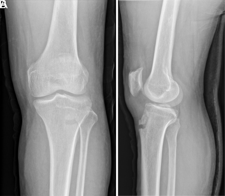
A 67-year-old man visited our emergency room with left knee pain after hitting his left knee on the edge of the stairs. A physical examination showed that active knee extension was impossible. A fracture of the tibial tuberosity was confirmed in the plain radiograph. Bone fragments with size of about 17 mm were displaced proximally 7 mm (Figure 1). The patient’s underlying diseases included osteoporosis.
Figure 1. A, B.
An isolated tibial tuberosity fracture without rotation of the fractured bone fragment was observed on preoperative x-rays (A-B).
Operative technique
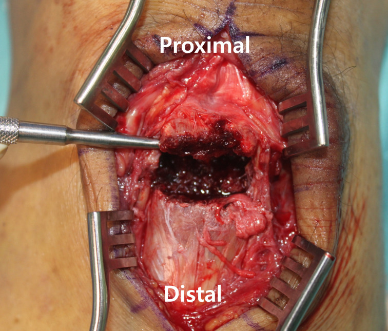
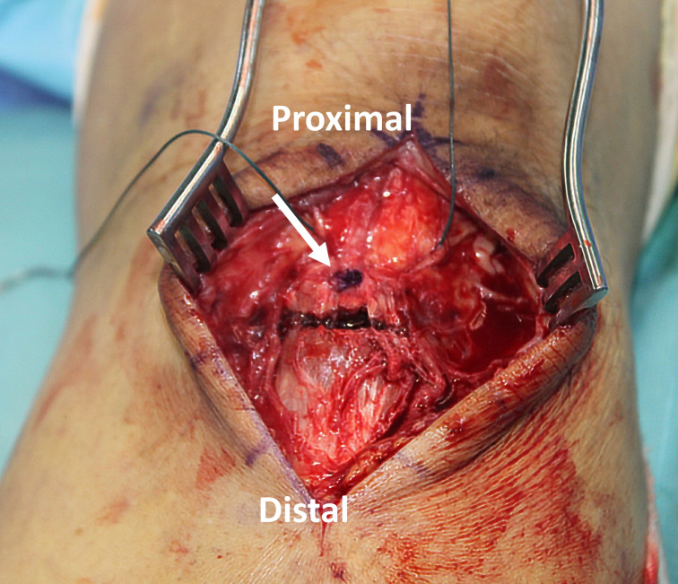
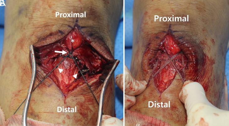
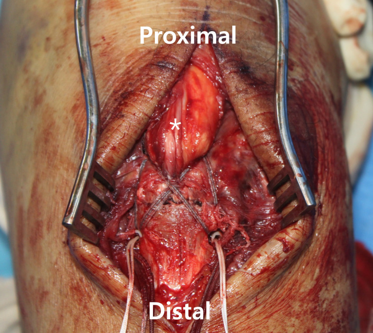
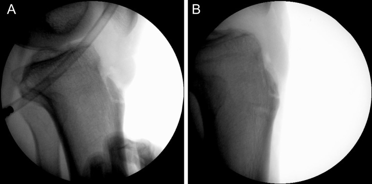
Under general anesthesia, the patient was placed in a supine position. A tourniquet was applied to the upper thigh. A 10-cm-long longitudinal midline incision of the knee was made, centered over the fracture site. When the fracture site was fully exposed, check for the bone fragments to be reduced (Figure 2). Tissues and hematomas impinged between the bone fragments were removed. Next, the part considered to be the center of the bone fragments was indicated with a marking pen (Figure 3). Ethibond 2-0 (Ethicon, Somerville, NJ, USA) was passed through the posterior aspect of the patellar tendon adjacent to the osteotendinous junction using a needle. Then, Ethibond was formed into a figure 8 shape and both ends of the thread were pulled in the distal direction. The Ethibond was pulled so that the crossing point of both threads passed through the center of the bone fragments marked with the marking pen. The reduction was performed while the knee joint was fully extended. The determination of whether the reduction state was satisfactory was performed using an image intensifier. In the place where the Ethibond passed, the locations that were 1-2 cm away from the fracture site were marked (Figure 4A). Marking was done in both the medial and lateral aspects. A suture anchor would be inserted here. By fixing the suture anchor in this position, the suture materials can stably press the anterior cortex of the bone fragments in an “X” shape after permanent fixation. The Ethibond was removed. Two Fibertapes (Arthrex, North Naples, Fla, USA) were passed through the posterior aspect of the patellar tendon at the location where the Ethibond was passed using a round needle. The reduction was performed using the 2 Fibertapes (Figure 4B). If the reduction was successful, the 2 Fibertapes were fixed with suture anchors at the locations previously marked. Two Swivelocks (Arthrex, North Naples, Fla, USA) were used as suture anchors. Using a drill, a guidewire was inserted at the marked position on the medial aspect of the tibia. A distance of about 2 cm was reamed along the guidewire using a reamer. A threaded channel was created using a bone tap. Both ends of the Fibertape were passed through the end of the suture anchor. While maintaining the traction of the Fibertape, the suture anchor was inserted. The suture anchor was fixed on the lateral side in the same way (Figure 5). After fixing both suture anchors completely, the reduction status was checked. We confirmed that the fixation was stable even when the knee joint was flexed by about 120˚ (Figure 6). After the surgical wound was sutured, a cylinder splint was applied with the knee fully extended.
Figure 2.
Exposed tibial tuberosity fracture site. Comminution is observed in the lateral aspect of the cortical bone.
Figure 3.
The center of the bone fragment is marked (arrow).
Figure 4. A, B.
Preliminary reduction with Ethibond was done (A). Ethibond wrapped around the patellar tendon (asterisk). The crossing point of both threads passes through the point previously marked at the center of the bone fragment (arrow). The location where the suture anchors will be inserted is marked at a distance of 1-2 cm distal from the fracture site among the locations where the thread passes (arrowheads). The preliminary reduction was done with 2 Fibertapes (B).
Figure 5.
The final fixation was done with 2 knotless suture anchors. Four strands of Fibertapes fix the bone fragments and patellar tendon (asterisk), preventing further displacement.
Figure 6. A, B.
Intraoperative fluoroscopic evaluation with the knee fully extended (A), flexed at 120˚ (B).
Postoperative rehabilitation
Adults show functionally worse recovery and slower rehabilitation than adolescents.8 In addition, since we confirmed intraoperatively that the fracture site was stable even when the knee was flexed, aggressive rehabilitation was performed. Range of motion (ROM) exercises were performed from the first day after surgery with the limitation of a motion brace applied. During the first week after surgery, joint motion was allowed from 0 to 30˚, and the ROM was increased by 30˚ after 1 week. At 4 weeks after surgery, the ROM was allowed from 0 to 120˚. After that, no braces were applied, and the ROM was not limited. Tolerable weight bearing was allowed until 4 weeks after surgery, but the brace was fixed at 0˚ during weight bearing. After 4 weeks postoperatively, full weight bearing was allowed without a brace.
Results
Four weeks after surgery, a straight leg raise was possible without an extension lag and the ROM was almost within the normal range (Figure 7). Five months after surgery, near-complete bone union was achieved without additional displacement of the bone fragments (Figure 8C and D). In addition, the patient did not complain of discomfort in daily life and was able to kneel. One year after surgery, the International Knee Documentation Committee score was 79.3, and bone union was achieved without complications (Figure 8E and F).
Figure 7. A, B.
At 4 weeks postoperatively, the patient achieved an active straight leg raise without an extension lag (A) and near full range of motion (B).
Figure 8. A-F.
Follow-up x-rays. Postoperative x-rays (A-B). x-rays 5 months postoperatively (C-D). Near-complete bone union was achieved. x-rays 1 year after surgery (E-F).
Discussion
Tibial tuberosity fractures in adults due to direct trauma have already been reported, and direct trauma may be one of the mechanisms that can cause fractures in adults.5,6,9 The fracture of our patient was also caused by direct trauma, and the characteristics of the fracture were different from avulsion fractures. It is known that when tibial tuberosity avulsion occurs, rotation of the bone fragment is followed.10 However, in this case, only the translation of the bone fragments was observed. This finding indirectly provides information that the fracture was caused by direct trauma rather than avulsion.
These types of fractures may need to be treated differently than fractures in adolescents because the pattern of fractures may be different. For example, in fractures of the patella, direct trauma often leads to minimally displaced comminuted fractures, while indirect trauma tends to be more displaced and associated with simple transverse or inferior pole sleeve fractures.11 Similarly, TTF resulting from direct trauma may be more likely to have comminution. In addition, fixation failure after fixation with 1 screw and washer has been reported in elderly patients.1
We attempted to fix TTF caused by direct trauma with 2 suture anchors. This surgery has several advantages over conventional methods that use screws or wires. First, this method fixes the bone fragments without directly inserting screws into the fragments. Therefore, damage to the bone fragments and bone loss can be minimized, so that it may be a suitable surgery for elderly or osteoporotic patients. And it is expected that complications after surgery may be reduced because irritation by the screw head does not occur. The second advantage is that our method fixes the bone fragments in the form of TBW plus cerclage wiring. Therefore, based on the theory of TBW, rigid fixation is possible by converting the tensile force into a compression force.12 Also, due to the augmentation of cerclage wiring, we think that it may be possible to prevent fixation failure by securing the bone fragments from both sides.
Parinyakhup et al13 suggested a fixation method using transosseous sutures, instead of suture anchors, with the same suture configuration as our method. This surgical technique is likely to share most of the advantages of our method. However, in studies related to patellar and quadriceps tendon repair, suture anchors showed superior mechanical properties compared to transosseous sutures.14 In addition, there was also a study that the suture anchor may be better even in the presence of osteoporosis.15 Therefore, our method may show higher stability than transosseous sutures. However, because of the use of anchors, our technique was not cost-effective compared to the transosseous technique. Therefore, suture anchors may be considered as an alternative to transosseous sutures in patients with high demands or who want rapid rehabilitation.
Our study has several limitations. Above all, there was only 1 patient included in this study. In addition, it is impossible to compare our surgical method with other methods. However, the purpose of this study was to demonstrate the surgical method and report the results. We believe that the accumulation of small-scale studies such as ours can serve as a basis for making a better treatment modality for this rare fracture.
Conclusion
Fixation with 2 knotless suture anchors seems to be a rational surgery to treat TTF in adults. This technique may provide rigid fixation while preventing complications after surgery.
Footnotes
Informed Consent: Written consent was obtained from the patient prior to the submission of this study.
Author Contributions: Concept - Y.H.C; Design - Y.H.C.; Supervision - D.P.; Materials - Y.H.C.; Data Collection and/or Processing - Y.H.C.; Analysis and/or Interpretation - D.P., Y.H.C.; Literature Review - D.P., Y.H.C.; Writing - D.P., Y.H.C.; Critical Review - D.P.
Declaration of Interests: The authors have no conflicts of interest to declare.
Funding: The authors declared that this study has received no financial support.
References
- 1. Brown E, Sohail MT, West J, Davies B, Mamarelis G, Sohail MZ. Tibial tuberosity fracture in an elderly gentleman: an unusual injury pattern. Case Rep Orthop. 2020;2020:8650927. 10.1155/2020/8650927) [DOI] [PMC free article] [PubMed] [Google Scholar]
- 2. Bolesta MJ, Fitch RD. Tibial tubercle avulsions. J Pediatr Orthop. 1986;6(2):186 192. 10.1097/01241398-198603000-00013) [DOI] [PubMed] [Google Scholar]
- 3. McKoy BE, Stanitski CL. Acute tibial tubercle avulsion fractures. Orthop Clin North Am. 2003;34(3):397 403. 10.1016/s0030-5898(02)00061-5) [DOI] [PubMed] [Google Scholar]
- 4. Levi JH, Coleman CR. Fracture of the tibial tubercle. Am J Sports Med. 1976;4(6):254 263. 10.1177/036354657600400604) [DOI] [PubMed] [Google Scholar]
- 5. Pires e Albuquerque R, Campos AS, de Araújo GC, Gameiro VS. Fracture of Tibial Tuberosity in an Adult. BMJ Case Rep. 2013. :1 3. 10.1097/01241398-198603000-00013) [DOI] [PMC free article] [PubMed] [Google Scholar]
- 6. Woolnough T, Lovsted G, MacDonald A, Johal H, Al-Asiri JA. Combined tibial tubercle fracture with patellar tendon avulsion in an adult: A rare case and novel fixation technique. Cureus. 2020;12(5):e7929. 10.7759/cureus.7929) [DOI] [PMC free article] [PubMed] [Google Scholar]
- 7. Formiconi F, D'Amato RD, Voto A, Panuccio E, Memeo A. Outcomes of surgical treatment of the tibial tuberosity fractures in skeletally immature patients: an update. Eur J Orthop Surg Traumatol. 2020;30(5):789 798. 10.1007/s00590-020-02629-y) [DOI] [PubMed] [Google Scholar]
- 8. Ceroni D, Martin X, Lamah L.et al. Recovery of physical activity levels in adolescents after lower limb fractures: a longitudinal, accelerometry-based activity monitor study. BMC Musculoskelet Disord. 2012;13:131. 10.1186/1471-2474-13-131) [DOI] [PMC free article] [PubMed] [Google Scholar]
- 9. Kang S, Chung PH, Kim YS, Lee HM, Kim JP. Bifocal disruption of the knee extensor mechanism: a case report and literature review. Arch Orthop Trauma Surg. 2013;133(4):517 521. 10.1007/s00402-013-1696-7) [DOI] [PubMed] [Google Scholar]
- 10. Wu K-C, Ding D-C. Tibial tubercle fracture with avulsion of patellar ligament. Formos J Musculoskelet Disord. 2013;4(1):15 17. 10.1016/j.fjmd.2013.01.005) [DOI] [Google Scholar]
- 11. Henrichsen JL, Wilhem SK, Siljander MP, Kalma JJ, Karadsheh MS. Treatment of patella fractures. Orthopedics. 2018;41(6):e747 e755. 10.3928/01477447-20181010-08) [DOI] [PubMed] [Google Scholar]
- 12. Baran O, Manisali M, Cecen B. Anatomical and biomechanical evaluation of the tension band technique in patellar fractures. Int Orthop. 2009;33(4):1113 1117. 10.1007/s00264-008-0602-3) [DOI] [PMC free article] [PubMed] [Google Scholar]
- 13. Parinyakhup W, Boonriong T. Tension band suture in isolated tibial tubercle avulsion: a case report and review literatures. Int J Surg Case Rep. 2020;71:1 5. 10.1016/j.ijscr.2020.04.029) [DOI] [PMC free article] [PubMed] [Google Scholar]
- 14. Onggo JR, Babazadeh S, Pai V. Smaller gap formation with suture anchor fixation than traditional transpatellar sutures in patella and quadriceps tendon rupture: a systematic review. Arthroscopy. 2022;38(7):2321 2330. 10.1016/j.arthro.2022.01.012) [DOI] [PubMed] [Google Scholar]
- 15. Pietschmann MF, Fröhlich V, Ficklscherer A.et al. Pullout strength of suture anchors in comparison with transosseous sutures for rotator cuff repair. Knee Surg Sports Traumatol Arthrosc. 2008;16(5):504 510. 10.1007/s00167-007-0460-3) [DOI] [PubMed] [Google Scholar]


 Content of this journal is licensed under a