Abstract
The prevalence of atria-related diseases increases exponentially with age and is associated with ATP supply-to-demand imbalances. Because evidence suggests that cAMP regulates ATP supply-to-demand, we explored aged-associated alterations in atrial ATP supply-to-demand balance and its correlation with cAMP levels. Right atrial tissues driven by spontaneous sinoatrial node impulses were isolated from aged (22–26 months) and adult (3–4 months) C57/BL6 mice. ATP demand increased by addition of isoproterenol or 3-Isobutyl-1-methylxanthine (IBMX) and decreased by application of carbachol. Each drug was administrated at the dose that led to a maximal change in beating rate (Xmax) and to 50% of that maximal change in adult tissue (X50). cAMP, NADH, NAD + NADH, and ATP levels were measured in the same tissue. The tight correlation between cAMP levels and the beating rate (i.e., the ATP demand) demonstrated in adult atria was altered in aged atria. cAMP levels were lower in aged compared to adult atrial tissue exposed to X50 of ISO or IBMX, but this difference narrowed at Xmax. Neither ATP nor NADH levels correlated with ATP demand in either adult or aged atria. Baseline NADH levels were lower in aged as compared to adult atria, but were restored by drug perturbations that increased cAMP levels. Reduction in Ca2+-activated adenylyl cyclase-induced decreased cAMP and prolongation of the spontaneous beat interval of adult atrial tissue to their baseline levels in aged tissue, brought energetics indices to baseline levels in aged tissue. Thus, cAMP regulates right atrial ATP supply-to-demand matching and can restore age-associated ATP supply-to-demand imbalance.
Keywords: Aging, Beating rate, cAMP, Energetics, Mitochondria
Introduction
According to World Health Organization estimates, by the year 2030, 1 in 6 people worldwide will be aged 60 years or older. The number of persons aged 80 years or older is expected to triple between 2020 and 2050, reaching 426 million. In parallel, the number of persons with cardiac diseases that affect atrial function, and specifically atrial fibrillation (AF), has reached epidemic proportions, with incidences increasing exponentially with increasing age [1]. Understanding changes in atrial physiology that accompany healthy aging will serve as a sound foundation for elimination of age-associated atrial diseases.
While the tight links between aging and electrophysiological [2] and structural remodeling [3] of the atria have long been described, more recent evidence suggests that imbalances in energetic metabolism that are tightly coupled with ion channel function and excitation–contraction coupling may also underlie age-associated deterioration of atrial function [4–6]. Of note, age-associated alterations in well-known ATP control mechanisms [7, 8], such as Ca2+ cycling [9], NAD+ [10], and post-translational modification of signaling molecules [11, 12], were documented in rapidly paced atria of aged animals and in aged patients who underwent surgery that suffer from heart disease. However, the link between post-translational modification signaling, specifically cAMP-associated protein phosphorylation, and ATP supply-to-demand matching in the atria of the healthy aged heart remains unknown.
Because the spontaneous beating rate of the sinoatrial node (SAN) regulates ATP demand and because the basal spontaneous beating rate and the beating rate responses to drug perturbations of isolated SAN are affected by age [13], we aimed to evaluate the energetics of atrial tissues spontaneously driven by SAN. We explored changes in ATP supply-to-demand balance and their relationship to changes in cAMP levels in response to drug perturbations that influence ATP demand (beating rate). We hypothesized that (i) changes in cAMP are directly related to changes in ATP demand (quantified by change in spontaneous beating rate) and that this link becomes altered in advanced age; (ii) no correlation exists between ATP demand and other energetic indexes (e.g., NADH, NAD + NADH and ATP); (iii) reduction in Ca2+-activated adenylyl cyclase (AC)-cAMP/PKA signaling and prolongation of the spontaneous beat interval to the level reported for aged tissue, can mimic the effect of age-associated energetics imbalances; and (iv) age-associated differences in cAMP, NADH, NAD + NADH, and ATP exist between right and left regions of the heart.
In this work, right atrial tissue from adult (2–4 months) and aged (22–26 months) C57/BL6 mice were exposed to drugs that alter ATP demand, after which, cAMP, NADH, NAD + NADH, and ATP levels were measured. We found that cAMP is an important regulator of right atrial ATP supply-to-demand matching, and that age-associated ATP demand–supply imbalance can be restored by increasing cAMP levels in aged right atria.
Methods
Tissue isolation
All animal studies were performed in accordance with the Guide for the Care and Use of Laboratory Animals published by the National Institutes of Health (NIH Publication no. 85–23, revised 1996). Experimental protocols were approved by the Animal Care and Use Committee of the National Institutes of Health (protocol #441LCS2013). Experiments were performed on 3–4-month-old (n = 35; adult) and 22–26-month-old (n = 36; aged) C57/BL6 mice, sedated with ketamine. Hearts were quickly excised and placed in HEPES-buffered solution (36 ± 0.5 °C) of the following composition (in mM): 137 NaCl, 4.9 KCl, 1 MgCl2, 20 HEPES, 1.2 NaH2PO4, 5 NaHCO3 1.8 CaCl2, and 16.6 glucose, titrated to pH 7.2 with NaOH and bubbled with O2. The right and left atria were dissected together with the SAN region. The right and left ventricles were quickly frozen in liquid nitrogen. The SAN was identified by its anatomic landmarks (the crista terminalis, the inter-atrial septum, and the superior and inferior vena cava) and, together with the right atria, was fixed in a heated bath (36 ± 0.5 °C) and superfused with HEPES-buffered solution at a rate of 8 ml/min.
Electrophysiological measurements
The average beat interval was quantified from extracellular electrograms collected using a Neurolog system NL900D (Digitimer, Hertfordshire, UK). An insulated custom-made stainless-steel electrode with a 0.15-mm-diameter tip was placed in the center of the SAN that was connected to the right atria. Extracellular electrograms were recorded for 10 min under control conditions (after 15 min of stabilization) and 20 min following drug application. Average beat intervals were calculated using PhysioZoo, as previously described [14, 15].
Tissue preparation
Left atrial tissue was dissected from the heart and immediately transferred to a liquid nitrogen tank. Right atrial tissue was dissected from the SAN and quickly transferred to a liquid nitrogen tank. Left and right ventricles were transferred to liquid nitrogen tank after the dissection of the atria. Each tissue was then transferred to a Precellys CK14 Lysing Kit tube (VWR) with beads for soft tissue homogenizing; atrial tissues were transferred to 0.5 ml tubes and ventricular tissues to 2 ml tubes. Tris–EDTA (TE) buffer (100 mM trizma base (Sigma-Aldrich) and 4 mM EDTA (Sigma-Aldrich), adjusted to pH 7.75 with HCl) was added to the tubes (200 µl for atrial tissues and 400 µl for ventricular tissues). The tissues underwent the first homogenization (3 cycles of 20 s at 5500 RPM with 120 s rest between cycles) using a Precellys tissue homogenizer (Bertin), then placed on ice for 5 min and then homogenized a second time (2-time protocol of 3 cycles of 10 s at 5000 RPM with 120 s rest between cycles). The tubes were heated for 5 min at 95 °C to release all ATP, cooled on ice for 5 min, and then centrifuged at 5000 RPM 4 °C for 5 min (Centrifuge 5430R, Eppendorf). Supernatants were transferred to − 80 °C. Pellets were resuspended in RIPA buffer (Thermo Fisher Scientific) supplemented with a protease inhibitor cocktail (PIC) (Sigma-Aldrich; 1:300) (100 µl for atrial tissues and 400 µl for ventricular tissues), and then vortexed for 10 s. The samples were left to cool on ice for 30 min and then centrifuged (5000 RPM, 4 °C, 5 min). The supernatants were collected and the total protein concentration was determined by a BCA Protein Assay (Pierce).
cAMP measurements
cAMP concentrations were determined using the LANCE Ultra cAMP 384 assay kit (PerkinElmer). The standard curve was prepared according to the manufacturer’s instructions. The tissue samples were thawed and kept on ice. Samples (5 µl) were dispensed into black 384-well microplates (Thermo Fisher), after which, 5 µl HEPES solution containing (in mM) NaCl 140, KCl 5.4, HEPES 5, Glucose 10, MgCl2 2, CaCl2 1 (pH 7.4 with NaOH), and cAMP antibody solution, was added. The microplates were incubated in the dark for 30 min at room temperature. Eu-cAMP tracer (7.5 µl) and ULight-anti-cAMP working solution (7.5 µl) were added to the cAMP standards and tissue samples, and plates were incubated again in the dark for 60 min, at room temperature. The time-resolved fluorescence (emission/excitation ratio: 340/665 nm) was measured in the Varioskan LUX 3020 (Thermo Fisher Scientific).
NADH and NAD + NADH measurements
NADH and NAD + NADH concentrations were measured using the fluorometric NAD/NADH Assay Kit (Abcam). Due to the small tissue volumes and because there is more NADH compared to NAD in healthy mammalian cells and tissue, we measured only the total NAD + NADH and NADH concentrations in the sample. The experiments were performed in black 96-chimney-well microplates (Thermo Fisher). The tissue samples were thawed and kept on ice. A standard curve was prepared per the manufacturer’s instructions. Following the addition of 25 µl of each tissue sample, 12.5 µl of NAD/NADH control solution or NADH extraction solution was dispensed over the tissue samples for measurements of the total NAD + NADH and NADH concentrations, respectively. Samples were then incubated for 15 min at 37 °C. NADH reaction mixture (37.5 µl) was then added into each well and the plates were incubated at room temperature for 2 h. Fluorescence increase was measured with a Varioskan LUX 3020 microplate reader (Thermo Fisher Scientific) at excitation/emission wavelengths of 540/590 nm.
ATP measurements
ATP concentrations were measured using the ATP Bioluminescence Assay Kit HS II (Roche). ATP standard solutions and the luciferase reagent were prepared per the manufacturer’s instructions. The measurements were performed in white 96-well NUNC optical-bottom microplates (Grenier). Tissue samples were thawed and kept on ice. Each standard or tissue sample (5 µl) was pipetted into the wells with 45 µl TE buffer (see above). Thereafter, 100 µl luciferase reagent was added to each well. ATP bioluminescence was measured by the microplate reader (Varioskan LUX 3020 (Thermo Fisher Scientific)) at a measurement time of 10 s and a lag time of 1 s.
Drugs
Isoproterenol (ISO), carbachol and 3-isobutyl-1-methylxanthin (IBMX) were purchased from Sigma.
Statistical analyses
All data are presented as mean ± SD. T-test was used to determine age and drug effects. Differences were considered statistically significant at p ≤ 0.05.
Results
Changes in ATP demand (i.e., spontaneous beating rate) were elicited by applying one of the following drugs: isoproterenol (ISO), which stimulates β-adrenergic receptors (AR) and subsequently upregulates cAMP; carbachol (CCh), which results in physiological suppression of cAMP through activation of cholinergic receptors; or 3-isobutyl-1-methylxanthin (IBMX), which inhibits phosphodiesterase (PDE) that degrades cAMP, leading to upregulation of cAMP. Each drug was tested at two concentrations: X50 (10 nM ISO, 50 nM CCh or 5 µM IBMX) and Xmax (100 nM ISO, 100 nM CCh, 100 µM IBMX). At X50 50% of max response of beat interval of adult SAN is achieved and at Xmax maximum change in beat interval in both adult and aged mice is achieved [16]. With the exception of experiments in which energetics was measured in different regions of aged and adult hearts, all experiments were performed on right atria.
Changes in cAMP level and ATP demand in aged vs. adult atrial tissue
Because changes in cAMP/PKA levels were suggested as one of the regulatory mechanisms of energetics, we first quantified their concentrations in aged vs. adult atrial tissue. Figure 1A shows that cAMP levels tended to be lower in aged atrial tissue as compared to their levels in adult atrial tissue, but this trend did not reach significant levels.
Fig. 1.
cAMP levels under different ATP-demand conditions in adult and aged atrial tissues. (A) cAMP levels in control versus in response to drug perturbation of adult and aged atrial tissue. Three drugs were applied (isoproterenol (ISO), carbachol (CCh), and 3-isobutyl-1-methylxanthin (IBMX)) at two different concentrations: at the dose that led to a 50% change in the beating rate in adult tissue (X50) and the dose that led to maximal change in beating rate (Xmax). (B) Correlation between spontaneous beat intervals and cAMP levels in adult and aged atrial tissue. #Compared to control (either adult or aged). *Compared to adult for the same treatment
ISO50 increased cAMP concentrations in both adult and aged atrial tissue, but to a greater magnitude in the former as compared to the latter (Fig. 1A). In contrast, ISOmax increased cAMP concentrations in both adult and aged atrial tissue by the same degree.
CCh50 did not change cAMP levels in adult or in aged atrial tissue (Fig. 1A), while CChmax decreased cAMP in both adult and aged atrial tissue by the same degree.
Effects similar to those measured following ISO treatment were documented for IBMX. IBMX50 increased cAMP levels in both adult and aged atria, but to a greater magnitude in the former (Fig. 1A). In contrast, IBMXmax increased cAMP levels in both adult and aged atrial tissue by the same degree.
We investigated how changes in ATP demand correlate with changes in cAMP levels. Increase in beat interval leads to increased work of the atria and thus to an increase in ATP demand and vice versa. Thus, change in beat interval is a direct measure of ATP demand. Figure 1B shows the relationship between the average beat interval and cAMP in adult and aged atria under basal conditions and in response to drug challenges. This tight correlation at adult age (− 0.96) was reduced (− 0.7) with advanced age.
Changes in NADH, NAD + NADH levels and ATP demand in aged vs. adult atrial tissue
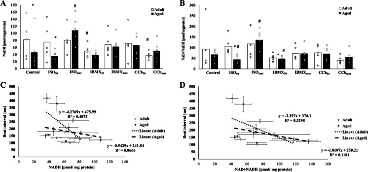
NADH and total NADH + NAD levels were quantified in the same tissues in which cAMP levels were quantified. NADH levels were lower in aged compared to adult atrial tissue (Fig. 2A). However, there was no difference in total NADH + NAD levels between adult and aged atrial tissue (Fig. 2B).
Fig. 2.
NAD and NAD + NADH levels under different ATP-demand conditions in adult and aged atrial tissues. (A) NADH and (B) NAD + NADH levels in control versus in response to drug perturbation of adult and aged atrial tissue. Three drugs were applied (isoproterenol (ISO), carbachol (CCh) and 3-isobutyl-1-methylxanthin (IBMX)) at two different concentrations: at the dose that led to a 50% change in the beating rate in adult tissue (X50) and the dose that led to maximal change in beating rate (Xmax). Correlation between spontaneous beat intervals and (C) NADH and (D) NAD + NADH levels in adult and aged atrial tissue. #Compared to control (either adult or aged). *Compared to adult for the same treatment
ISO50 did not change NADH levels in either adult or aged tissue, but NADH was higher in adult than aged atrial tissue (Fig. 2A). In response to ISO50, the total NADH + NAD concentration was lower in the aged tissue as compared to aged basal and was also higher in adult compared to aged atrial tissue (Fig. 2B). In contrast, following treatment with ISOmax, there were no differences measured in either NADH (Fig. 2A) or total NAD + NADH (Fig. 2B) concentrations in adult vs. aged atrial tissue.
CCh50 did not impact NADH levels in either adult or aged atrial tissue (Fig. 2A). The total NADH + NAD concentrations were not different in adult as compared to aged samples (Fig. 2B). Similarly, following treatment with CChmax, there was no difference in either NADH (Fig. 2A) or total NAD + NADH (Fig. 2B) in adult versus aged atrial tissue. However, the NADH level in CChmax-treated adult atrial tissue was lower than its adult basal.
Following IBMX50 treatment, NADH levels in adult atrial tissue were lower than in basal adult, which were not different than the NADH levels measured in aged atrial tissues (Fig. 2A). The total NAD + NADH content was lower in aged atrial tissue compared to basal aged, but was not different than the total NAD + NADH measured in adult atrial tissue (Fig. 2B). Following treatment with IBMXmax, there was no difference in either NADH (Fig. 2A) or total NAD + NADH (Fig. 2B) levels in adult versus aged atrial tissue.
To investigate how changes in ATP demand correlate with NAD or NAD + NADH levels, the relationship between the average beat interval and NAD or NAD + NADH levels in adult and aged atria under basal conditions and in response to drug challenges was calculated. There was no correlation between average beat interval and either NADH (− 0.6 for adult and − 0.3 for aged; Fig. 2C) or NAD + NADH (− 0.6 for adult and − 0.3 for aged; Fig. 2D) levels.
Changes in ATP levels and ATP demand in aged vs. adult atrial tissue
ATP levels quantified in the same tissues in which NAD, total NADH + NAD, and cAMP levels were measured, were not affected by ISO50, ISOmax, CCh50, CChmax, IBMX50, or IBMXmax (Fig. 3A). To investigate how changes in ATP demand correlate with ATP levels, the relationship between the average beat interval and ATP in adult and aged atria under basal conditions and in response to drug challenges was calculated. No correlation (− 0.3 for both adult and aged) was found between the average beat interval and ATP levels (Fig. 3B).
Fig. 3.
ATP levels at different ATP-demand conditions in adult and aged atrial tissues. (A) ATP levels in control versus in response to drug perturbations of adult and aged atrial tissue. Three drugs were applied (isoproterenol (ISO), carbachol (CCh), and 3-isobutyl-1-methylxanthin (IBMX)) at two different concentrations: at the dose that led to a 50% change in the beating rate in adult tissue (X50) and the dose that led to maximal change in beating rate (Xmax). (B) Correlation between spontaneous beat intervals and ATP levels in adult and aged atrial tissue
The effect of prolonged beat interval on energetic indices in adult atrial tissue
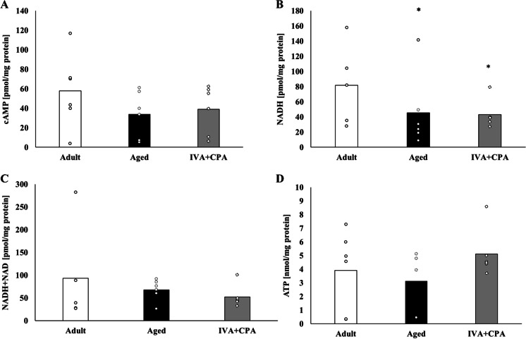
It has been shown that Ca2+ activates adenylyl cyclase (AC)-cAMP/protein kinase A (PKA) in the atria [17]. Thus, a reduction in cytosolic Ca2+ concentrations can potentially reduce cAMP levels to those of aged tissue. To test whether changes in the levels of Ca2+-activated AC-cAMP/PKA can mimic the effect of aging on energetic balance, 1 µM CPA, a SERCA2A blocker, was applied to adult atrial tissues. To physiologically prolong the beat interval (an index of ATP demand) in adult atrial tissue to the same interval observed in aged atrial tissue, 3 µM IVA was applied to block HCN4 in par, which is downregulated by aging [18]. The combination of these two drugs prolonged the average beat interval by 24.8 ± 8% (n = 6), confirming previous results for untreated control-aged tissue compared to adult [13]. cAMP level was similar to those measured in aged atrial tissue (Fig. 4A) as were NADH levels. NADH levels were lower in both untreated aged controls and in IVA- and CPA-treated atrial tissue as compared to untreated adult atrial tissue. Total NADH + NAD content following IVA and CPA treatment was similar to that measured in untreated aged atrial tissue (Fig. 3C). ATP levels were similar in adult atrial tissue treated with IVA and CPA as compared to its levels in both untreated adult and aged atrial tissue (Fig. 4D).
Fig. 4.
Energetics indices in response to reduced Ca.2+. (A) cAMP, (B) NADH, (C) NAD + NADH, and (D) ATP levels in adult and aged atrial tissues and adult atrial tissues treated with SERCA pump blocker (1 µM CPA) and HCN4 blocker (3 µM IVA). *Compared to adult
cAMP levels and energetics indices in different regions of aged and adult hearts
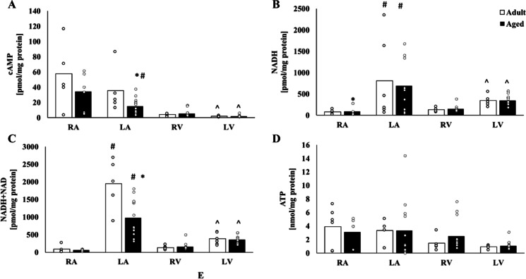
We next compared cAMP and the energetic indices quantified above in different regions of the heart. Figure 5A shows that cAMP levels were lower in aged left atria compared to adult. The cAMP level in adult left atrial tissue was similar to the level in adult right atrial tissue. In contrast, the cAMP levels in aged left atria were lower than those measured in aged right atrial tissue. cAMP levels in aged left ventricles were similar to their levels in adult left ventricles (Fig. 5A). cAMP levels in aged right ventricular and adult right ventricular tissues were similar. cAMP levels in both adult and aged samples were lower in the left ventricles as compared their right ventricles.
Fig. 5.
Energetics balance in different heart regions. (A) cAMP, (B) NADH, (C) NAD + NADH, and (D) ATP levels in adult and aged right atrial (RA), left atrial (LA), right ventricular (RV), and left ventricular (LV) tissues. Note, that RV and LV were not paced. #Compared to RA (either adult or aged LA). ^Compared to RV (either adult or aged RV). *Compared to adult for the same tissue type
NADH levels were not different in aged compared to adult left atrial tissues (Fig. 5B). In contrast, total NAD + NADH levels were lower in aged as compared to adult right atrial tissues (Fig. 5C). NADH levels in aged right ventricles were similar to their levels in adult right ventricles (Fig. 5B). Similarly, NAD + NADH levels were comparable in aged right ventricles and adult right ventricles (Fig. 5C). NADH levels in aged left ventricles were similar to their levels in adult left ventricles (Fig. 5B), as were NAD + NADH levels versus adult left ventricles (Fig. 5C). NADH and total NAD + NADH levels in both adult and aged left ventricles were lower as compared to their levels in right ventricles.
ATP levels were similar in adult and aged left atrial tissues and were similar to their levels measured in counterpart right atrial regions (Fig. 5D). Similarly, ATP levels in aged left ventricles were comparable to their levels in adult left ventricles. Similar ATP levels were measured in aged as compared to adult right ventricles.
Discussion
Here we report on the change in energetics indices in healthy adult and aged right atrial tissues under basal conditions and in response to drugs that affect ATP demand. The right atrium was connected to the sinoatrial node to ensure a biological pace. A tight correlation was found between atrial cAMP levels and the beating rate driven by the SAN, which was altered in advanced age. ATP, NADH, or NADH + NAD were not corelated with beating rate. Reduced NADH levels were documented at advanced age, but were restored by drug-induced perturbations that increased cAMP levels. Pharmacological perturbations that decrease intracellular Ca2+ of adult atrial tissue and prolonged the spontaneous beat interval to the level reported for aged tissue modified energetics indices to achieve those of aged mice. Finally, differences in energetics indices were found between aged right and left atria and between aged right and left ventricles.
The difference between cAMP levels in adult versus aged tissue was not significant at baseline. Upon treatment at X50 of ISO or IBMX, cAMP level was lower in aged vs. adult (Fig. 1A), suggesting that increased PDE amount or activity in aged atrial cells or disbalance in intracellular Ca2+ limits cAMP activity in response to these interventions. These differences were abolished, however, following treatment with maximal concentrations of ISO and IBMX. Thus, cAMP production reserve capacity is restored to match ATP demand at Xmax. Restoration of cAMP activity at Xmax in aged atrial can be due to increased cAMP production or decreased cAMP degradation. Future experiments will be needed to clarify this point. We also showed (Fig. 1B) a tight connection between cAMP levels and average spontaneous beat intervals, with different correlations measured for aged versus adult tissue. Age-dependent change in drug sensitivity may explain the differences between these correlations.
In addition, we showed here an age-associated reduction in NADH levels under basal conditions (Fig. 2). Both aging and cardiac diseases are associated with reduced NAD+ in the heart (reviewed in [10]). Note that NAD+ is also connected to other energy-related signaling pathways. For example, sirtuins, which are conserved proteins of NAD+-dependent deacylases, serve as important energy status sensors and their levels correlate with cardiac protection and incident onset of cardiac diseases [19]. AMP-activated protein kinase (AMPK) regulates energy balance by modulating NAD+ metabolism and SIRT1 activity [20]. Reduced NAD+ implies that activation of AMPK that switch on catabolic pathways that produce ATP is reduced. Thus, the reduced NADH documented here in aging tissues implies altered energetics. This conclusion is supported by the experimental evidence that showed alterations in other energetic parameters in aged atria [4, 6]. The fact that NADH levels were similar in aged and adult tissue on treatment with maximal concentrations of ISO, IBMX, or CCh, implies supply-to-demand matching under these conditions. It is possible that the increase in cAMP increases PKA activity, which directly or indirectly (e.g., through Ca2+ signaling) restores the energy balance. Thus, increasing the level of cAMP/PKA activity may be an effective means of restoring aged-associated deterioration in energetics signaling. As marked overexpression of AC8 leads to incident of heart failure and reduced lifespan [21], only short-term increases in cAMP may be the solution for age-associated energy imbalance.
NADH was suggested as a possible substrate ensuring matching of ATP supply to demand [22]. However, in the current study, no correlation was found between NADH levels and ATP demand (which correlated with beat interval). Similar conclusions were reached when analyzing isolated mitochondria [23] and whole hearts [24]. ATP was also suggested as a regulator of ATP supply to demand matching. However, in our study, no such correlation was identified, which aligned with a previous report of constant ATP levels at different ATP demands [22]. Considering the results showing that only cAMP correlated with ATP demand, it can be concluded that cAMP/PKA is an important regulator of ATP supply-to-demand matching. cAMP/PKA signaling phosphorylates several mitochondrial proteins as well as complexes I–V [25]. Reduced cAMP/PKA-dependent phosphorylation of these complexes in the electron transport chain would thereby eliminate the proton flux that drives complex V and decrease the rate of ATP production. In parallel, the rate of ATP delivery from the mitochondria to the cytosol via voltage-dependent anion channels that are phosphorylated by PKA, would be decreased [26, 27]. Because Ca2+ activates AC-cAMP/PKA signaling in atrial cells [17], the reduction in cAMP in advanced age may be stimulated by a reduction in Ca2+. Such a decrease was indeed documented in human right atria [28]. Because a reduction in Ca2+ deactivates mitochondrial enzymes and leads to decreased ATP production [29], Ca2+ acting either directly or indirectly, via Ca2+-activated AC-cAMP/PKA signaling, may affect the ATP supply-to-demand balance. Indeed, when we downregulated Ca2+ in adult atrial tissue to prolong the beat interval to the basal level reported in aging hearts (by also blocking HCN4), cAMP, NADH, NAD + NADH, and ATP levels shifted to those observed in the aged model (Fig. 4). Thus, Ca2+ may be an additional energy balance control mechanism that becomes downregulated with aging. Additional experiments are still necessary to characterize its role (see “Limitations”).
Finally, we compared the energetics indices between heart tissues of different ages. Although the cAMP level decrease in aged right atria was not significant, it was significantly reduced in the aged left atria compared to adult. In parallel, aged left atria showed lower NAD levels. The decrease in cAMP and possibly associated alterations in Ca2+ cycling, may lead to age-associated ATP supply-to-demand imbalances in the left atrium. Lower levels of both cAMP and ATP were documented in the ventricular tissue as compared to the atrium, which may have been due to the fact that the ventricular tissues were quiescent during isolation as was documented previously [30]. Note that no energy difference was documented between aged and adult ventricular tissues. However, the relative cAMP, NADH, and NAD + NADH levels were lower in the left as compared to the right ventricle. These results are consistent with literature reporting on constant NADH levels in aged ventricular myocytes that were not electrically stimulated and reduced levels after 10 min of continuous electrical stimulation [31]. Moreover, oxygen consumption and ATP production were reduced in aged ventricular tissues extracted from isolated working hearts [32]. Thus, differences in cAMP levels and energetics indexes between adult and aged ventricles depend on the workload.
Clinical insight
The exact mechanisms of initiation and maintenance of AF are still a topic of debate. Understanding energetics balances in healthy aging tissue may be beneficial for future treatment of AF, whose prevalence exponentially increases with age. Recent evidence suggests that perturbations in energetics metabolism are tightly coupled with ion channel function and membrane excitability, and may be associated with short or chronic AF. For example, stretch-induced AF in a rabbit model led to a decrease in adenine nucleotide concentrations, and to an increase in phosphocreatine levels, with no change in mitochondrial ATPase activity [33]. Changes in ATP supply-to-demand control mechanisms, i.e., abnormal intracellular Ca2+ handling [34, 35] and altered post-translational protein modification [36–38], also occur in AF. A mismatch between energy transduction, transfer, and consumption processes reduces the tolerance of atrial tissue to hemodynamic and metabolic demands and can lead to irreversible cell and tissue damage.
Limitations
This work quantified the steady state level of energetics indexes, which allow measurement and comparison of all indexes at the same tissues. Yet, future experiments will be necessary to quantify cAMP and energetics index kinetics to determine the possible existence of age-dependent differences in kinetics. While parallel in vivo experiments may enable quantification of NAD/NAD + NADH and ATP levels, no in vivo method exists for cAMP and, thus, not all energetic indexes can be measured in the same tissue.
Intracellular Ca2+ levels were not quantified, and thus, potential age-associated changes in Ca2+ in our tissues could not be evaluated. However, in our study when intracellular Ca2+ of adult tissues was altered by CPA, similar changes in energetic indexes were documented as aged atrial tissue. Note that use of a Ca2+ indicator interferes with the cAMP and ATP measurements and, therefore, the three cannot be quantified in parallel.
All the experiments were performed ex vivo, a setup which allows for rapid freezing of the entire tissue without contamination from other tissues. Because we documented here constant ATP and NADH in response to change in demand and these results were similar with levels measured in vivo [24], it can be assumed that the ex vivo environment accurately represents the in vivo environment.
Author contribution
YY and KT conceived and designed the research. NKP, SS, RS, KT, and YY did the experiments. NKP and OS did the analysis. YY, YL, IB, and EGL designed experimental protocols. EGL contributed reagents. YY drafted the manuscript. YY and NKP edited and revised the manuscript. NKP, SS, RS, OS, YL, IB, EGL, KT, and YY approved the final version.
Funding
The work was supported by ISF 330/19 (YY) and partially by the Intramural Research Program of the NIH, National Institute on Aging. K.T was supported by Japan Society for the Promotion of Science Research Fellowship for Japanese Biomedical and Behavioral Researchers at NIH. The funders had no role in study design, data collection and analysis, decision to publish, or preparation of the manuscript.
Declarations
Conflict of interest
The authors declare competing interests.
Footnotes
Publisher's Note
Springer Nature remains neutral with regard to jurisdictional claims in published maps and institutional affiliations.
Kenta Tsutsui and Yael Yaniv contributed equally to this work.
Contributor Information
Kenta Tsutsui, Email: knt22e@gmail.com.
Yael Yaniv, Email: yaely@bm.technion.ac.il.
References
- 1.Andrade J, Khairy P, Dobrev D, Nattel S. The clinical profile and pathophysiology of atrial fibrillation. Circ Res. Lippincott Williams & WilkinsHagerstown, MD; 2014;114:1453–68. [DOI] [PubMed]
- 2.Roberts-Thomson K, Kistler P, Sanders P, Morton J, Haqqani H, Stevenson I, et al. Fractionated atrial electrograms during sinus rhythm: relationship to age, voltage, and conduction velocity. Hear Rhythm Heart Rhythm. 2009;6:587–591. doi: 10.1016/j.hrthm.2009.02.023. [DOI] [PubMed] [Google Scholar]
- 3.Allessie M, Boyden P, Camm A, Kléber A, Lab M, Legato M, et al. Pathophysiology and prevention of atrial fibrillation. Circulation. 2001;103:769–777. doi: 10.1161/01.CIR.103.5.769. [DOI] [PubMed] [Google Scholar]
- 4.Emelyanova L, Preston C, Gupta A, Viqar M, Negmadjanov U, Edwards S, et al. Effect of aging on mitochondrial energetics in the human atria. J Gerontol A Biol Sci Med Sci. J Gerontol A Biol Sci Med Sci 2018;73:608–16. [DOI] [PMC free article] [PubMed]
- 5.Nemutlu E, Gupta A, Zhang S, Viqar M, Holmuhamedov E, Terzic A, Jahangir A, Dzeja P. Decline of phosphotransfer and substrate supply metabolic circuits hinders ATP cycling in aging myocardium. PLoS ONE. 2015;10:e0136556. doi: 10.1371/journal.pone.0136556. [DOI] [PMC free article] [PubMed] [Google Scholar]
- 6.Emelyanova L, Ashary Z, Cosic M, Negmadjanov U, Ross G, Rizvi F, et al. Selective downregulation of mitochondrial electron transport chain activity and increased oxidative stress in human atrial fibrillation. Am J Physiol Heart Circ Physiol. 2016;311:H54–63. doi: 10.1152/ajpheart.00699.2015. [DOI] [PMC free article] [PubMed] [Google Scholar]
- 7.Yaniv Y, Juhaszova M, Nuss HBB, Wang S, Zorov DBB, Lakatta EGG, et al. Matching ATP supply and demand in mammalian heart in vivo, in vitro, and in silico perspectives. Anal Card Dev From Embryo Old Age. 2010;1188:133–142. doi: 10.1111/j.1749-6632.2009.05093.x. [DOI] [PMC free article] [PubMed] [Google Scholar]
- 8.Yaniv Y, Juhaszova M, Sollott SJJ. Age-related changes of myocardial ATP supply and demand mechanisms. Trends Endocrinol Metab. 2013;24:495–505. doi: 10.1016/j.tem.2013.06.001. [DOI] [PMC free article] [PubMed] [Google Scholar]
- 9.Yan J, Zhao W, Thomson JK, Gao X, DeMarco DM, Carrillo E, et al. Stress signaling JNK2 crosstalk with CaMKII underlies enhanced atrial arrhythmogenesis. Circ Res Lippincott Williams and Wilkins. 2018;122:821–835. doi: 10.1161/CIRCRESAHA.117.312536. [DOI] [PMC free article] [PubMed] [Google Scholar]
- 10.Xu W, Li L, Zhang L. NAD+ metabolism as an emerging therapeutic target for cardiovascular diseases associated with sudden cardiac death. Front Physiol. Frontiers Media SA; 2020;11. [DOI] [PMC free article] [PubMed]
- 11.Nattel S. Aging and protein kinase activation. Circ Res. Lippincott Williams & WilkinsHagerstown, MD; 2018;122:799–801. [DOI] [PubMed]
- 12.Niemann B, Pan R, Teschner M, Boening A, Silber R, Rohrbach S. Age and obesity-associated changes in the expression and activation of components of the AMPK signaling pathway in human right atrial tissue. Exp Gerontol Exp Gerontol. 2013;48:55–63. doi: 10.1016/j.exger.2012.04.005. [DOI] [PubMed] [Google Scholar]
- 13.Liu J, Sirenko S, Juhaszova M, Sollott SJ, Shukla S, Yaniv Y, et al. Age-associated abnormalities of intrinsic automaticity of sinoatrial nodal cells are linked to deficient cAMP-PKA-Ca(2+) signaling. Am J Physiol Hear Circ Physiol. 2014;306:H1385–H1397. doi: 10.1152/ajpheart.00088.2014. [DOI] [PMC free article] [PubMed] [Google Scholar]
- 14.Behar JA, Rosenberg AA, Weiser-Bitoun I, Shemla O, Alexandrovich A, Konyukhov E, et al. PhysioZoo: a novel open access platform for heart rate variability analysis of mammalian electrocardiographic data. Front Physiol. 2018;9:1390. doi: 10.3389/fphys.2018.01390. [DOI] [PMC free article] [PubMed] [Google Scholar]
- 15.Shemla O, Tsutsui K, Behar JA, Yaniv Y. Beating rate variability of isolated mammal sinoatrial node tissue: insight into its contribution to heart rate variability. Front Neurosci. Frontiers Media SA; 2021;14. [DOI] [PMC free article] [PubMed]
- 16.Yaniv Y, Ahmet I, Tsutsui K, Behar J, Moen JM, Okamoto Y, et al. Deterioration of autonomic neuronal receptor signaling and mechanisms intrinsic to heart pacemaker cells contribute to age-associated alterations in heart rate variability in vivo. Aging Cell. 2016;15. [DOI] [PMC free article] [PubMed]
- 17.Collins TP, Terrar DA. Ca(2+)-stimulated adenylyl cyclases regulate the L-type Ca(2+) current in guinea-pig atrial myocytes. J Physiol. 2012;590:1881–1893. doi: 10.1113/jphysiol.2011.227066. [DOI] [PMC free article] [PubMed] [Google Scholar]
- 18.Tellez JO, Mączewski M, Yanni J, Sutyagin P, Mackiewicz U, Atkinson A, et al. Ageing-dependent remodelling of ion channel and Ca 2+ clock genes underlying sino-atrial node pacemaking. Exp Physiol. 2011;96:1163–1178. doi: 10.1113/expphysiol.2011.057752. [DOI] [PubMed] [Google Scholar]
- 19.Chang H, Guarente L. SIRT1 and other sirtuins in metabolism. Trends Endocrinol Metab. 2014;25:138–145. doi: 10.1016/j.tem.2013.12.001. [DOI] [PMC free article] [PubMed] [Google Scholar]
- 20.Cantó C, Gerhart-Hines Z, Feige JN, Lagouge M, Noriega L, Milne JC, et al. AMPK regulates energy expenditure by modulating NAD+ metabolism and SIRT1 activity. Nature. 2009;458:1056–1060. doi: 10.1038/nature07813. [DOI] [PMC free article] [PubMed] [Google Scholar]
- 21.Mougenot N, Mika D, Czibik G, Marcos E, Abid S, Houssaini A, et al. Cardiac adenylyl cyclase overexpression precipitates and aggravates age-related myocardial dysfunction. Cardiovasc Res. 2019;115:1778–1790. doi: 10.1093/cvr/cvy306. [DOI] [PMC free article] [PubMed] [Google Scholar]
- 22.Katz LA, Koretsky AP, Balaban RS. Respiratory control in the glucose perfused heart: A 31P NMR and NADH fluorescence study. FEBS Lett. No longer published by Elsevier; 1987;221:270–6. [DOI] [PubMed]
- 23.Moreno-Sánchez R, Hogue BA, Hansford RG. Influence of NAD-linked dehydrogenase activity on flux through oxidative phosphorylation. Biochem J. Portland Press Ltd; 1990;268:421. [DOI] [PMC free article] [PubMed]
- 24.Zhou L, Cabrera ME, Okere IC, Sharma N, Stanley WC. Regulation of myocardial substrate metabolism during increased energy expenditure: insights from computational studies. Am J Physiol-Heart Circ Physiol. 2006;291:H1036–H1046. doi: 10.1152/ajpheart.01382.2005. [DOI] [PubMed] [Google Scholar]
- 25.Covian R, Balaban RS. Cardiac mitochondrial matrix and respiratory complex protein phosphorylation. Am J Physiol-Heart Circ Physiol. 2012;303:H940–H966. doi: 10.1152/ajpheart.00077.2012. [DOI] [PMC free article] [PubMed] [Google Scholar]
- 26.Bera AK, Ghosh S, Das S. Mitochondrial VDAC can be phosphorylated by cyclic AMP-dependent protein kinase. Biochem Biophys Res Commun. Academic Press; 1995;209:213–7. [DOI] [PubMed]
- 27.Sheldon KL, Maldonado EN, Lemasters JJ, Rostovtseva TK, Bezrukov SM. Phosphorylation of voltage-dependent anion channel by serine/threonine kinases governs its interaction with tubulin. PLoS ONE. 2011;6:e25539. doi: 10.1371/journal.pone.0025539. [DOI] [PMC free article] [PubMed] [Google Scholar]
- 28.Herraiz-Martínez A, Álvarez-García J, Llach A, Molina C, Fernandes J, Ferrero-Gregori A, et al. Ageing is associated with deterioration of calcium homeostasis in isolated human right atrial myocytes. Cardiovasc Res. 2015;106:76–86. doi: 10.1093/cvr/cvv046. [DOI] [PMC free article] [PubMed] [Google Scholar]
- 29.Balaban RS. The role of Ca2+ signaling in the coordination of mitochondrial ATP production with cardiac work. Biochim Biophys Acta - Bioenerg. 2009;1787:1334–1341. doi: 10.1016/j.bbabio.2009.05.011. [DOI] [PMC free article] [PubMed] [Google Scholar]
- 30.Glancy B, Hartnell LM, Malide D, Yu Z-X, Combs CA, Connelly PS, et al. Mitochondrial reticulum for cellular energy distribution in muscle. Nature. 2015;523:617–620. doi: 10.1038/nature14614. [DOI] [PMC free article] [PubMed] [Google Scholar]
- 31.Barton GP, de Lange WJ, Ralphe JC, Aiken J, Diffee G. Linking metabolic and contractile dysfunction in aged cardiac myocytes. Physiological Reports. 2017;5:e13485. doi: 10.14814/phy2.13485. [DOI] [PMC free article] [PubMed] [Google Scholar]
- 32.Preston CC, Oberlin AS, Holmuhamedov EL, Gupta A, Sagar S, Syed RHK, et al. Aging-induced alterations in gene transcripts and functional activity of mitochondrial oxidative phosphorylation complexes in the heart. Mech Ageing Dev Elsevier. 2008;129:304–312. doi: 10.1016/j.mad.2008.02.010. [DOI] [PMC free article] [PubMed] [Google Scholar]
- 33.Kalifa J, Maixent J-M, Chalvidan T, Dalmasso C, Colin D, Cozma D, et al. Energetic metabolism during acute stretch-related atrial fibrillation. Mol Cell Biochem. 2008;317:69–75. doi: 10.1007/s11010-008-9832-3. [DOI] [PMC free article] [PubMed] [Google Scholar]
- 34.Dobrev D, Voigt N, Wehrens XHT. The ryanodine receptor channel as a molecular motif in atrial fibrillation: pathophysiological and therapeutic implications. Cardiovasc Res. 2011;89. [DOI] [PMC free article] [PubMed]
- 35.Dobrev D, Nattel S. Calcium handling abnormalities in atrial fibrillation as a target for innovative therapeutics. J Cardiovasc Pharmacol. 2008;52:293–299. doi: 10.1097/FJC.0b013e318171924d. [DOI] [PubMed] [Google Scholar]
- 36.Vest JA, Wehrens XHT, Reiken SR, Lehnart SE, Dobrev D, Chandra P, et al. Defective cardiac ryanodine receptor regulation during atrial fibrillation. Circulation Circulation. 2005;111:2025–2032. doi: 10.1161/01.CIR.0000162461.67140.4C. [DOI] [PubMed] [Google Scholar]
- 37.El-Armouche A, Boknik P, Eschenhagen T, Carrier L, Knaut M, Ravens U, et al. Molecular determinants of altered Ca2+ handling in human chronic atrial fibrillation. Circulation. 2006;114:670–680. doi: 10.1161/CIRCULATIONAHA.106.636845. [DOI] [PubMed] [Google Scholar]
- 38.Greiser M, Neuberger HR, Harks E, El-Armouche A, Boknik P, de Haan S, et al. Distinct contractile and molecular differences between two goat models of atrial dysfunction: AV block-induced atrial dilatation and atrial fibrillation. J Mol Cell Cardiol. 2009;46:385–394. doi: 10.1016/j.yjmcc.2008.11.012. [DOI] [PubMed] [Google Scholar]


