Abstract
Hydrogels, also known as three-dimensional, flexible, and polymer networks, are composed of natural and/or synthetic polymers with exceptional properties such as hydrophilicity, biocompatibility, biofunctionality, and elasticity. Researchers in biomedicine, biosensing, pharmaceuticals, energy and environment, agriculture, and cosmetics are interested in hydrogels. Hydrogels have limited adaptability for complicated biological information transfer in biomedical applications due to their lack of electrical conductivity and low mechanical strength, despite significant advances in the development and use of hydrogels. The nano-filler-hydrogel hybrid system based on supramolecular interaction between host and guest has emerged as one of the potential solutions to the aforementioned issues. Black phosphorus, as one of the representatives of novel two-dimensional materials, has gained a great deal of interest in recent years owing to its exceptional physical and chemical properties, among other nanoscale fillers. However, a few numbers of publications have elaborated on the scientific development of black phosphorus hybrid hydrogels extensively. In this review, this review thus summarized the benefits of black phosphorus hybrid hydrogels and highlighted the most recent biological uses of black phosphorus hybrid hydrogels. Finally, the difficulties and future possibilities of the development of black phosphorus hybrid hydrogels are reviewed in an effort to serve as a guide for the application and manufacture of black phosphorus -based hydrogels.
Graphical Abstract
Recent applications of black phosphorus hybrid hydrogels in biomedicine.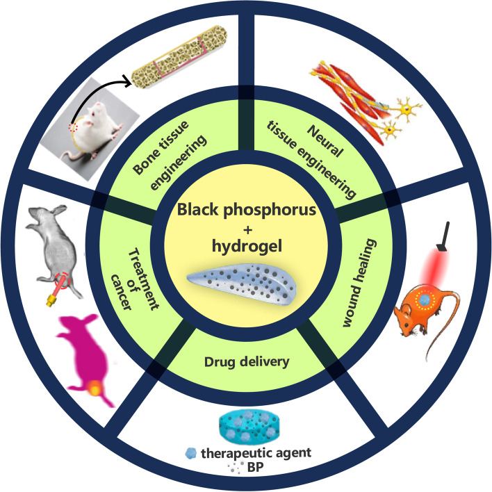
Keywords: Black phosphorus (BP), Hybrid hydrogels, Nanomaterials, Biomedical applications, Tissue engineering
Introduction
Hydrogels resembling the tissue structure of natural extracellular matrix are among the most well-established materials for biomedical applications. Hydrogels are defined as three-dimensional (3D) networks with hydrophilic groups cross-linked by natural and/or synthetic polymers such that they absorb and retain a significant volume of liquid components while keeping their shape and 3D structure after swelling [1, 2]. The highly hydrous 3D porous network of hydrogels can imitate the microenvironment of natural tissues. Hydrogels are often engineered to retain, release, or trap materials, which have been applied to many biomedical fields including tissue engineering, wound healing, medication administration, bio-imaging, and diagnostics [3, 4]. For the preparation of hydrogels, it is necessary to choose materials based on a range of essential properties, including swelling, mechanical properties, diffusion rate, and chemical functionality. These properties are dependent on the cross-link density, the distance between cross-links, the gel’s macromolecular structure, and the residual chemicals (such as monomers, initiators, and others substances) [5]. Traditional hydrogels have mediocre mechanical characteristics and unspectacular characteristic functions, limiting their potential applications. The fact that nanocomposite hydrogels combine the properties of polymers and nanoparticles to provide excellent functionalities draws greater attention to them. Among the nano-fillers of many hydrogels, two-dimensional (2D) nanomaterials play an increasingly important role [6]. Black phosphorus is an outstanding example due to its biodegradability, biocompatibility, photothermal performance, electrical conductivity, and high surface area mechanical strength [7, 8]. As with other nanocomposite hydrogels, the preparation of black phosphorus-hybrid hydrogels includes blending method, grafting method, in-situ precipitation, and freeze-thaw method [9, 10]. The use of physical crosslinking, chemical crosslinking, and the principle of electrostatic interactions in aforementioned methods will be discussed further in the following section, along with their biomedical applications.
Synthesis and properties of black phosphorus
In the last decade, novel 2D nanomaterials with diverse characteristics have garnered exploding interest in the domains of biomedicine and other disciplines, where they play an increasingly crucial role [11–13]. These single or multilayer nano-crystalline materials with planar shape exhibit greater interatomic interactions than stacking interactions. More than a thousand 2D material structures have been found and categorized into a variety of monolayer or multilayer structures, each of which exhibits unique physicochemical properties. They cover the complete periodic table, from transition metals through boron −/ carbon −/ nitrogen −/ sulfur groups, as well as graphene and its derivatives (e.g., graphene oxide or reduced graphene oxide, carbon nano tube), single element materials (e.g., Xene materials including phosphorene, silicene, germanene), metal carbides and nitrides (e.g., MXenemarterials including Ta4C3 Tx), transition metal disulphide (TMDCs materials including MoS2, WS2, WSe2), transition metal oxides (TMOs including TiO2, MnO2), layered double hydroxide (LDH), hexagonal boron nitride (hBN), graphite carbon nitride (gC3N4) or silicate clay (nanoclay), and other materials [7, 14, 15]. Due to their distinctive shape, 2D nanomaterials possess a huge surface area and anisotropic physical/chemical properties in addition to distinct mechanical/chemical/optical properties, biocompatibility, and degradability. However, several of these materials have intrinsic limitations, such as graphene’s lack of band gap and MoS2’s very poor carrier mobility [14–17]. An ideal 2D material should have a high charge mobility and a sufficient and tunable bandgap, and the emergence of black phosphorus satisfies these scientific conditions. This band structure provides a much-needed gap for field-effect transistor (FET) applications in two-dimensional materials such as graphene, while the thickness-dependent direct band gap, which permits wide-band absorption from the visible to mid-infrared range, could lead to applications in optoelectronics, particularly in the infrared field. In addition, black phosphorene can offer the optimal trade-off between mobility and switching current ratio, which is highly desirable for the development of high-speed, flexible electronic systems that can function in the tens of megahertz frequency range [16].
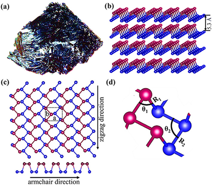
Phosphorene, sometimes referred to as black phosphorus (BP) when layered, is a 2D substance. Since Bridgman successfully obtained the first massive BP crystals in 1914, there has been little interest in BP research for over a century. Since two independent teams successfully stripped BP of its monolayer in 2014 [17, 18], however, researchers have been uncovering its unique combination of properties, such as high surface activity, adjustable band gap, favorable on/off current ratio, excellent carrier mobility, near-infrared photo responsiveness (NIR), biocompatibility, and non-toxic biodegradation products [19–21]. These properties cannot be separated from those of its fundamental structure (see Fig. 1). Phosphorus is one of the most plentiful elements in the earth’s crust, making up around 0.1% of the total volume [22]. Phosphorus occurs primarily as four allotropes: white phosphorus (WP), red phosphorus (RP), violet phosphorus (VP), and black phosphorus (BP). Under ambient circumstances, WP is the most reactive and unstable allotrope, whereas BP is the most stable and least reactive [23, 24]. BP is a kind of highly purified black sheet basic crystal. Chemical bonds bind the phosphorus atoms in BP to three phosphorus atoms in the vicinity. The several BP layers are joined by van der Waals’ force. BP has a layer-dependent band gap that is adjustable from 0.3 eV (bulk) to 2.0 eV (monolayer) and has stronger absorption in the UV and NIR areas than the 2D materials discussed before. Also shown modest carrier mobility is BP. BP has been developed as saturable absorbers for solid-state lasers to create short pulses because to its high potential for pulse generation in the 3 μm spectral region of the mid-infrared [25]. The excellent biocompatibility and biodegradability of BP in vivo imply that it is more likely than other materials to be acceptable for interdisciplinary biological applications [8, 26, 27]. Due to edge states and quantum confinement phenomena, BP nanoribbons and zero-dimensional quantum dots with unique properties can be produced by morphological engineering. BP quantum dots were produced by ultrasonic liquid phase stripping, solvent heat treatment, and electrochemical stripping on 2D BP nanosheets. BPQD features an extremely tiny dimension, a broad band gap that is tunable, good edge states, and a greater surface area to volume ratio [28].
Fig. 1.
(a): image of bulk BP crystal; (b) Perspective side view of multi-layer BP; (c) Top and side views of single-layer phosphorene where ‘a’ and ‘b’ represent lattice parameters; (c) A representation of the arrangement of atomic structures, illustrating dihedral/hinge angles and bond lengths, where different colored P atoms in each image represent P atoms in different planes. Reproduced with permission from Ref [16]. 2019IOP Publishing Ltd. All rights reserved.
The combination of these prosperities makes BP and its derivatives attractive and promising solutions for biological applications when used as a nano-filler of hydrogels to form a system.
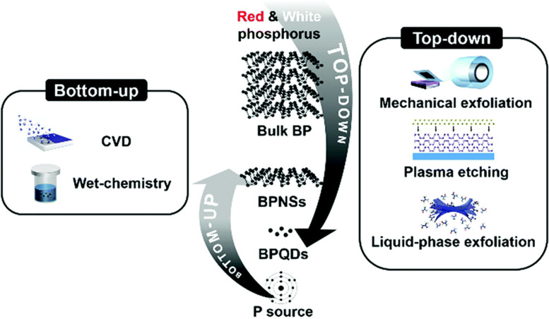
Due to the recent invention of technologies to peel bulk BP powders or crystals into nanostructures, its promise for biological applications has only been examined in the last decade. The fabrication of ultra-thin 2D nanomaterials is often separated into two fundamental methods, the “top-down approach” and the “bottom-up approach,” and the preparation of BP is no exception. Typically, top-down approach concentrates on the exfoliation of bulk materials with the aid of a driving force (e.g., mechanical exfoliation, chemical exfoliation, and liquid exfoliation), resulting in the disintegration of the material into nanoparticles. The approach comprises chemical synthesis and extraction from certain starting materials. Top-down approach typically consist of mechanical exfoliation, liquid exfoliation, plasma-assisted techniques such as plasma etching, ultrasound-assisted exfoliation, and electrochemical techniques [29, 30] (see Fig. 2). It is highly recommended to read the following reviews on the synthesis of black phosphorus [26, 31–35]. The hunt for ever-simpler and economic mass synthetic production technologies will usher in an expansion of applications.
Fig. 2.
Scheme of the synthetic method for nano-BP .Reproduced with permission from Ref [26]. Copyright 2019Wiley Ltd. All rights reserved
Biomedical application of black phosphorus hybrids hydrogel
Despite the fact that research on BP started in 2014, the research results of this novel 2D material has achieved a resounding success. This section illustrates the significance of BP in biological applications such as bone tissue engineering (see Table 1), nerve tissue engineering, immunological and cancer treatment, skin wound healing, and drug delivery.
Table 1.
Application of black phosphorus composite hydrogel in bone tissue engineering
| Hydrogel matrix | Black phosphorus form | Scaffold performance | Animal model | References |
|---|---|---|---|---|
| GelMA and U-Arg-PEA | BPNs | Strong mechanical properties, capturing calcium ions, accelerating biomineralization, and enhancing osteogenic differentiation of HDPSCS | Rabbit skull defect model | [36] |
| GEL and DFO | BPNs | Excellent swelling, degradation and release rate, satisfactory biocompatibility, capable of promoting the proliferation and osteogenesis of hBMSC in vitro, improving bone regeneration and neovascularization in vivo | Rat model of acute ischemic tibial defect | [37] |
| GEL | BPNs | Excellent NIR photothermal antibacterial, eliminating cancer cell properties, and enhancing bone regeneration | Rat model of skull defect | [38] |
| Agarose | BPNs | Providing phosphorus source and nucleation site, accelerating PO 4 3− and Ca 2+ reactions to promote biomineralization; The mechanical properties and bio-mineralization capabilities are customized by adjusting the timing and location of NIR light | – | [39] |
| Chitosan/ collagen | MSC membrane coated BPNs | Activating the heat shock response of osteoblasts, stimulating downstream responses, enhancing osteoblast migration/differentiation, and stimulating biomineralization processes to promote bone healing upon remote NIR activation | Rat model of skull defect | [40] |
| PLGA | BPNs | Strategies for heat-stimulated bone regeneration, ranging from inefficient external hyperthermia to more effective self-hyperthermia with “smart” bone implants under remote control | Rat model of tibial defect | [41] |
| CNTpega -OPF | BPNs | Injectable, good conductivity combined with electrical stimulation, improving the adhesion, proliferation, filament and focal adhesion development, and osteogenic differentiation of pre-osteoblast cells | Rabbit model of defect at the fusion site of femur, vertebral cavity and posterolateral spine | [42] |
| OPF | BPNs | Controlled degradation rate, improving the spread, distribution, proliferation and differentiation of MC3T3 cells on hydrogels, and controling the cytotoxicity | – | [43] |
| OPF/ Collagen | BPNs | The appropriate 3D microenvironment for MSC cell culture, providing clues for osteogenic differentiation | – | [44] |
| OPF | BPQDs | The smallest BPQDs, promoting the spread, distribution, proliferation and differentiation of MC3T3 cells | – | [45] |
| WW/RSF | BPQDs packaged with PLGA | Strong mechanical properties, inhibiting osteoclast differentiation, and showing photothermal effects on spinal metastases | Femur defect in rat model and tumor-bearing nude mouse model | [46] |
| DNA and 3D-printed PCL | Vegf-engineered BPNs | Sustainable delivery of growth factors, promoting the growth of mature blood vessels, and inducing osteogenesis | Rat model of cranial defect of critical size | [47] |
| GelMA | BP@Mg | Biomimetic periosteal structures, significantly promoting angiogenesis by inducing endothelial cell migration, and upregulating the expression of neuro-associated proteins in neural stem cells (NSCs) | Rat model of skull defect | [48] |
| PVA and Chitosan | MgO blended BPNs | Excellent antibacterial effect, promoting the recruitment, osteogenic differentiation and biologic mineralization of MSCs | Rat model of skull defect | [49] |
| PLGA | BP-SrCl2 | Excellent biodegradability, and photo-controlled Sr release | Rat model of femur defect | [50] |
Bone tissue engineering
In addition to the participation of osteoblasts and osteoclasts, bone mineralization induced by appropriate calcium and phosphorus metabolism is essential for bone remodeling. BP is highly homologous to the inorganic constituent of bone [50, 51]. The degradation product of BP is non-toxic phosphates. Phosphate is essential for bone regeneration [52] and may be utilized as a biomineralization inducer. Phosphate ions (PO43−) serve as anionic ligands for positive calcium ions (Ca2+), which may stimulate the attraction, binding, and accumulation of free calcium ions in the physiological environment of bone tissue [53], resulting in the formation of calcium phosphate (CaP) nanoparticles. Calcium phosphate may facilitate the formation of hydroxyapatite (HAP), an essential structural component of bone and tooth enamel. BP regulates osteogenesis, osseointegration, and fracture repair due to its capacity to release phosphate ions and trap metal ions such as calcium [54].
Based on the above properties, Huang et al. [36] developed a BP nanosheets-based hydrogel platform by photo-crosslinking of gelatin methacrylamide, BPNs, and cationic arginine-based unsaturated poly (ester amide)s. Prior to ultraviolet (UV) irradiation, BP nanosheets (BPNs) were introduced to and mixed with the precursor solution. In addition to their gelling effect, cationic unsaturated arginine-based poly (ester amide) s [U-Arg-PEA] offered a positive charge to facilitate the hydrogel and BPNs’ strong electrostatic contact. Complementing one another, the surface charge and large surface area of BPNs fostered robust interactions with polymer chains and further reinforced the cross-linking network generated by UV irradiation. The addition of BPNs raised the hydrogel’s compressive modulus by three to four times, and it was biocompatible. hDPSCs cultured on the hydrogel surface were able to multiply and spread properly. This sustained supply of calcium-free phosphorus hydrogel approach was capable of releasing phosphate in response to light, accelerating mineralization in vitro, and promoting the osteogenic differentiation of hDPSCs through the BMP-RUNX2 pathway. In vivo studies from a model of a skull lesion in a rabbit shown that BPNs accelerated bone repair. Xu et al. [37] prepared scaffolds by loading BPNs and deferoxamine (BPN-DFO) into gelatin hydrogels using a similar approach. In ischemic tibial areas of SD (Sprague Dawley) rats with acute femoral artery blockage, the scaffolds displayed excellent swelling, degradation, and release rates, and significantly enhanced osteogenesis and blood vessel development.
In addition to BP/Gel-like nanocomposite hydrogels, Miao et al. [38] devoted particular emphasis to the exceptional photothermal performance of BPNs in comparison to GO and nanosilicate. The photothermal ablation of bacteria and tumor cells is enabled by the high photothermal conversion efficiency of BPNs [26, 55, 56]. The findings of their experiments indicated that BP/Gel nanocomposite hydrogels had excellent near-NIR photothermal property and the capacity to destroy cancer cells and germs. In vitro, the nanocomposite matrix may sustain cell proliferation and induce osteogenic differentiation of human mesenchymal stem cells (hMSCs) in the absence of bone inducible factor. In addition to the bactericidal and cancer cell-killing capabilities of BP, NIR irradiation enhanced the degradation of BP to PO 4 3−, increased the chemical activity, expedited the interaction between PO 4 3− and Ca 2+, and promoted in situ biomeralization. In BP/agarose hydrogel [39], BP/chitosan/collagen hydrogel [40], and BP/PLGA hydrogel [41], it was shown that this property may be exploited to remotely regulate activation to accelerate biomineralization during bone tissue regeneration. This property may also be exploited for remote infrared controlled medication release. The mechanism of osteogenesis has also been examined further. BPNs may generate moderate photothermal effects induced by NIR light, and promote osteoblast recruitment via activating matrix metalloproteinase (MMP) and ERK-Wnt/β-catenin-RUNX2 axis mediated by heat shock proteins (HSPs) [57]. Some researchers in the field of bone tissue engineering have focused on the conductivity of BP [58]. Liu et al. [42] reported a new injectable carbon nanotube (CNT) and BP gel for tissue engineering with improved mechanical strength, conductivity, and continuous phosphate ion release. As a crosslinking matrix, a biodegradable oligomeric (poly (ethylene glycol) dimethyl fumarate) (OPF) polymer was used, and a crosslinked CNT-poly (ethylene glycol) -acrylate (CNTpega) was included to offer mechanical support and conductivity. However, CNT inclusion was primarily responsible for the hydrogel’s conductivity, and the influence of BPNs is minimal.
The appropriate concentration and size of BPNs in these novel nanocomposite hydrogel materials for bone regeneration need more investigation. In research, the addition of BPNs into a cross-linked OPF hydrogel improved the adhesion, distribution, proliferation, and differentiation of MC3T3-E1 cells. Optimal cell development was observed at BP doses up to 500 ppm [43]. In an additional research of 3D mesenchymal stem cell (MSC) preparations, Li et al. [44] examined the osteogenic response of MSC spheroids by a novel combination of collagen and BP. The stiff, wrinkly structure of BP offered mechanical signals for MSCs, which created a favorable microenvironment for osteogenic differentiation [59].
With 6 μg/mL collagen and/or BP concentration gradients (0 μg/mL, 4 μg/mL, 8 μg/mL,and 16 μg/mL), MSC spheres were effectively constructed. Runx2, osteopontin, and alkaline phosphatase were expressed at greater levels in the BP group with 4 μg/mL and 8 μg/mL concentrations than in the control group. Furthermore, the aforementioned findings showed that BP hybrid hydrogel might serve many functions in the cell treatment of bone regeneration, including precursor cell culture in vitro and carrier in vivo. In terms of BP size, the nanosheet form of BP is cytotoxic under certain circumstances [60], which restricts its biological applicability to some degree. The BP quantum dot is a novel kind of BP nanostructure that was initially created using the liquid exfoliation approach in 2015 and has a nanometer-scale dimension [61]. This ultra-small nanomaterial exhibited reduced cytotoxicity and more biocompatibility than BPNs [62]. Xu et al. [45] examined the osteogenic potential of BPNs of various sizes and quantum dots in BP/OPF hydrogels, where BP quantum dots was obtained by the combination of bath-sonication and probe-sonication. BPNs, particularly BP quantum dots, increased the behavior of MC3T3 cells on OPF hydrogels, including their spreading, distribution, proliferation, and differentiation. However, the intrinsic instability of BP quantum dots (particularly after being exposed to complicated blood circulation and physiological settings) is a primary barrier to clinical use. In order to solve this problem, Hu et al. [46] used the oil-in-water emulsion solvent evaporation method to encapsulated BPQDs into PLGA (poly (lactic-co-glycolic acid) to prepare BPQD/PLGA NS. Due of the hydrophobic structure’s special encapsulating effect, PLGA prevented the oxidation and degradation of BPQD by isolating internal BPQD, hence enhancing photothermal stability [63]. The combination of BPQD/PLGA NS and highly strong regenerated silk fibroin based delignified wood hydrogel increased the proliferation, migration, and osteogenic differentiation of BMSCs significantly. They were also surprisingly discovered that BPQD/PLGA NS inhibited osteoclast differentiation. Moreover, BPQD in hydrogels shown remarkable performance in vitro and in vivo for photothermal tumor ablation, giving evidence of potential therapeutic uses for both bone regeneration and bone metastasis ablation. These results give crucial references for the future selection of BP concentration and size in hydrogel scaffolds for bone tissue creation.
Due to their large specific surface area, BPNs are good drug carrier (drug loading efficiency up to 95%), and medicines are adsorbed on BPNs through non-chemical bonding (this property preserves drug activity and purity) [50]. Miao et al. [47] developed a dynamic DNA hydrogel modified with BPNs and VEGF to impart mechanical strength, where BPNs were modified by VEGF. Due of the non-covalent interaction between VEGF and BPNS, the integrated nanogel scaffold structure displayed a sustained VEGF release. In addition, BPNs were paired with a 3D-printed PCL scaffold to create a bioactive gel scaffold structure, in which BPNS attached to macromolecular DNA chains to impart function to the hydrogel and tighten the DNA cross-linking network. In vitro and in vivo, the whole gel scaffold system stimulated the growth of mature blood vessels and induced osteogenesis to encourage new bone formation. The capacity of BP to capture metal ions enabled it to transport helpful metal ions for bone rebuilding, such as magnesium.
Mg-doped biomaterials trigger bone regeneration by releasing Mg2+, therefore attracting MSCs and boosting osteogenic differentiation [64]. Recently, it has been reported that the modification of metal ion also considerably increased the stability of BP [65]. Xu et al. [48] developed a bilayer hydrogel that resembles periosteum to increase the efficacy of vascularized bone healing. The magnesium ion-modified BPNs (BP@Mg) was mixed into gelatin methacryloyl (GelMA) hydrogels to generate the top hydrogel as the periosteal repair layer, and the bottom hydrogel (GelMA-PEG/β-TCP) as the bone healing layer. The top hydrogel considerably increased angiogenesis by inducing endothelial cell migration and offerd many benefits for up-regulating the expression of neural-related proteins in neural stem cells (NSCs). This bilayer hydrogel approach opens the path for the development of biomaterials for neurovascular networks for bone regeneration. Magnesium may also be contained in hydrogels made by freezing and thawing processes by co-mixing with BPNs [49]. The results described above imply an optimistic future for BP-based nanocomposite hydrogels as therapeutic platforms for bone tissue engineering applications.
Nerve tissue engineering
BP is mostly utilized for bone tissue engineering in the area of tissue engineering since its composition is close to the inorganic constituent of natural bone. However, investigations have shown that phosphate is also a crucial neurogenic differentiation signal promoter that may induce neurite outgrowth [66]. The availability of phosphorus ions, which are critical for regulating cell signaling, and the high conductivity of multiple electron pairs from phosphorus atoms in the BP fold layer may contribute to increased neuronal development [67, 68]. BP also has the potential to regulate the neural differentiation lineage of stem cells in stem cell therapies for neurological disorders. Strong electrical conductivity up to 300 S m− 1 [69] and good thermal conductivity may be exploited as a support to facilitate biological signal transduction, which may explain why BPNs are advantageous for promoting neural differentiation of stem cells. Xu et al. [70] have proven that the incorporation of polydopamine-modified BPNs into a hydrogel matrix significantly accelerated the differentiation of bone marrow-derived mesenchymal stem cells (BMSCs) into neurons in response to electrical stimulation. The rigidity of hydrogels and the conductivity of BP are regarded as optimal physical clues for regulating neuronal lineage control by MSCs. Modification or functionalization of BP is also a crucial element in stem cell differentiation regulation [71].
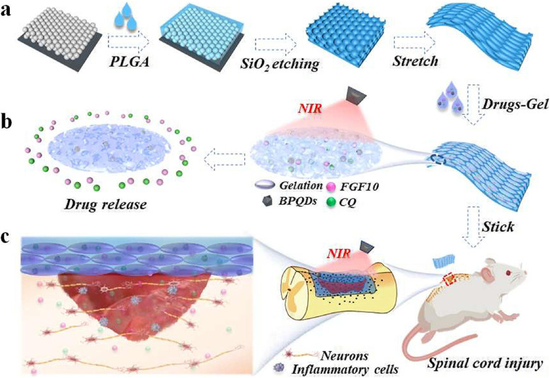
When designing engineered scaffolds for spinal cord injury healing, fundamental criteria such as structural stability, local microenvironment modeling, and cellular or biomolecular transmissibility should be taken into account. Under NIR irradiation, Wu et al. [72] employed BPQDs as a responsive drug release medium to produce hydrogels containing agarose, gelatin, and hyaluronic acid. Then, they permeated a temperature-sensitive hydrogel containing BPQD and drugs (fibroblast growth factor 10 (FGF10) and chloroquine phosphate (CQ)) into the pores of stretched inverse opal film (Drugs-Gel@SIOF), achieving Nir-controlled drug delivery and to a certain extent site-specific SCI repair in a rat model of SCI (see Fig. 3).
Fig. 3.
Schematic diagram of the fabrication of SIOF and its application in SCI repair. .Reproduced with permission from Ref [72]. Copyright 2019 Elsevier Ltd. All rights reserved
Recent research shown that BP promoted nerve regeneration by targeting copper ions, which are responsible for neuron degeneration in the blood-brain barrier (BBB). In addition to acting as nano-traps to control Cu2+ concentration, BPNs also lowered cellular reactive oxygen species (ROS) and protected cells from the toxicity associated with Cu2+ dysregulation [73]. Besides, the photothermal properties of BP confer exceptional permeability at the BBB, allowing the BP hybrid hydrogel with mechanical strength comparable to neural tissue to be utilized as a drug delivery and controlled release carrier in the central nervous system [73].
Treatment of cancer
BP has a high absorption when exposed to visible light. The capacity of BPNs to generate ROS by absorbing light makes them good candidates for NIR-based photothermal therapy (PTT) and photodynamic therapy (PDT) for tumor. In addition to phototherapy, it has been shown that BP itself has an inherent chemotherapeutic effect [74] The photothermal antimicrobial capabilities and bacterial toxicity of BP are also advantageous in adjuvant antimicrobial therapy for the direct treatment of cancer-related infections [75, 76]. Besides, owing to their relatively wide surface area, BPNs have been used to load therapeutic agents in order to accomplish various therapeutic combinations, such as combined chemical/photothermal therapy, gene/photodynamic therapy, and others. Combined therapy for cancer has shown reduced adverse effects and increased therapeutic effectiveness [77]. The important property of bare BP is its propensity to oxidize (or deteriorate naturally) and precipitate in the tumor microenvironment, leading in a short treatment cycle and an uneven photothermal effect. Therefore, the development of anticancer nanocomposites based on BP remains difficult. Due to the benefits of minimally invasive, high local concentration, low systemic toxicity, local drug toxicity, and sustained release exclusively at the tumor site, appropriate injectable hydrogel carrier system forms have been the subject of intense study [78, 79]. Various smart hydrogel delivery methods, such as heat-sensitive, pH-sensitive, photosensitive, and dual-sensitive hydrogels [80], have been created in response to the various kinds and stages of cancer. This review discussed the use of BP composite hydrogels in the treatment of cancer (see Table 2).
Table 2.
Application of black phosphorus composite hydrogel in cancer treatment
| Hydrogel matrix | Type of tumor | Highlights | Application type | References |
|---|---|---|---|---|
| PLEL | HeLa tumor | High PTT efficacy and antimicrobial activity | PTT | [81] |
| Cellulose | Liver cancer. Melanoma | Excellent photothermal response, enhanced stability and good flexibility and biocompatibility | PTT | [82] |
| Agarose | Liver cancer | NIR controlled release of Emetine used to regulate SG formation in tumor tissues during PTT and improve tumor sensitivity to PTT | PTT/ Drug delivery | [83] |
| Polypeptide | Liver cancer | The temperature-sensitive system releases bufalin by light control, reducing its side effects | PTT/ Chemotherapy | [84] |
| OSA and AHA | Gastric caner | Photo-controlled release of paclitaxel effectively inhibits tumor cell proliferation | PTT/ Chemotherapy | [85] |
| Pluronic F127 | Breast cancer | Photo-controlled release of gemcitabine with good photothermal efficiency and good biodegradability. | PTT/ Chemotherapy | [86] |
| PAHy | HeLa tumor | Excellent gel properties, pH sensitivity and NIR responsiveness. | PTT/ Chemotherapy | [87] |
| Pluronic F127 | Breast cancer | DTX with particle size dominance is continuously released to generate ROS synergistic injury to tumors | PDT/ Chemotherapy | [88] |
| DNA | Breast cancer | Photo-controlled DOX release system with positive charge has high permeability, reduces drug resistance and improves survival | PTT/PDT/ Chemotherapy | [89] |
| Chitosan | Lung cancer, glioblastoma, liver cancer | Temperature-sensitive cavernous system, hemostasis, antimicrobial, high penetration of the hematologic tumor barrier, capturing CuNPs, utilizing CDT and PTT of CuNPs, synergistic with aPD-L1 | PTT/CDT/IT | [90] |
| pNIPAM | Breast cancer, bladder cancer | Photo-controlled release of zoledronate as a scaffold for γδT cell proliferation and activation | IT/ Drug delivery | [91] |
| p (AAm-co-AAc) | Breast cancer, lung cancer | Pollen grains with their spike structure, high surface area, releasing cytokines and antibodies to proliferate and activate T cells | IT/ Drug delivery | [92] |
| Pluronic F-127 | Lung cancer | Heat-sensitive, controlled release GM-CSF and LPS, personalized cancer vaccine, tumor antigen carrier, binding PD-1 antibody | IT/ Drug delivery | [93] |
PTT is a novel kind of cancer treatment that destroys cancerous cells by the thermal action of exogenous light absorbers under NIR irradiation. Compared with other traditional treatment strategies, researchers have observed that PTT is easy, minimally invasive, has a low complication rate, and has a high spatial and temporal accuracy of near-infrared light, and diverse photothermal absorbers have been studied for PTT [94, 95]. Hydrogel platforms based on BP have also been employed for PTT. Shao et al. [81] exploited poly(d,l-lactide)-poly (ethyleneglycol)-poly(d,l-lactide)(PDLLA-PEG-PDLLA: PLEL) as heat-sensitive hydrogel matrix to develop a sprayable PTT system (BP@PLEL) by adding BPNs. BP@PLEL exhibited the potential to expedite the rapid transformation of sol-gel under NIR irradiation, obtain high PTT efficiency and antibacterial activity, which can be used to treat cancer postoperatively. Natural polymers may also be employed as host material for BP photothermal agent-based hydrogels. Xing et al. [82] developed cellulose and BPNs-based green injectable composite hydrogels (BPNSs). The green injectable composite hydrogel demonstrated an exceptional photothermal response, increased stability, and high flexibility on the basis of being fully non-toxic and biocompatible. In the PTT, a further rise in temperature causes in significant tissue damage. Inadequate heating of deep tumor tissue might result in tumor recurrence. Changing the sensitivity of tumor cells to PTT might potentially resolve this issue. Xie et al. [83] discovered that stress particles (SG) played a crucial role in the integration of internal and external stressors to control cell viability. SGs promoted and contributed in tumor resistance to PTT through a PTT-dependent mechanism reliant on eukaryotic initiation factor 2α. On the basis of this, they developed a hydrogel with BP as the photothermal agent for tumor-specific delivery and photo controlled release of NIR (SG inhibitor Emetine). Under near-infrared illumination, photothermal conversion of BPNs resulted in photothermal therapy (PTT) for tumor. Simultaneously, the photo-controlled release of Emetine in tumor tissues effectively blocked the synthesis of SG caused by PTT, rendering tumors susceptible to PTT, and so augmenting the tumor-inhibiting effect of PTT.
Due to their ability to absorb anticancer medications, BPNs can also be used to combine photothermal therapy with chemotherapy. Simultaneously, the intelligent co-delivery system of hydrogel can address the fundamental issues of drug toxicity, local sustained release, and photo-controlled release. Myocardial toxicity, for instance, restricts the therapeutic usage of Bufalin in cancer. In order to address this issue, He et al. [84] developed a BP hybrid polypeptide thermosensitive hydrogel (BP-bufalin@SH), which exhibited quick and significant temperature increase and released bufalin by photocontrol, therefore significantly minimizing the adverse effects of bufalin. Destruction of mitochondrial transmembrane potential may result in the irreversible apoptosis of cancer cells.
Sang et al. [85] prepared OSA/AHA/BP/PTX hydrogel by combining oxidized sodium alginate (OSA), amylated hyaluronic acid (AHA), black phosphorus (BP), and paclitaxel (PTX) under physiological conditions utilizing the formation of Scheff base bonds, and the results demonstrated excellent photothermal effect and sustained release ability of PTX. In brief, photothermal therapy combined with chemotherapy has shown more antitumor activity than chemotherapy alone [86, 87].
BPNs has been discovered to be an effective photosensitizer. Under NIR laser irradiation, BPNs generate a substantial quantity of ROS to destroy tumors, and have been employed in photodynamic therapy (PDT) [96–98]. PDT may also be used with chemotherapy, which has been extensively studied for its ability to boost anti-cancer effectiveness and inhibit tumor development. Li et al. [88] developed an injectable thermoreversible hydrogel (BPNs/ DTX-M-Hydrogel) employing F127 as the hydrogel matrix to encapsulate BPNs and docetaxel (DTX) micelles in order to promote the accumulation of medicines in tumor tissues and enhance anticancer activity. The combination of PTT with PDT is a potential strategy for enhancing treatment effectiveness. Zhou et al. [89] developed highly permeable, photothermal, injectable, and positively charged biodegradable nucleic acid hydrogel (DNA-gel) nanoparticles to deliver the anti-cancer medication azithromycin by combining cationic polymer PEI with negatively charged BPQD to generate PEI@BPQD. In mice with orthotopic breast cancers, DNA-gel treatment significantly decreased drug resistance and enhanced overall survival (83.3%, 78 days) compared to DOX chemotherapy alone. In addition, the beneficial photothermal effect of BP may increase the permeability of the blood tumor barrier (BTB) and facilitate therapy [99]. Utilizing this property, Wang et al. [90] developed a new therapeutic nanocomposite from chitosan (CS) hydrogel coupled with BPNs and copper nanoparticles produced in situ (CuNPs). The hydrogels (CS@BPNSs@CuNPs) were obtained with a temperature-sensitive spongy condition and a coagulation index of 24.98%. The released BPNs@CuNPs generated ROSto kill infected invasive bacteria (98.1%) and inhibit local residual tumor cell regeneration (11.3%), and demonstrated a 19.6% penetration rate across the BTB for the treatment of brain tumors. Combining the hydrogel platform with aPD-L1 immunotherapy (IT) produced a synergistic therapeutic effect for tumor prevention.
In addition to the aforementioned strategies, BP hybrid hydrogels may be used in cancer immunotherapy [100]. IT refers to processes that enhance the host immune system’s ability to react to tumors and create long-lasting antitumor responses [101]. Diverse immunotherapies for cancer, including immune checkpoint blockade (ICB) therapies, cytokine therapies, cancer vaccines, and adoptive T-cell therapies, have been developed and have shown promising clinical efficacy [102]. Emerging interest is particularly focused on manipulating nanocomposites as immune adjuvants to form unique drug delivery systems required for the combination of phototherapy and immunotherapy to eliminate primary and metastatic tumor cells by inducing dendritic cell (DC) maturation and cytotoxic T-lymphocyte infiltration [103]. For example, Shou et al. [91] employed the excellent biocompatibility of BPQDs-doped pNIPAM hydrogel particles to prepare scaffolds for the proliferation of γδT cells. In terms of γδT cell activation and growth, BPQDs-doped pNIPAM hydrogel particles loaded with zoledronate performed very well. Their team developed hydrogel-integrated natural pollen grains as artificial antigen presentation scaffolds for T cell activation and proliferation in vitro 2 years later. BP was in the scaffolds and enabled NIR to stimulate T cells by triggering the release of cytokines and antibodies. In addition, the pollen’s characteristic spike shape and large surface area led to the formation of T-cell clusters and increased their local proliferation. After in vitro growth, activated T cells exhibited significant anticancer activity [92]. Additionally, BP hybrid hydrogels might be utilized to deliver cancer vaccinations. Ye et al. [93] prepared BP quantum dot nanovesicles (BPQD-CCNVs) covered with surgically excised tumor cell membranes. Due of the presence of patient-specific tumor antigens in the surgically excised tumors, they were loaded onto heat-sensitive hydrogels containing GM-CSF and LPS. Sustained release of GM-CSF from subcutaneously injected Gel-BPQD-CCNVs effectively recruited dendritic cells to capture tumor antigens. LPS stimuli and NIR irradiation enhanced the growth and activation of DCs, which subsequently migrated to lymph nodes to deliver antigen to CD8+ T lymphocytes. In addition, the combination of PD-1 antibody significantly increased the eradication of surgical residual and metastatic lung tumors by tumor-specific CD8+ T cells. Therefore, it indicates that in cancer immunotherapy, in vitro cultivation of therapeutic T cells and personalization of cancer vaccines should give more attention to the BP composite hydrogel material.
In brief, BP composite hydrogels show significant promise as a novel way to enhance postoperative treatment (antibacterial property and tissue regeneration) and different therapeutic approaches for a wide range of malignancies (including but not limited to PTT, PDT, chemotherapy, drug delivery, immunotherapy, and vaccines).
Wound healing
The treatment of skin incisions, particularly chronic complicated incisions such as diabetic infection incisions, remains a significant challenge in regenerative medicine [104]. Single dressings are utilized in the clinic. There is a shortage of systemic, multifunctional wound dressings with high absorbability, form customization, quick self-healing, directing tissue regeneration, and restoring physiological function. Hydrogels offer better flexibility, exceptional elasticity, outstanding biocompatibility, a high-water content, and physiological environment sensitivity compared to other materials. Therefore, researchers have paid particular attention to hydrogels [105, 106]. In the hydrogel repair scaffold for wound healing, the antioxidant capacity of BP itself and the antibacterial ability of photodynamic treatment, which generates a significant quantity of ROS under NIR, have also been investigated in more detail [107, 108].
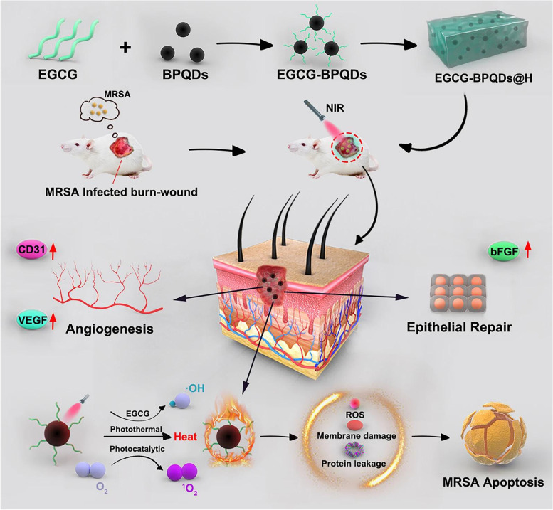
Mao et al. [109] discovered that the hybrid hydrogel prepared based on the basic electrostatic interaction between BP and chitosan (CS) generated a substantial quantity of singlet oxygen (1O2) when exposed to NIR light, which killed 98.90% of escherichia coliand 99.51% pearl and 99.51% staphylococcus aureus within 10 minutes. In addition to enhancing the synthesis of early fibrinogen and accelerating the formation of scabs during tissue regeneration, the hybrid hydrogel was capable of repeatability. BPNSs progressively degraded to phosphate, activating the PI3K/Akt and ERK1/2 signaling pathways stimulating wound healing and bacterial infection, and enhancing cell proliferation and differentiation. Xu et al. [110] used EGCG (Epigallocatechin gallate) modified BP quantum dots to load onto berberine nanohydrogel (BNH) to form EGCG-BPQDs@H in the selection of healing platform for MRSA (methicillin-resistant Staphylococcus aureus) -infected deep burn wounds (MIDBW) in diabetic patients (see Fig. 4).EGCG-BPQDs@H was more essential than BPQDs@H for photocatalytic singlet oxygen generation, according to the findings of electron spin resonance. Inhibition data indicated that the GCG-BPQDs@H sterilizing rate against MRSA was 88.6%. According to molecular biological investigation, EGCG-BPQDs significantly raised CD31 nearly 4 times and basic fibroblast growth factor (bFGF) nearly 2 times, which was advantageous for promoting the proliferation of vascular endothelial cells and skin epidermal cells. Under NIR irradiation, the MIDBW area treated with GCG-BPQDs@Hwas promptly sterilized by heating to 55 °C. After 21 days of therapy, the MIBDW closure rate was 92.4%, which was significantly higher than the control group (61.1%). Huang et al. [111] attached BPQDs to PVA (polyvinyl alcohol) and Alg (sodium alginate) matrices to generate BPQDs@NH using the same MIDBW model. Under NIR irradiation, BPQDs@NH generatedROS, lipid peroxidation, glutathione, adenosine triphosphate buildup, and bacterial membrane breakdown, all of which killed MRSA in a synergistic manner. In addition, animal experiments showed that BPQDs@NH effectively closed 95% of MIDWs after 12 days by lowering the inflammatory response and modifying the production of vascular endothelial growth factor (VEGF) and basic fibroblast growth factor (bFGF). To improve the antibacterial and antioxidant properties of BP composite hydrogels, researchers have also explored synergistic effects with other substances with excellent antibacterial properties, such as antimicrobial peptides, metal ions (e.g., zinc ions and copper ions), anthocyanins with potent antioxidant properties, and 4-octyl itaconate (4OI), as well as with immunotherapy [112–115]. Trace zinc, for instance, inhibited the polarization of macrophages towards the M2 phenotype. A significant number of M2 macrophages release anti-inflammatory proteins and cytokines to decrease inflammation and promote neovascularization [114]. An additional instance is the production of dendrimer-modified X+ Y-type DNA joint structural hydrogel based on the base pairing principle, which supported the change of macrophages from the pro-inflammatory M1 to the repair M2 phenotype and maintained a stable wound remodeling state. Surprisingly, DNA-hydrogel dressings induced neurons to enter a repair state, hence speeding nerve regeneration and angiogenesis in the skin. In addition, it might recruit bone marrow cells to trigger adaptive immune responses and enhance DNA-hydrogel dressings’ potential to induce tissue regeneration, ultimately enhancing hair follicle and hair regeneration [115].
Fig. 4.
Schematic diagram of synthesis of EGCG-BPQDs@H nanocomposites and the process of bactericidal and stimulating cell behavior. Reproduced with permission from Ref [110] Copyright 2019 BMC Ltd. All rights reserved
BP can also be loaded onto microneedle hydrogel tips for use in other wound healing attempts. The photothermal effect of BP is projected to dissociate oxygen from hemoglobin, therefore inducing local oxygen supply to increase skin cell proliferation and tissue remodeling [116]. It has also been shown that BP composite hydrogels generated in conjunction with gelatin or collagen matrix are a multifunctional and effective platform for the simultaneous treatment of melanoma and skin defect regeneration, which may cooperate with drug delivery and chemotherapy [117, 118].
Other biological applications
As a new type of drug delivery carrier, 2D BP nanomaterial demonstrates a high drug loading capacity owing to its rippled crystallization and structural properties, and BPNs have the ability to generate ROS by absorbing light, implying a significant development potential in other biomedical fields. Pan et al. [119] developed a unique therapeutic platform for the treatment of rheumatoid arthritis by mixing BPNs with platelet-rich plasma (PRP) -chitosan thermos-responsive hydrogel. Under NIR irradiation, BPNs created local heat while providing ROS to the inflamed joint in order to eliminate hyperplastic synovial tissue. Moreover, PRP efficiently increased the adherence of MSCS to thermosensitive chitosan hydrogels. This heat-responsive hydrogel also preserved articular cartilage by minimizing tissue friction. Assays of methotrexate release and absorption revealed the drug’s delayed release qualities, and the whole system significantly decreased the degree of edema in mice with arthritic inflammation.
Intelligent drug delivery nano-systems react to tiny changes in the environment’s physical and/or chemical signals by significantly altering their physical and/or chemical properties and subsequently releasing medications suited to illness development at proper paces. Due to their unique properties in the use of intelligent drug delivery nano systems [120, 121], BP and other 2D materials have a major place in many scientific and technical domains. In a diabetic mouse model, Dong et al. [122] reported a novel real-time bidirectional blood glucose regulatory drug delivery system (BDRS) comprised of glucose-loaded pressure responsive nanovesicles (Glu@PRNV), insulin-loaded BNPs (Insulin@BPNs), hydrogels, and painless glucose monitoring patches. BDRS could monitor glucose levels in real time by observing color changes. Later, depending on the needs, BDRS could release glucose in response to external pressure or replenish insulin in response to NIR irradiation to precisely regulate blood glucose levels in diabetic patients within reasonable fluctuations, thereby reducing the likelihood of hyperglycemia or hypoglycemia. By incorporating BP hydrogel microspheres into microne (MN) arrays, Lu et al. [123] developed a novel approach to achieve multifunctional and controlled drug delivery. Solid MN arrays comprising BP and poly (N-isopropyl acrylamide) (pNIPAM) packed with porous ethoxylated trimethylol propane triacrylate (ETPTA) modulated blood glucose levels in streptozotocin (STZ) -induced diabetic mice through subcutaneously regulated insulin release. These findings suggest that with more study and in vivo testing in the future, the use of 2D stratified materials such as BP and its derivatives in intelligent drug delivery systems will be used in clinical practice.
Concluding comments and future prospects
In comparison to other established nanomaterials based on hydrogels [124], the use of BP-based hydrogels remains in its infancy, and there are many research gaps. For instance, the study on the safety of 2D nanomaterials, particularly over the long term, is inadequate for biological uses. In addition, it remains difficult to develop easy, large-scale, cost-effective, and environmentally friendly production of BP composite hydrogel materials [125]. At present, bulk BP is produced mostly by converting white or red phosphorus under high temperature and pressure, with no water or oxygen required for the conversion. Consequently, the production of BP in bulk is restricted to the laboratory. In addition, the BP production is insufficient to fulfill the needs of future industrial applications. Therefore, there is an urgent need for a technique that can accurately manage BP characteristics, including size, number of layers, and surface modification, for the production of uniform BP. The particle size and its modification are crucial for controlling the biological behavior of BP [8]. The defining properties of bare BP is its vulnerability to oxidation (or natural degradation) and precipitation in the tumor microenvironment, which results in short-term therapy and uneven photothermal effects. Therefore, it is often required to develop a number of techniques to change and passivate the surface of BP in order to enhance its photothermal stability. During the antibacterial treatment of skin wounds, a substantial quantity of ROS generated by the photothermal effect of BP are toxic to the DNA of normal cells, leading to progressive oxidative damage and final cell death. Consequently, determining the appropriate dosage is essential. The behavior of BP in vivo and the method by which it interacts with diverse biomolecules also need further research. As regards toxicity, as research on BP is still in its infancy, theoretical and experimental studies on BP, such as the degradation process of BP in vivo, need to be enhanced. The key to the effective clinical use of nanomedicine is its in vivo safety. The primary component of BP material is phosphorus, which is an abundant element in the human body. Therefore, BP may be broken down into phosphoric acid in the human body and is non-toxic. In mice, the safety of BP was also shown. Long-term research is required to determine whether long-term usage may produce excessive poisoning of phosphate ions, such as loss of metal ions (such as calcium and magnesium) in the body. Modern biomedical technology, physicochemical technology, and precision manufacturing technology are required for inter-disciplinary research into the development of safe, dependable, effective, and low-toxic 2D materials with clinical applications.
Opportunities exist with challenges. Extensive experimental and theoretical research is currently ongoing to fully unveil the potentials of BP. Based on new technology, BP has been employed in many biomedical domains, however it has not been further investigated in combination with hydrogels. Examples include highly sensitive and specific medical sensors for diagnostic and prognostic monitoring, biological imaging based on the strong interaction between electromagnetic waves and BP crystal structures (photoacoustic imaging, thermal imaging, fluorescence imaging), drug delivery by multiple pathways, gene therapy, and others [126–130]. These directions may be used to hydrogels in the future. We believe that BP-based hydrogel scaffolds will become an important method for medical research and clinical treatment in the near future, bringing benefits to the diagnosis and treatment of patients with a variety of diseases, as a result of the in-depth study of their application in regenerative medicine and the resolution of in vivo stability and biosafety concerns.
Authors’ contributions
Haoxuan Li and Qingsan Zhu wrote the main manuscript text and Kunchi Zhao and Jiajia Jiang prepared Figs. 1, 2, 3 and 4. All authors reviewed the manuscript. The author(s) read and approved the final manuscript.
Funding
This work was supported by Jilin Province Health Research Talents Special Project (Grant No: 2019scz061).
Availability of data and materials
All data generated or analyzed during this study are included in this published article and its supplementary information files.
Declarations
Ethics approval and consent to participate
Not applicable.
Consent for publication
Not applicable.
Competing interests
There are no conflicts of interest to declare.
Footnotes
Publisher’s Note
Springer Nature remains neutral with regard to jurisdictional claims in published maps and institutional affiliations.
Contributor Information
Hao-xuan Li, Email: haoxuan20@mailis.jlu.edu.cn.
Kun-chi Zhao, Email: zhaokunchi@jlu.edu.cn.
Jia-jia Jiang, Email: jiangjiajia@jlu.edu.cn.
Qing-san Zhu, Email: zhuqs@jlu.edu.cn.
References
- 1.Wang J, Zhu M, Hu Y, Chen R, Hao Z, Wang Y, et al. Exosome-hydrogel system in bone tissue engineering: a promising therapeutic strategy. Macromol Biosci. 2022:e2200496-e. [DOI] [PubMed]
- 2.Xing Y, Zeng B, Yang W. Light responsive hydrogels for controlled drug delivery. Front Bioeng Biotechnol. 2022:10. [DOI] [PMC free article] [PubMed]
- 3.Hu Z-C, Lu J-Q, Zhang T-W, Liang H-F, Yuan H, Su D-H, et al. Piezoresistive MXene/silk fibroin nanocomposite hydrogel for accelerating bone regeneration by re-establishing electrical microenvironment. Bioactive Mater. 2023;22:1–17. doi: 10.1016/j.bioactmat.2022.08.025. [DOI] [PMC free article] [PubMed] [Google Scholar]
- 4.Zhu W, Zhou Z, Huang Y, Liu H, He N, Zhu X, et al. A versatile 3D-printable hydrogel for antichondrosarcoma, antibacterial, and tissue repair. J Mater Sci Technol. 2023;136:200–211. [Google Scholar]
- 5.Rafieian S, Mirzadeh H, Mahdavi H, Masoumi ME. A review on nanocomposite hydrogels and their biomedical applications. Sci Eng Composite Mater. 2019;26(1):154–74.
- 6.Song HS, Kwon OS, Kim J-H, Conde J, Artzi N. 3D hydrogel scaffold doped with 2D graphene materials for biosensors and bioelectronics. Biosens Bioelectron. 2017;89:187–200. doi: 10.1016/j.bios.2016.03.045. [DOI] [PubMed] [Google Scholar]
- 7.Huang H, Feng W, Chen Y. Two-dimensional biomaterials: material science, biological effect and biomedical engineering applications. Chem Soc Rev. 2021;50(20):11381–11485. doi: 10.1039/d0cs01138j. [DOI] [PubMed] [Google Scholar]
- 8.Xiong S, Chen X, Liu Y, Fan T, Wang Q, Zhang H, et al. Black phosphorus as a versatile nanoplatform: from unique properties to biomedical applications. J Innov Opt Health Sci. 2020;13(5).
- 9.Du C, Huang W. Progress and prospects of nanocomposite hydrogels in bone tissue engineering. Nanocomposites. 2022;8(1):102–124. [Google Scholar]
- 10.Vashist A, Kaushik A, Vashist A, Sagar V, Ghosal A, Gupta YK, et al. Advances in carbon nanotubes–hydrogel hybrids in nanomedicine for therapeutics. Adv Healthc Mater. 2018;7(9):1701213. [DOI] [PMC free article] [PubMed]
- 11.Liu J, Zhao C, Chen WR, Zhou B. Recent progress in two-dimensional nanomaterials for cancer theranostics. Coord Chem Rev. 2022;469.
- 12.Mustafar S, Yusuf AKNM, Borines LM, Kusumawati EN, Kamari A, Ali NM, et al. Metal-organic framework Nanosheets (MONs): a review on interfacial syntheses and applications of coordination Nanosheets. Biointerface Res Appl Chem. 2023;13(2).
- 13.Naikoo GA, Arshad F, Almas M, Hassan IU, Pedram MZ, Aljabali AAA, et al. 2D materials, synthesis, characterization and toxicity: a critical review. Chem Biol Interact. 2022;365:110081. doi: 10.1016/j.cbi.2022.110081. [DOI] [PubMed] [Google Scholar]
- 14.Wu M, Niu X, Zhang R, Xu ZP. Two-dimensional nanomaterials for tumor microenvironment modulation and anticancer therapy. Adv Drug Deliv Rev. 2022;187. [DOI] [PubMed]
- 15.Peera SG, Koutavarapu R, Chao L, Singh L, Murugadoss G, Rajeshkhanna G. 2D MXene nanomaterials as Electrocatalysts for hydrogen evolution reaction (HER): A Review. Micromachines. 2022;13(9). [DOI] [PMC free article] [PubMed]
- 16.Chaudhary V, Neugebauer P, Mounkachi O, Lahbabi S, El Fatimy A. Phosphorene—an emerging two-dimensional material: recent advances in synthesis, functionalization, and applications. 2D Mater. 2022;9(3):032001. [Google Scholar]
- 17.Li L, Yu Y, Ye GJ, Ge Q, Ou X, Wu H, et al. Black phosphorus field-effect transistors. Nat Nanotechnol. 2014;9(5):372–377. doi: 10.1038/nnano.2014.35. [DOI] [PubMed] [Google Scholar]
- 18.Liu H, Neal AT, Zhu Z, Luo Z, Xu X, Tománek D, et al. Phosphorene: an unexplored 2D semiconductor with a high hole mobility. ACS Nano. 2014;8(4):4033–4041. doi: 10.1021/nn501226z. [DOI] [PubMed] [Google Scholar]
- 19.Liang J, Hu Y, Zhang K, Wang Y, Song X, Tao A, et al. 2D layered black arsenic-phosphorus materials: synthesis, properties, and device applications. Nano Res. 2022;15(4):3737–3752. [Google Scholar]
- 20.Peng L, Abbasi N, Xiao Y, Xie Z. Black phosphorus: degradation mechanism, passivation method, and application for in situ tissue regeneration. Adv Mater Interfaces. 2020;7(23).
- 21.Wang H, Yu X-F. Few-layered black phosphorus: from fabrication and customization to biomedical applications. Small. 2018;14(6). [DOI] [PubMed]
- 22.Hu Z, Niu T, Guo R, Zhang J, Lai M, He J, et al. Two-dimensional black phosphorus: its fabrication, functionalization and applications. Nanoscale. 2018;10(46):21575–21603. doi: 10.1039/c8nr07395c. [DOI] [PubMed] [Google Scholar]
- 23.Eswaraiah V, Zeng Q, Long Y, Liu Z. Black phosphorus Nanosheets: synthesis, characterization and applications. Small. 2016;12(26):3480–3502. doi: 10.1002/smll.201600032. [DOI] [PubMed] [Google Scholar]
- 24.Miao J, Zhang L, Wang C. Black phosphorus electronic and optoelectronic devices. 2d Mater. 2019;6(3).
- 25.Dinh KN, Zhang Y, Sun W. The synthesis of black phosphorus: from zero- to three-dimensional nanostructures. J Phys Energy. 2021;3(3).
- 26.Luo M, Fan T, Zhou Y, Zhang H, Mei L. 2D black phosphorus–based biomedical applications. Adv Funct Mater. 2019;29(13):1808306.
- 27.Huang X, Zhou Y, Woo CM, Pan Y, Nie L, Lai P. Multifunctional layered black phosphorene-based nanoplatform for disease diagnosis and treatment: a review. Front Optoelectron. 2020;13(4):327–351. doi: 10.1007/s12200-020-1084-1. [DOI] [PMC free article] [PubMed] [Google Scholar]
- 28.Wei H, Fan W, Dong Y, Wang Y, Zhou L, Wang Y, et al. Black phosphorus quantum dots: Nonlinear optical modulation material with ultraviolet saturable absorption.Frontiers in Physics.2022;10.
- 29.Anju S, Ashtami J, Mohanan PV. Black phosphorus, a prospective graphene substitute for biomedical applications. Mater Sci Eng C. 2019;97:978–993. doi: 10.1016/j.msec.2018.12.146. [DOI] [PubMed] [Google Scholar]
- 30.Yi Y, Yu X-F, Zhou W, Wang J, Chu PK. Two-dimensional black phosphorus: synthesis, modification, properties, and applications. Mater Sci Eng. 2017;120:1–33. [Google Scholar]
- 31.Gusmao R, Sofer Z, Pumera M. Black phosphorus rediscovered: from bulk material to monolayers. Angewandte Chemie-Int Edition. 2017;56(28):8052–8072. doi: 10.1002/anie.201610512. [DOI] [PubMed] [Google Scholar]
- 32.Liu H, Du Y, Deng Y, Ye PD. Semiconducting black phosphorus: synthesis, transport properties and electronic applications. Chem Soc Rev. 2015;44(9):2732–2743. doi: 10.1039/c4cs00257a. [DOI] [PubMed] [Google Scholar]
- 33.Thurakkal S, Feldstein D, Perea-Causin R, Malic E, Zhang X. The art of constructing black phosphorus Nanosheet based Heterostructures: from 2D to 3D. Adv Mater. 2021;33(3). [DOI] [PMC free article] [PubMed]
- 34.Yuan Z, Liu D, Tian N, Zhang G, Zhang Y. Structure, preparation and properties of phosphorene. Acta Chim Sin. 2016;74(6):488–497. [Google Scholar]
- 35.Qiu M, Ren WX, Jeong T, Won M, Park GY, Sang DK, et al. Omnipotent phosphorene: a next-generation, two-dimensional nanoplatform for multidisciplinary biomedical applications. Chem Soc Rev. 2018;47(15):5588–5601. doi: 10.1039/c8cs00342d. [DOI] [PubMed] [Google Scholar]
- 36.Huang K, Wu J, Gu Z. Black phosphorus hydrogel scaffolds enhance bone regeneration via a sustained supply of calcium-free phosphorus. ACS Appl Mater Interfaces. 2019;11(3):2908–2916. doi: 10.1021/acsami.8b21179. [DOI] [PubMed] [Google Scholar]
- 37.Xu D, Gan K, Wang Y, Wu Z, Wang Y, Zhang S, et al. A composite Deferoxamine/black phosphorus Nanosheet/gelatin hydrogel scaffold for ischemic Tibial bone repair. Int J Nanomedicine. 2022;17:1015–1030. doi: 10.2147/IJN.S351814. [DOI] [PMC free article] [PubMed] [Google Scholar]
- 38.Miao Y, Shi X, Li Q, Hao L, Liu L, Liu X, et al. Engineering natural matrices with black phosphorus nanosheets to generate multi-functional therapeutic nanocomposite hydrogels. Biomater Sci. 2019;7(10):4046–4059. doi: 10.1039/c9bm01072f. [DOI] [PubMed] [Google Scholar]
- 39.Shao J, Ruan C, Xie H, Chu PK, Yu X-F. Photochemical activity of black phosphorus for near-infrared light controlled in situ biomineralization. Advanced Science. 2020;7(14). [DOI] [PMC free article] [PubMed]
- 40.Tan L, Hu Y, Li M, Zhang Y, Xue C, Chen M, et al. Remotely-activatable extracellular matrix-mimetic hydrogel promotes physiological bone mineralization for enhanced cranial defect healing. Chem Eng J. 2022;431:133382. [Google Scholar]
- 41.Tong L, Liao Q, Zhao Y, Huang H, Gao A, Zhang W, et al. Near-infrared light control of bone regeneration with biodegradable photothermal osteoimplant. Biomaterials. 2019;193:1–11. doi: 10.1016/j.biomaterials.2018.12.008. [DOI] [PubMed] [Google Scholar]
- 42.Liu X, George MN, Li L, Gamble D, Miller Ii AL, Gaihre B, et al. Injectable electrical conductive and phosphate releasing gel with two-dimensional black phosphorus and carbon nanotubes for bone tissue engineering. ACS Biomater Sci Eng. 2020;6(8):4653–4665. doi: 10.1021/acsbiomaterials.0c00612. [DOI] [PMC free article] [PubMed] [Google Scholar]
- 43.Xu H, Liu X, George MN, Miller AL, 2nd, Park S, Xu H, et al. Black phosphorus incorporation modulates nanocomposite hydrogel properties and subsequent MC3T3 cell attachment, proliferation, and differentiation. J Biomed Mater Res A. 2021;109(9):1633–1645. doi: 10.1002/jbm.a.37159. [DOI] [PMC free article] [PubMed] [Google Scholar]
- 44.Li L, Liu X, Gaihre B, Li Y, Lu L. Mesenchymal stem cell spheroids incorporated with collagen and black phosphorus promote osteogenesis of biodegradable hydrogels. Mater Sci Eng C. 2021;121:111812. doi: 10.1016/j.msec.2020.111812. [DOI] [PubMed] [Google Scholar]
- 45.Xu H, Liu X, Park S, Terzic A, Lu L. Size-dependent osteogenesis of black phosphorus in nanocomposite hydrogel scaffolds. J Biomed Mater Res A. 2022;110(8):1488–1498. doi: 10.1002/jbm.a.37382. [DOI] [PMC free article] [PubMed] [Google Scholar]
- 46.Hu Z, Lu J, Hu A, Dou Y, Wang S, Su D, et al. Engineering BPQDs/PLGA nanospheres-integrated wood hydrogel bionic scaffold for combinatory bone repair and osteolytic tumor therapy. Chem Eng J. 2022;446:137269. [Google Scholar]
- 47.Miao Y, Chen Y, Luo J, Liu X, Yang Q, Shi X, et al. Black phosphorus nanosheets-enabled DNA hydrogel integrating 3D-printed scaffold for promoting vascularized bone regeneration. Bioactive Mater. 2023;21:97–109. doi: 10.1016/j.bioactmat.2022.08.005. [DOI] [PMC free article] [PubMed] [Google Scholar]
- 48.Xu Y, Xu C, He L, Zhou J, Chen T, Ouyang L, et al. Stratified-structural hydrogel incorporated with magnesium-ion-modified black phosphorus nanosheets for promoting neuro-vascularized bone regeneration. Bioactive Mater. 2022;16:271–284. doi: 10.1016/j.bioactmat.2022.02.024. [DOI] [PMC free article] [PubMed] [Google Scholar]
- 49.Qing Y, Wang H, Lou Y, Fang X, Li S, Wang X, et al. Chemotactic ion-releasing hydrogel for synergistic antibacterial and bone regeneration. Mater Today Chem. 2022;24:100894. [Google Scholar]
- 50.Wang X, Shao J, Abd El Raouf M, Xie H, Huang H, Wang H, et al. Near-infrared light-triggered drug delivery system based on black phosphorus for in vivo bone regeneration. Biomaterials. 2018;179:164–174. doi: 10.1016/j.biomaterials.2018.06.039. [DOI] [PubMed] [Google Scholar]
- 51.Yang B, Yin J, Chen Y, Pan S, Yao H, Gao Y, et al. 2D-black-phosphorus-reinforced 3d-printed scaffolds:a stepwise countermeasure for osteosarcoma. Adv Mater (Deerfield Beach, Fla). 2018;30(10). [DOI] [PubMed]
- 52.Ling X, Wang H, Huang S, Xia F, Dresselhaus MS. The renaissance of black phosphorus. Proc Natl Acad Sci U S A. 2015;112(15):4523–30. [DOI] [PMC free article] [PubMed]
- 53.Chen S, Guo R, Liang Q, Xiao X. Multifunctional modified polylactic acid nanofibrous scaffold incorporating sodium alginate microspheres decorated with strontium and black phosphorus for bone tissue engineering. J Biomater Sci Polym Ed. 2021;32(12):1598–1617. doi: 10.1080/09205063.2021.1927497. [DOI] [PubMed] [Google Scholar]
- 54.Liu X, Miller AL, 2nd, Park S, George MN, Waletzki BE, Xu H, et al. Two-dimensional black phosphorus and graphene oxide Nanosheets synergistically enhance cell proliferation and osteogenesis on 3D printed scaffolds. ACS Appl Mater Interfaces. 2019;11(26):23558–23572. doi: 10.1021/acsami.9b04121. [DOI] [PMC free article] [PubMed] [Google Scholar]
- 55.Chen H, Jin Y, Wang J, Wang Y, Jiang W, Dai H, et al. Design of smart targeted and responsive drug delivery systems with enhanced antibacterial properties. Nanoscale. 2018;10(45):20946–20962. doi: 10.1039/c8nr07146b. [DOI] [PubMed] [Google Scholar]
- 56.Wu Q, Wei G, Xu Z, Han J, Xi J, Fan L, et al. Mechanistic insight into the light-irradiated carbon capsules as an antibacterial agent. ACS Appl Mater Interfaces. 2018;10(30):25026–25036. doi: 10.1021/acsami.8b04932. [DOI] [PubMed] [Google Scholar]
- 57.Shui C, Scutt A. Mild heat shock induces proliferation, alkaline phosphatase activity, and mineralization in human bone marrow stromal cells and mg-63 cells. In Vitro. 2001;16(4):731–741. doi: 10.1359/jbmr.2001.16.4.731. [DOI] [PubMed] [Google Scholar]
- 58.Xia F, Wang H, Jia Y. Rediscovering black phosphorus as an anisotropic layered material for optoelectronics and electronics. Nat Commun. 2014;5:4458. doi: 10.1038/ncomms5458. [DOI] [PubMed] [Google Scholar]
- 59.Qu G, Xia T, Zhou W, Zhang X, Zhang H, Hu L, et al. Property–activity relationship of black phosphorus at the nano–bio interface: from molecules to organisms. Chem Rev. 2020;120(4):2288–2346. doi: 10.1021/acs.chemrev.9b00445. [DOI] [PubMed] [Google Scholar]
- 60.Zhang X, Zhang Z, Zhang S, Li D, Ma W, Ma C, et al. Size effect on the cytotoxicity of layered black phosphorus and underlying mechanisms. Small. 2017;13(32):1701210. [DOI] [PubMed]
- 61.Zhang X, Xie H, Liu Z, Tan C, Luo Z, Li H, et al. Black phosphorus quantum dots. Angewandte Chemie (International ed in English) 2015;54(12):3653–3657. doi: 10.1002/anie.201409400. [DOI] [PubMed] [Google Scholar]
- 62.Sun Z, Xie H, Tang S, Yu X-F, Guo Z, Shao J, et al. Ultrasmall Black Phosphorus Quantum Dots: Synthesis and Use as Photothermal Agents. Angew Chem Int Ed. 2015;54(39):11526–30. [DOI] [PubMed]
- 63.Shao J, Xie H, Huang H, Li Z, Sun Z, Xu Y, et al. Biodegradable black phosphorus-based nanospheres for in vivo photothermal cancer therapy. Nat Commun. 2016;7(1):12967. doi: 10.1038/ncomms12967. [DOI] [PMC free article] [PubMed] [Google Scholar]
- 64.Lin S, Yang G, Jiang F, Zhou M, Yin S, Tang Y, et al. A magnesium-enriched 3D culture system that mimics the bone development microenvironment for vascularized bone regeneration. Adv Sci 2019;6(12). [DOI] [PMC free article] [PubMed]
- 65.Park J-W, Hanawa T, Chung J-H. The relative effects of ca and mg ions on MSC osteogenesis in the surface modification of microrough Ti implants. Int J Nanomed. 2019;14:5697–5711. doi: 10.2147/IJN.S214363. [DOI] [PMC free article] [PubMed] [Google Scholar]
- 66.Vaudry D, Stork PJS, Lazarovici P, Eiden LE. Signaling pathways for PC12 cell differentiation: making the right connections. Science. 2002;296(5573):1648–1649. doi: 10.1126/science.1071552. [DOI] [PubMed] [Google Scholar]
- 67.Cheng L, Cai Z, Zhao J, Wang F, Lu M, Deng L, et al. Black phosphorus-based 2D materials for bone therapy. Bioactive Mater. 2020;5(4):1026–1043. doi: 10.1016/j.bioactmat.2020.06.007. [DOI] [PMC free article] [PubMed] [Google Scholar]
- 68.Kang MS, Song S-J, Cha JH, Cho Y, Lee HU, Hyon S-H, et al. Increased neuritogenesis on ternary nanofiber matrices of PLCL and laminin decorated with black phosphorus. J Ind Eng Chem. 2020;92:226–235. [Google Scholar]
- 69.Sun J, Zheng G, Lee H-W, Liu N, Wang H, Yao H, et al. Formation of stable phosphorus–carbon bond for enhanced performance in black phosphorus nanoparticle–graphite composite battery anodes. Nano Lett. 2014;14(8):4573–4580. doi: 10.1021/nl501617j. [DOI] [PubMed] [Google Scholar]
- 70.Xu C, Xu Y, Yang M, Chang Y, Nie A, Liu Z, et al. Black-phosphorus-incorporated hydrogel as a conductive and biodegradable platform for enhancement of the neural differentiation of mesenchymal. Stem Cells. 2020;30(39):2000177. [Google Scholar]
- 71.Halim A, Qu K-Y, Zhang X-F, Huang N-P. Recent advances in the application of two-dimensional nanomaterials for neural tissue engineering and regeneration. ACS Biomater Sci Eng. 2021;7(8):3503–3529. doi: 10.1021/acsbiomaterials.1c00490. [DOI] [PubMed] [Google Scholar]
- 72.Wu F, Zu Y, Weng W, Yang Y, Hu J, Mao Y, et al. Multifunctional inverse opal film as a responsive drug carrier for spinal cord injury repair. Chem Eng J. 2022;436:135256. [Google Scholar]
- 73.Chen W, Ouyang J, Yi X, Xu Y, Niu C, Zhang W, et al. Black phosphorus nanosheets as a neuroprotective nanomedicine for neurodegenerative disorder therapy. Adv Mater .2018;30(3):1703458. [DOI] [PubMed]
- 74.Zhou W, Pan T, Cui H, Zhao Z, Chu PK, Yu X-F. Black phosphorus: bioactive nanomaterials with inherent and selective chemotherapeutic effects. Angew Chem Int Ed. 2019;58(3):769–74. [DOI] [PubMed]
- 75.Xiong Z, Zhang X, Zhang S, Lei L, Ma W, Li D, et al. Bacterial toxicity of exfoliated black phosphorus nanosheets. Ecotoxicol Environ Saf. 2018;161:507–514. doi: 10.1016/j.ecoenv.2018.06.008. [DOI] [PubMed] [Google Scholar]
- 76.Alibek K, Bekmurzayeva A, Mussabekova A, Sultankulov B. Using antimicrobial adjuvant therapy in cancer treatment: a review. Infect Agents Cancer. 2012;7(1). [DOI] [PMC free article] [PubMed]
- 77.Qin L, Jiang S, He H, Ling G, Zhang P. Functional black phosphorus nanosheets for cancer therapy. J Control Release. 2020;318:50–66. doi: 10.1016/j.jconrel.2019.12.013. [DOI] [PubMed] [Google Scholar]
- 78.Norouzi M, Nazari B, Miller DW. Injectable hydrogel-based drug delivery systems for local cancer therapy. Drug Discov Today. 2016;21(11):1835–1849. doi: 10.1016/j.drudis.2016.07.006. [DOI] [PubMed] [Google Scholar]
- 79.Xiao Y, Gu Y, Qin L, Chen L, Chen X, Cui W, et al. Injectable thermosensitive hydrogel-based drug delivery system for local cancer therapy. Colloids Surf B-Biointerfaces. 2021:200. [DOI] [PubMed]
- 80.Fan D-y, Tian Y, Liu Z-j. Injectable hydrogels for localized cancer therapy. Front Chem. 2019:7. [DOI] [PMC free article] [PubMed]
- 81.Shao J, Ruan C, Xie H, Li Z, Wang H, Chu PK, et al. Black-phosphorus-incorporated hydrogel as a Sprayable and biodegradable Photothermal platform for postsurgical treatment of. Cancer. 2018;5(5):1700848. doi: 10.1002/advs.201700848. [DOI] [PMC free article] [PubMed] [Google Scholar]
- 82.Xing C, Chen S, Qiu M, Liang X, Liu Q, Zou Q, et al. Conceptually novel black phosphorus/cellulose hydrogels as promising photothermal agents for effective cancer therapy. Adv Healthc Mater. 2018;7(7):1701510. [DOI] [PubMed]
- 83.Xie J, Fan T, Kim JH, Xu Y, Wang Y, Liang W, et al. Emetine-loaded black phosphorus hydrogel sensitizes tumor to photothermal therapy through inhibition of stress granule formation. Adv Funct Mater. 2020;30(43):2003891.
- 84.He J, Chen G, Zhao P, Ou C. Near-infrared light-controllable bufalin delivery from a black phosphorus-hybrid supramolecular hydrogel for synergistic photothermal-chemo tumor therapy. Nano Res. 2021;14(11):3988–3998. [Google Scholar]
- 85.Sang S, Jiang Z, Xie N, Rao H, Liao K, Hu Q, et al. Black phosphorus nanosheets and paclitaxel encapsulated hydrogel for synergistic photothermal-chemotherapy. Nanophotonics. 2021;10(10):2625–37.
- 86.Qin L, Ling G, Peng F, Zhang F, Jiang S, He H, et al. Black phosphorus nanosheets and gemcitabine encapsulated thermo-sensitive hydrogel for synergistic photothermal-chemotherapy. J Colloid Interface Sci. 2019;556:232–238. doi: 10.1016/j.jcis.2019.08.058. [DOI] [PubMed] [Google Scholar]
- 87.Wu R-S, Lin J, Xing Y-M, Dai Z-L, Wang L-W, Zhang X-P. pH-sensitive black phosphorous–incorporated hydrogel as novel implant for cancer treatment. J Pharm Sci. 2019;108(8):2542–2551. doi: 10.1016/j.xphs.2019.03.003. [DOI] [PubMed] [Google Scholar]
- 88.Li R, Shan L, Yao Y, Peng F, Jiang S, Yang D, et al. Black phosphorus nanosheets and docetaxel micelles co-incorporated thermoreversible hydrogel for combination chemo-photodynamic therapy. Drug Deliv Transl Res. 2021;11(3):1133–1143. doi: 10.1007/s13346-020-00836-y. [DOI] [PubMed] [Google Scholar]
- 89.Zhou L, Pi W, Hao M, Li Y, An H, Li Q, et al. An injectable and biodegradable nano-photothermal DNA hydrogel enhances penetration and efficacy of tumor therapy. Biomater Sci. 2021;9(14):4904–4921. doi: 10.1039/d1bm00568e. [DOI] [PubMed] [Google Scholar]
- 90.Wang W, Zhang Q, Zhang M, Lv X, Li Z, Mohammadniaei M, et al. A novel biodegradable injectable chitosan hydrogel for overcoming postoperative trauma and combating multiple tumors. Carbohydr Polym. 2021;265:118065. doi: 10.1016/j.carbpol.2021.118065. [DOI] [PubMed] [Google Scholar]
- 91.Shou X, Liu Y, Wu D, Zhang H, Zhao Y, Sun W, et al. Black phosphorus quantum dots doped multifunctional hydrogel particles for cancer immunotherapy. Chem Eng J. 2021;408:127349. [Google Scholar]
- 92.Shou X, Yu Y, Wu D, Wang F, Sun W, Duan P, et al. Spiny pollen-based antigen-presenting clusters for promoting T cells expansion. Chem Eng J. 2022;437:135374. [Google Scholar]
- 93.Ye X, Liang X, Chen Q, Miao Q, Chen X, Zhang X, et al. Surgical tumor-derived personalized Photothermal vaccine formulation for Cancer immunotherapy. ACS Nano. 2019;13(3):2956–2968. doi: 10.1021/acsnano.8b07371. [DOI] [PubMed] [Google Scholar]
- 94.Li J, Zhang W, Ji W, Wang J, Wang N, Wu W, et al. Near infrared photothermal conversion materials: mechanism, preparation, and photothermal cancer therapy applications. J Mater Chem B. 2021;9(38):7909–7926. doi: 10.1039/d1tb01310f. [DOI] [PubMed] [Google Scholar]
- 95.Zhang Y, Ang CY, Zhao Y. Polymeric nanocarriers incorporating near-infrared absorbing agents for potent photothermal therapy of cancer. Polym J. 2016;48(5):589–603. [Google Scholar]
- 96.Wang H, Yang X, Shao W, Chen S, Xie J, Zhang X, et al. Ultrathin black phosphorus Nanosheets for efficient singlet oxygen generation. J Am Chem Soc. 2015;137(35):11376–11382. doi: 10.1021/jacs.5b06025. [DOI] [PubMed] [Google Scholar]
- 97.Qi F, Ji P, Chen Z, Wang L, Yao H, Huo M, et al. Photosynthetic Cyanobacteria-hybridized black phosphorus Nanosheets for enhanced tumor photodynamic therapy. Small. 2021;17(42). [DOI] [PubMed]
- 98.Zhang M, Wang W, Cui Y, Zhou N, Shen J. Near-infrared light-mediated photodynamic/photothermal therapy nanoplatform by the assembly of Fe3O4 carbon dots with graphitic black phosphorus quantum dots. Int J Nanomed. 2018;13:2803–2819. doi: 10.2147/IJN.S156434. [DOI] [PMC free article] [PubMed] [Google Scholar]
- 99.Chen W, Ouyang J, Liu H, Chen M, Zeng K, Sheng J, et al. Black phosphorus Nanosheet-based drug delivery system for synergistic photodynamic/Photothermal/chemotherapy of. Cancer. 2017;29(5):1603864. doi: 10.1002/adma.201603864. [DOI] [PubMed] [Google Scholar]
- 100.Guo R, Wang S, Zhao L, Zong Q, Li T, Ling G, et al. Engineered nanomaterials for synergistic photo-immunotherapy. Biomaterials. 2022;282:121425. doi: 10.1016/j.biomaterials.2022.121425. [DOI] [PubMed] [Google Scholar]
- 101.Hu ZJ. The future of immune checkpoint blockade immunotherapy: towards personalized therapy or towards combination therapy. J Thorac Dis. 2017;9(11):4226–9. [DOI] [PMC free article] [PubMed]
- 102.Chen Q, Chen M, Liu Z. Local biomaterials-assisted cancer immunotherapy to trigger systemic antitumor responses. Chem Soc Rev. 2019;48(22):5506–5526. doi: 10.1039/c9cs00271e. [DOI] [PubMed] [Google Scholar]
- 103.Liu H, Mei Y, Zhao Q, Zhang A, Tang L, Gao H, et al. Black phosphorus, an emerging versatile nanoplatform for cancer immunotherapy. Biopharm. 2021;13(9):1344. [DOI] [PMC free article] [PubMed]
- 104.Uppuluri VNVA, Sathanantham ST, Bhimavarapu SK, Elumalai L. Polymeric hydrogel scaffolds: skin tissue engineering and regeneration. Adv Pharm Bull. 2022;12(3):437–448. doi: 10.34172/apb.2022.069. [DOI] [PMC free article] [PubMed] [Google Scholar]
- 105.Maleki A, He J, Bochani S, Nosrati V, Shahbazi M-A, Guo B. Multifunctional photoactive hydrogels for wound healing acceleration. ACS Nano. 2021;15(12):18895–18930. doi: 10.1021/acsnano.1c08334. [DOI] [PubMed] [Google Scholar]
- 106.Zhang L, Yin H, Lei X, Lau JNY, Yuan M, Wang X, et al. A systematic review and Meta-analysis of clinical effectiveness and safety of hydrogel dressings in the management of skin wounds. Front Bioeng Biotechnol. 2019:7. [DOI] [PMC free article] [PubMed]
- 107.Ran P, Chen W, Zheng H, Zhou J, Qiu B, Cao W, et al. Surface decoration of black phosphorus nanosheets to generate oxygen and release O-1(2) for photodynamic killing of bacteria. Nanoscale. 2021;13(31):13506–13518. doi: 10.1039/d1nr02605d. [DOI] [PubMed] [Google Scholar]
- 108.Hou J, Wang H, Ge Z, Zuo T, Chen Q, Liu X, et al. Treating acute kidney injury with Antioxidative black phosphorus Nanosheets. Nano Lett. 2020;20(2):1447–1454. doi: 10.1021/acs.nanolett.9b05218. [DOI] [PubMed] [Google Scholar]
- 109.Mao C, Xiang Y, Liu X, Cui Z, Yang X, Li Z, et al. Repeatable photodynamic therapy with triggered signaling pathways of fibroblast cell proliferation and differentiation to promote Bacteria-accompanied wound healing. ACS Nano. 2018;12(2):1747–1759. doi: 10.1021/acsnano.7b08500. [DOI] [PubMed] [Google Scholar]
- 110.Xu S, Chang L, Hu Y, Zhao X, Huang S, Chen Z, et al. Tea polyphenol modified, photothermal responsive and ROS generative black phosphorus quantum dots as nanoplatforms for promoting MRSA infected wounds healing in diabetic rats. J Nanobiotechnol. 2021;19(1):362. doi: 10.1186/s12951-021-01106-w. [DOI] [PMC free article] [PubMed] [Google Scholar]
- 111.Huang S, Xu S, Hu Y, Zhao X, Chang L, Chen Z, et al. Preparation of NIR-responsive, ROS-generating and antibacterial black phosphorus quantum dots for promoting the MRSA-infected wound healing in diabetic rats. Acta Biomater. 2022;137:199–217. doi: 10.1016/j.actbio.2021.10.008. [DOI] [PubMed] [Google Scholar]
- 112.Ding Q, Sun T, Su W, Jing X, Ye B, Su Y, et al. Bioinspired multifunctional black phosphorus hydrogel with antibacterial and antioxidant properties: a stepwise countermeasure for diabetic skin wound healing. Adv Healthc Mater. 2022;11(12):2102791. [DOI] [PubMed]
- 113.Zhang H, Zhang Z, Zhang H, Chen C, Zhang D, Zhao Y. Protein-based hybrid responsive microparticles for wound healing. ACS Appl Mater Interfaces. 2021;13(16):18413–18422. doi: 10.1021/acsami.0c19884. [DOI] [PubMed] [Google Scholar]
- 114.Zhou L, Zhou L, Wei C, Guo R. A bioactive dextran-based hydrogel promote the healing of infected wounds via antibacterial and immunomodulatory. Carbohydr Polym. 2022;291:119558. doi: 10.1016/j.carbpol.2022.119558. [DOI] [PubMed] [Google Scholar]
- 115.Zhou L, Pi W, Cheng S, Gu Z, Zhang K, Min T, et al. Multifunctional DNA hydrogels with hydrocolloid-cotton structure for regeneration of diabetic infectious wounds. Adv Funct Mater. 2021;31(48):2106167.
- 116.Zhang X, Chen G, Liu Y, Sun L, Sun L, Zhao Y. Black phosphorus-loaded separable microneedles as responsive oxygen delivery carriers for wound healing. ACS Nano. 2020;14(5):5901–5908. doi: 10.1021/acsnano.0c01059. [DOI] [PubMed] [Google Scholar]
- 117.Xue C, Sutrisno L, Li M, Zhu W, Fei Y, Liu C, et al. Implantable multifunctional black phosphorus nanoformulation-deposited biodegradable scaffold for combinational photothermal/ chemotherapy and wound healing. Biomaterials. 2021;269:120623. doi: 10.1016/j.biomaterials.2020.120623. [DOI] [PubMed] [Google Scholar]
- 118.Sutrisno L, Chen H, Yoshitomi T, Kawazoe N, Yang Y, Chen G. PLGA–collagen–BPNS bifunctional composite mesh for photothermal therapy of melanoma and skin tissue engineering. J Mater Chem B. 2022;10(2):204–213. doi: 10.1039/d1tb02366g. [DOI] [PubMed] [Google Scholar]
- 119.Pan W, Dai C, Li Y, Yin Y, Gong L, Machuki JO, et al. PRP-chitosan thermoresponsive hydrogel combined with black phosphorus nanosheets as injectable biomaterial for biotherapy and phototherapy treatment of rheumatoid arthritis. Biomaterials. 2020;239:119851. doi: 10.1016/j.biomaterials.2020.119851. [DOI] [PubMed] [Google Scholar]
- 120.Zhang H, Fan T, Chen W, Li Y, Wang B. Recent advances of two-dimensional materials in smart drug delivery nano-systems. Bioactive Mater. 2020;5(4):1071–1086. doi: 10.1016/j.bioactmat.2020.06.012. [DOI] [PMC free article] [PubMed] [Google Scholar]
- 121.Yang G, Wan X, Gu Z, Zeng X, Tang J. Near infrared photothermal-responsive poly (vinyl alcohol)/black phosphorus composite hydrogels with excellent on-demand drug release capacity. J Mater Chem B. 2018;6(11):1622–1632. doi: 10.1039/c7tb03090h. [DOI] [PubMed] [Google Scholar]
- 122.Dong L, Ren Y, Zhang W, Liu Y, Liu M, Hong C, et al. A painless and flexible bi-directional blood glucose-regulating system inspired by an inverter air conditioner. Biomater Sci. 2022;10(18):5318–5325. doi: 10.1039/d2bm00920j. [DOI] [PubMed] [Google Scholar]
- 123.Fan L, Zhang X, Liu X, Sun B, Li L, Zhao Y. Responsive hydrogel microcarrier-integrated microneedles for versatile and controllable drug delivery. Adv Healthc Mater. 2021;10(9). [DOI] [PubMed]
- 124.Sui B, Li Y, Yang B. Nanocomposite hydrogels based on carbon dots and polymers. Chin Chem Lett. 2020;31(6):1443–1447. [Google Scholar]
- 125.Pandey A, Nikam AN, Fernandes G, Kulkarni S, Padya BS, Prassl R, et al. Black phosphorus as multifaceted advanced material nanoplatforms for potential biomedical applications. Nanomaterials.2021;11(1):13. [DOI] [PMC free article] [PubMed]
- 126.Chen L, Chen C, Chen W, Li K, Chen X, Tang X, et al. Biodegradable black phosphorus nanosheets mediate specific delivery of hTERT siRNA for synergistic cancer therapy. ACS Appl Mater Interfaces. 2018;10(25):21137–21148. doi: 10.1021/acsami.8b04807. [DOI] [PubMed] [Google Scholar]
- 127.Nawaz MAH, Akhtar MH, Ren J, Akhtar N, Hayat A, Yu C. Black phosphorus nanosheets/poly (allylamine hydrochloride) based electrochemical immunosensor for the selective detection of human epididymis protein 4. Nanotechnology. 2022;33(48). [DOI] [PubMed]
- 128.Shi F, Wang B, Yan L, Wang B, Niu Y, Wang L, et al. In-situ growth of nitrogen-doped carbonized polymer dots on black phosphorus for electrochemical DNA biosensor of Escherichia coli O157: H7. Bioelectrochemistry. 2022;148. [DOI] [PubMed]
- 129.Liu W, Xiao D, Tao Z, Dong A. Europium and Glycopolymer-modified silica nanoparticles on black phosphorus Nanosheets for bacterial targeting, imaging, and ablation. Acs Appl Nano Mater. 2022;5(8):11948–11955. [Google Scholar]
- 130.Wang D, Qin J, Zhang C, Li Y. Facile synthesis of black phosphorus Nanosheet@NaReF4 nanocomposites for potential bioimaging. Nanomaterials (Basel, Switzerland). 2022;12(19). [DOI] [PMC free article] [PubMed]
Associated Data
This section collects any data citations, data availability statements, or supplementary materials included in this article.
Data Availability Statement
All data generated or analyzed during this study are included in this published article and its supplementary information files.


