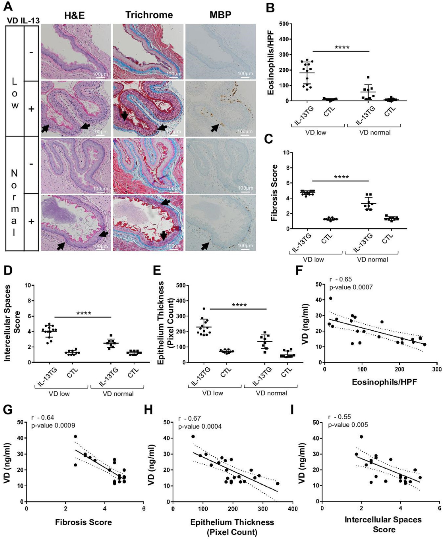
Figure 4.

Effect of vitamin D (VD) on allergic oesophageal inflammation in vivo. (A–I) Wildtype controls (CTL) and IL-13 transgenic (IL-13TG) mice were maintained on low or normal VD diets (see the Methods section). (A) Mouse oesophageal tissue samples were stained for H&E, trichrome and anti-MBP (eosinophil marker). Arrows indicate hyperproliferation (H&E), fibrosis (trichrome), intercellular spaces (H&E; trichrome) and eosinophils (anti-MBP). (B–E) Quantification of eosinophils (B), fibrosis (C), intercellular spaces (D), and hyperproliferation (epithelial thickness; E). (F–I) Spearman correlation of serum VD levels (Supplemental Figure 2) with oesophageal histopathological markers of EoE (IL-13 TG group): eosinophil counts (F), fibrosis (G), hyperproliferation (epithelium thickness; H) and intercellular spaces (I). Data are representative (A) or a summary (B–I) of n=3 independent experiments. Each data point is a mean of a technical duplicate ±SD of in vivo (individual mouse tissue sample) assays. Statistics by one-way ANOVA with Tukey’s multiple comparisons test (B–E): ****p≤0.0001; Spearman correlation coefficient (r) and p values are as indicated (F–I). ANOVA, analysis of variance; EoE, eosinophilic oesophagitis.
