Abstract
Context:
Laboratory-based biomechanical analyses of sport-relevant movements such as landing and cutting have classically been used to quantify kinematic and kinetic factors in the context of injury risk, which are then used to inform targeted interventions designed to improve risky movement patterns during sport. However, the noncontextual nature of standard assessments presents challenges for assessing sport-relevant skill transfer.
Objective:
To examine injury-risk biomechanical differences exhibited by athletes during a jump-landing task performed as part of both a standard biomechanical assessment and a simulated, sport-specific virtual reality (VR)-based assessment.
Design:
Observational study.
Setting:
Medical center laboratory.
Participants:
Twenty-two female adolescent soccer athletes (age = 16.0 [1.4] y, height = 165.6 [4.9] cm, and weight = 60.2 [11.4] kg).
Interventions:
The landing performance was analyzed for a drop vertical jump task and a VR-based, soccer-specific corner-kick scenario in which the athletes were required to jump to head a virtual soccer ball and land.
Main Outcome Measures:
Hip, knee, and ankle joint kinematic differences in the frontal and sagittal planes.
Results:
Athletes exhibited reduced hip and ankle flexion, hip abduction, and frontal plane ankle excursion during landing in realistic sport scenario compared with the standard drop vertical jump task.
Conclusion:
VR-based assessments can provide a sport-specific context in which to assess biomechanical deficits that predispose athletes for lower-extremity injury and offer a promising approach to better evaluate skill transfer to sport that can guide future injury prevention efforts.
Keywords: sport biomechanics, virtual reality, injury risk
Biomechanical analyses of sport movements have classically been used to quantify kinematic (ie, joint angular motion) and kinetic (ie, joint moments of force) factors in the context of task performance, injury risk, or pathology. Traditional biomechanical assessments include indices of walking and running gait, as well as sport-relevant tasks, such as landing and cutting, to identify risk factors for musculoskeletal injuries in sport, particularly at the knee1,2 and ankle.3 The prospective determination of risk factors for injury in sport using standard biomechanical tasks can identify athletes at high risk for injury,4 which can then be used to inform training interventions to ameliorate aberrant biomechanics to reduce injury risk.5 Although neuromuscular training interventions can lead to task-specific biomechanical improvements in the performance of classic laboratory tasks,6-9 these interventions do not consistently reduce injury risk (see Sugimoto et al10 for a review). This may be due, in part, to the lack of adequate knowledge on how to optimize interventions to maximize the potential for sport-relevant skill transfer. It also may indicate that the laboratory tasks do not adequately correspond to the movement demands placed on athletes during more dynamic sport contexts. Subsequently, researchers rely on these traditional biomechanical assessments as proxies for the evaluation of this transfer. Given that traditional assessments evaluate biomechanical performance on a set of standard, noncontextual tasks, these assessments may not accurately represent movement patterns (and thereby, injury risk) exhibited during actual sport competition and may be limited in their generalizability to sport-specific performance. Therefore, it may be more effective to examine injury-risk biomechanics on individuals performing sport-specific tasks within the real-world sport environment itself either exclusively or in conjunction with, and as a validation of, standard assessments.
Analysis of real-world, sport-specific injury risk biomechanics presents logistical challenges that limit the feasibility of such investigations aimed to understand realistic sport demands, such as issues with equipment, weather, compliance with teams and regulatory staff, lack of experimental conditions.11 Accordingly, studies have attempted to evaluate the movement patterns of sport-specific tasks within the laboratory, including shot anticipation in handball, sprinting,12-14 kicking in soccer, blocking in volleyball, and skillful performance (ie, passing, dribbling, and shooting) in basketball.15 However, given that these skills were examined outside of an actual sport environment, the associated analyses are still limited in their generalizability to sport. Moreover, limited data exist that examine specific injury risk biomechanics that have previously been identified using classical biomechanical assessment, such as external knee abduction moment.1 A promising solution that may overcome the limitations of classical assessments involves simulating sport-specific environments through virtual reality (VR), which can effectively present simulated scenarios that facilitate real-world athletic performance and competition.16,17 VR-based assessments may provide a more accurate profile of injury risk and adaption from training by examining the biomechanics of athletes performing fundamental tasks that occur in sport, although they are immersed in a well-controlled sport-specific scenario that accounts for the complex environments of actual competition, including interactions with other players, objects, and sport implements. VR technology has been implemented in the biomechanical analysis of handball throwing kinematics, rugby strategy,18 and cutting in soccer11; however, only the latter examined movement patterns specific to injury risk.
Traditional biomechanical assessments are useful in identifying biomechanical factors for injury, screening athletes based on these factors, and informing targeted interventions to improve the factors that are modifiable; however, it is unclear how the biomechanical profile of an at-risk athlete varies between a standard, noncontextual environment, such as that of a traditional biomechanical assessment and an actual sport (or simulated sport) environment. In the case of the latter, a sport movement context might provide an indication of the true nature of an athlete’s risk for injury while participating in a given sport. Therefore, the purpose of this study was to examine movement patterns exhibited by athletes during a jump-landing task performed as part of both a standard biomechanical assessment (ie, drop vertical jump [DVJ]) and a sport-specific VR-based assessment to examine potential differences in assessed injury risk biomechanical factors. It was hypothesized that the VR scenario would elicit increased magnitude of injury risk biomechanics compared with the standard biomechanical assessment.
Methods
Design
The present study was an observational study undertaken in a medical center biomechanics laboratory.
Participants
Thirty-eight healthy, female adolescent soccer athletes (mean [SD]; age = 16.0 [1.3] y, height = 1.65 [5.5] m, and weight = 59.5 [9.9] kg) participated in this study. All athletes had no recent history of lower-extremity injury or other neuromuscular contraindications that would prevent them from normal athletic activity. Prior to the initial data collection, the study protocol was approved by the Cincinnati Children’s Hospital Medical Center’s Institutional Review Board, and informed, written consent was obtained from each participant and legal guardian.
Procedures
Each athlete underwent both a standard biomechanical assessment, which included a series of dynamic landing and jumping tasks, followed by a VR-based assessment of jump-landing performance during a soccer-specific corner-kick scenario in which athletes moved dynamically to head a virtual soccer ball. For both the standard laboratory and VR assessments, each athlete was instrumented with 37 retroreflective markers (9 mm in diameter) with a minimum of 3 tracking markers per segment. Markers were placed on the sternal notch and sacrum between the S5 and T1 vertebrae and bilaterally on the acromioclavicular joint, lateral epicondyle of the elbow, midwrist, anterior superior iliac spine, greater trochanter, midthigh, medial and lateral femoral condyles, tibial tubercle, lateral and distal aspects of the shank, and medial and lateral malleoli. Participants also wore a standardized shoe (Supernova Glide 2; Adidas AG, Herzogenaurach, Germany) with markers embedded at the heel, the dorsal surface of the midfoot, the lateral foot (fifth metatarsal), and central forefoot (between the second and third metatarsals). For the VR assessment, athletes also wore a custom-built, head-mounted display with 5 additional markers (20 mm in diameter) attached for tracking of head position and orientation, to which the virtual environment was streamed wirelessly with a 1:1 mapping of head movement in the laboratory to the resultant head motion in the environment (see Kiefer et al11 for additional technical specifications). The 3-dimensional marker trajectories were collected using a 39-camera passive optical motion capture system (Motion Analysis Corp, Santa Rosa, CA) that sampled at 240 Hz for the standard assessment and 120 Hz for the VR assessment.
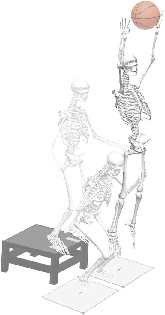
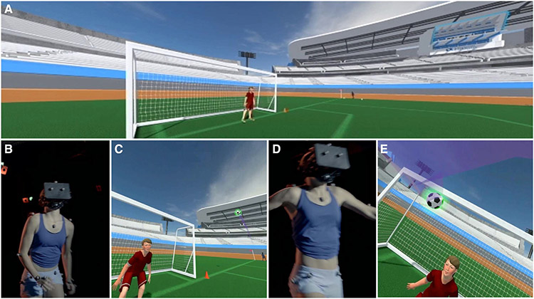
A separate static trial was collected at the start of each assessment with the subject in anatomical pose and with foot direction and placement standardized to the laboratory’s coordinate system to define each subject’s neutral kinematic posture. The standard biomechanical assessment included 3 successful trials of a DVJ task. During the DVJ, participants stood on top of a 31-cm box with their feet aligned with tape at the edge of the box situated approximately shoulder-width apart. Participants were then instructed to drop off the box with both feet at the same time, land on the floor immediately in front of the box, and perform a maximum vertical leap to reach a hanging overhead target (Figure 1). The VR assessment began with the participants completing an acclimation scenario in which they performed various walking, jogging, and running tasks designed to acclimate them to move naturally through the virtual environment. After completing this scenario, the participants then entered a simulated soccer corner-kick scenario, which consisted of 2 practice trials followed by 4 recorded trials. For each trial, participants were required to jump and head a virtual soccer ball that was kicked by a computer-controlled nonplayer character toward a virtual goal (Figure 2). The trials were counter-balanced so that the athlete performed 1 practice trial and 2 recorded trials each in which they approached the ball from the left and right side of the goal, respectively. The scenario was programmed such that the kicked ball followed a parabolic trajectory, during which the ball passed directly in front of the virtual goal as it was descending. The height of the arc was standardized according to participants’ height so that the inclination angle was approximately the same for each athlete as it approached the area in front of the virtual goal, and such that each participant would be required to jump roughly the same amount—corresponding to a maximum effort jump—to successfully make head contact with the virtual ball. Before the scenario began, participants were instructed to attempt to jump and head the ball toward the virtual goal to the maximum of their ability.
Figure 1 —
A representative participant and applied skeletal model performing the drop vertical jump task.
Figure 2 —
The corner-kick scenario. Panel A shows the beginning of the scenario from the perspective of the participant with the nonplaying character in position to kick the ball. Panel B shows the participant wearing the head-mounted display in the laboratory space getting ready to head the ball, and Panel C shows the participant view of the approaching ball. Panel D shows the participant heading the ball in the laboratory space, and Panel E shows the participant view of the ball being headed.
Each trial was visually inspected to determine the effort by which the participant performed the trial; only trials in which the athlete achieved an acceptable level of effort were assessed. Effort was indicated through a qualitative inspection of each recorded trial and determined acceptable if the athlete made a substantial effort at heading the ball (ie, the athlete’s head position during the attempt was close in proximity to the ball in the simulated scenario). Participants who did not perform at least 2 acceptable trials from either side were excluded from the final analysis, leaving 22 athletes (mean [SD]; age = 16.0 [1.4] y, height = 165.6 [4.9] cm, and weight = 60.2 [11.4] kg) for analysis.
Due to the difference in sampling frequencies between the standard and VR conditions, the marker data collected during performance on the DVJ task were down sampled to match that of the frequency of the VR condition (ie, 120 Hz). Marker trajectories were filtered using a low-pass, fourth-order Butterworth filter at a cutoff frequency of 12 Hz, and lower-extremity Cardan joint angles were calculated using a 6-degree-of-freedom skeletal model in Visual3D (C-Motion Inc, Germantown, MD). Kinematic waveforms were exported and time-normalized to 101 points (representing 0%–100% of the given analysis phase) during the landing period after the jump (ie, when participants made contact with the ground after jumping to either reach for the overhead target or head the virtual soccer ball) using custom MATLAB software (The Math-Works Inc, Natick, MA). The landing phase of a jump-landing task has been shown previously to exhibit differential movement patterns and increased magnitude of kinematic differences with respect to the stance phase before the jump,19 and subsequently, it was examined in the present study because it was a feature of the movement goals in both the standard (ie, DVJ) and VR (ie, header task in the soccer corner-kick scenario) conditions. The second landing phase was determined as the period of time between the first heel strike after the jump to maximum knee flexion. Heel strike and toe-off events were determined using a proprietary algorithm in Visual3D.20
Statistical Analysis
Hip, knee, and ankle joint angular motions were extracted from the time-normalized landing phase in both conditions and averaged across trials and sides for all athletes for statistical analysis. Specifically, the joint angular motions during the landing phase were compared by computing the statistical distance between the mean waveforms in the standard and VR conditions using Cohen d.21 Meaningful differences (ie, constituting a large effect size) between kinematic waveforms were postulated as having an effect size d > 0.8, whereas a trend (ie, constituting a medium effect size) was postulated as having an effect size 0.5 > d ≤ 0.8.21,22
To further assess the groups in terms of statistical significance, discrete joint angles at specified time points during the landing phase were compared between conditions. Specifically, hip angles in the sagittal and frontal planes at the time of peak knee flexion, peak knee angle in the sagittal and frontal planes, and ankle angle in the sagittal plane at the time of peak knee flexion were extracted and compared across the standard and VR conditions using Wilcoxon signed-rank tests. The P value was selected to be .05 to indicate statistical significance and adjusted using Holm-Bonferroni corrections for multiple comparisons.
Results
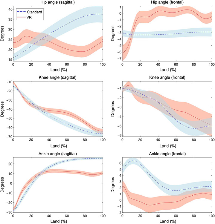
Figure 3 shows the mean kinematic waveforms for the 3 lower-extremity joints during the landing phase for both the standard and VR conditions during the landing phase, and Table 1 presents the summarized meaningful differences and trends between kinematic waveforms across conditions. In general, athletes in the VR condition exhibited reduced sagittal plane range of motion, particularly in the latter half of the landing phase (ie, approaching peak knee flexion) as indicated by meaningful differences at the ankle and trends at the hip and knee and meaningful differences at the ankle and hip joints in the frontal plane during the beginning and middle of the landing phase, respectively.
Figure 3 —
Kinematic time series for the hip, knee, and ankle joints during the landing phase (ie, heel strike to peak knee flexion) of the jump-landing task in the standard condition (ie, DVJ, light grey; blue in online) and the VR (ie, header task in the soccer corner-kick scenario, dark grey; red in online). Shaded regions denote standard error regions of each condition.
Table 1.
Summary of the Meaningful Differences (ie, Large Effect Size) and Trends (ie, Medium Effect Size) Between Kinematic Waveforms for Each of the Lower-Extremity Joints in the Sagittal and Frontal Planes and the % of the Land Phase in Which the Differences Occurred
| Meaningful |
Trend |
|||
|---|---|---|---|---|
| Joint angle | Mean d | %Land | Mean d | %Land |
| Hip (sagittal) | – | – | 0.66 | 59–101 |
| Hip (frontal) | −0.84 | 41–50, 62–80 | −0.66 | 18–40, 51–61, 81–89 |
| Knee (sagittal) | – | – | −0.58 | 52–81 |
| Knee (frontal) | – | – | – | – |
| Ankle (sagittal) | 1.31 | 53–101 | 0.65 | 45–52 |
| Ankle (frontal) | 1.04 | 5–30 | 0.66 | 1–4, 31–41 |
Table 2 presents the summarized hip, knee, and ankle kinematic measures for the tested athletes during the landing phase in both conditions. The cohort exhibited reduced sagittal plane flexion at the hip and ankle at the point of peak knee flexion in the VR condition compared with the standard condition (both adj P < .05). In addition, during the standard condition, the athletes exhibited increased hip abduction at the point of peak knee flexion and increased ankle inversion at initial contact (both adj P < .05).
Table 2.
Mean (SD) of the Hip, Knee, and Ankle Joint Kinematic Measures During the Landing Phase of a Jump-Landing Task in Both the Standard (ie, DVJ) and VR (ie, Header Task in the Soccer Corner-Kick Scenario) Conditions
| Kinematic measure | Standard | VR | Adj P |
|---|---|---|---|
| Hip flexion at peak knee flexion, deg | 37.4 (19.5) | 25.2 (15.7) | .04* |
| Hip abduction at peak knee flexion, deg | −2.9 (4.8) | 0.5 (5.6) | .02* |
| Peak knee flexion, deg | −67.0 (14.5) | −63.7 (13.3) | > .99 |
| Peak knee abduction, deg | −6.4 (3.9) | −7.0 (6.3) | > .99 |
| Ankle dorsiflexion at peak knee flexion, deg | 25.6 (5.4) | 12.6 (10.3) | <.001* |
| Ankle inversion at initial contact, deg | 4.9 (3.7) | 1.7 (5.7) | .01* |
Abbreviations: DVJ, drop vertical jump; VR, virtual reality.
Statistical significance.
Discussion
The purpose of this study was to examine how the biomechanical profiles of soccer athletes vary between traditional biomechanical assessments using standard, noncontextual tasks, and VR-based assessments that simulate real-world sport scenarios (and thus, facilitate movement patterns more similar to actual sport performance). Specifically, we examined kinematic factors previously shown to be associated with future musculoskeletal injury—in particular, anterior cruciate ligament (ACL) injury—during the landing phase of a jump-landing task in both a standard (ie, DVJ) condition and in a VR (ie, header task in the soccer corner-kick scenario) condition. Interestingly, athletes performing a soccer-specific task in VR landed with reduced hip, knee, and ankle flexion, as well as slightly decreased hip abduction and ankle inversion. The sport-specific nature of the header task assessed in VR may more accurately reflect the biomechanical risk profile of soccer athletes performing a jump-landing maneuver during sport-specific scenarios.
Because the landing phase of a jump-landing task has been shown previously to elicit increased magnitude of biomechanical deficits compared with the stance phase prior to jumping19 and landing from a jump was a common feature of task execution in both the standard condition (ie, landing from a DVJ) and VR condition (landing after jumping to head a virtual soccer ball), analyzing the landing phase provided a suitable comparison for assessing the kinematic profile of the athletes across both conditions. Reduced sagittal plane joint motion during landing and jumping in the DVJ has been associated with an increased risk of ACL injury, due to the decrease in muscular energy absorption23 and increase in magnitude of ground reaction force (and subsequently, higher joint moments that excessively stress the passive structures of the knee).1,2 Although it appears that, in general, athletes in both conditions made contact with the ground in approximately the same sagittal plane joint configuration, athletes in the VR condition exhibited reduced sagittal plane range of motion during landing that result in stiffer landings (ie, ones that likely result in higher ground reaction forces). In addition, athletes in the VR condition may have exhibited a decreased ability to attenuate the ground reaction forces on landing as evidenced by reduced frontal plane range of motion at the ankle joint. In general, when loaded during dynamic movement, the foot attenuates ground reaction forces through ankle frontal plane excursion that is coupled with rotation of the shank and thigh segments.24 Landing “toe first” (ie, with a plantar flexed foot)—along with inversion of the foot—allows for a similar force-attenuating mechanism through eccentric contraction and energy absorption by the plantar flexors and passive absorption by the structures of the foot via rearfoot pronation, respectively. A reduction in the frontal plane ankle excursion on landing, as exhibited by the athletes in the VR condition, may inhibit this mechanism, leading to unmitigated ground reaction forces that may increase the risk for injury; moreover, this may induce compensatory (and possibly risky) kinematic strategies onto the proximal kinematic chain, such as that at the hip in the frontal and transverse planes.25 Subsequently, the reduced sagittal plane joint range of motion, hip abduction, and frontal plane ankle motion during landing after a header task in a soccer corner-kick scenario assessed in VR, in contrary to the standard condition, are consistent with a kinematic profile of an athlete at increased risk for ACL injury.
It is possible that the landing in the VR condition more closely reflects that of a landing after a jumping task in a sport-specific scenario than of other standard assessments. Although not experimentally controlled for in the present study, the variation in participants’ movement patterns (and subsequently, the increased magnitude of the elicited injury risk factors) in the VR condition may have been due to the fact that their attention was explicitly focused on a goal-relevant behavior (ie, heading a moving soccer ball). Participants were not explicitly instructed to focus on their movement patterns during the DVJ and were given an external focus of attention during this task by instructing them to reach for an overhead target during the laboratory-based task. It can be reasonably assumed that heading a moving soccer ball in an immersive, VR environment is more attentionally demanding than reaching to grasp a static target at a constant position within a controlled, laboratory environment. As such, it may be that the resultant movement patterns exhibited in the VR condition was illustrative of the more naturalistic self-organization and coordination of the musculoskeletal system that emerges during jumping in a sport-specific environment and that perhaps those patterns exhibited in the standard condition were more constrained due to the controlled, noncontextual nature of the environment.26 Thus, movement patterns in a sport-specific environment might elicit a more accurate movement response based on foundational coordination processes that arise in service of the sport-specific task goal as the athlete’s attention is directed to sport-specific informational variables (ie, other players, objects, sport implements, or the environment itself). As such, the inclusion of more naturalistic or sport-specific movements may enhance existing biomechanical screening, analysis, and intervention paradigms for prevention and rehabilitation purposes.
There are several limitations in the present study that should be taken into consideration. A significant limitation is the loss of data from excluded participants (ie, the final analysis only retained 22 of 38 athletes) indicated as not having put forth an acceptable effort level during the header task. Some participants may not felt fully comfortable in the simulated environment, which may have precluded their full participation in the task. It may also be possible that our operational definition of effort was too stringent and that, for some excluded athletes, their effort more closely resembled their actual sport effort; however, this is difficult to confirm. Future investigations that examine VR in sport should focus on ensuring that participants are comfortable in the simulated environment, such as with the use of a longer “acclimation” scenario, taking necessary steps to ensure full immersion (eg, limiting ambient sound and light), and emphasizing the importance of giving full effort to complete a given task in conjunction with providing additional feedback that motivates a more urgent movement performance. Similarly, it is also possible that the landing phase as defined in the present study did not represent a suitable kinematic mapping across the standard and VR conditions. It is possible that the differences in coordination strategies between a drop landing and a countermovement of reduced magnitude (as in the VR task) could have obfuscated the results. However, the timing, magnitude, and ranges of motion of the drop-landing task appear to be reasonably comparable in both conditions (Figure 3). Another limitation was the failure to capture joint moments of force during the jump-landing task in the VR condition. Although standard biomechanical tasks such as the DVJ allow for consistent ground reaction force calculation due to the placement of task execution over embedded force platforms, it is difficult to consistently gather ground reactions forces in the VR condition due to the lack of clean foot strikes on the force platforms. Not having joint moment of force data perhaps increases the difficulty in accurately assessing risk for injuries that are biomechanical in nature, such as ACL tear. Given the development of portable force sensors, it will be necessary to assess their viability for this type of analysis in the future. Finally, it is also possible that the effort level in executing the jump-landing task across conditions or even within athletes was not consistent, and similarly, the effort level exhibited by the individuals in the VR condition may not have approached that which is actually exhibited in a real-world environment.
Conclusion
The VR-based assessments can provide a sport-specific context in which to assess movement patterns that, while comparable with standard assessments, exhibit increased magnitudes of biomechanical deficits, particularly in the sagittal and frontal planes that predispose athletes for lower-extremity injury. Based on the current results, untethered VR simulations may provide a promising approach to better evaluate training adaptation transfer to sport that can guide future developments in injury prevention.
Acknowledgments
The authors would like to acknowledge funding support from the National Institutes of Health/NIAMS Grant U01AR067997.They also would like to thank Kim Barber Foss, Staci Thomas, Katie Kitchen, and Brooke Gadd for their assistance with data collection; Bernard Casey, Ian Anderson, and Chris Collins of the University of Cincinnati Simulation and Virtual Environments for the development of the VR scenario; and Matt Batie for the development of the head-mounted display.
Contributor Information
Christopher A. DiCesare, Division of Sports Medicine, the SPORT Center, Cincinnati Children’s Hospital Medical Center, Cincinnati, OH, USA.
Adam W. Kiefer, Division of Sports Medicine, the SPORT Center, Cincinnati Children’s Hospital Medical Center, Cincinnati, OH, USA.; Department of Pediatrics, University of Cincinnati, Cincinnati, OH, USA.; Department of Psychology, Center for Cognition, Action, & Perception, University of Cincinnati, OH, USA.
Scott Bonnette, Division of Sports Medicine, the SPORT Center, Cincinnati Children’s Hospital Medical Center, Cincinnati, OH, USA..
Gregory D. Myer, Division of Sports Medicine, the SPORT Center, Cincinnati Children’s Hospital Medical Center, Cincinnati, OH, USA.; Department of Pediatrics, University of Cincinnati, Cincinnati, OH, USA.; Department of Orthopaedic Surgery, University of Cincinnati, Cincinnati, OH, USA; The Micheli Center for Sports Injury Prevention, Waltham, MA, USA.
References
- 1.Hewett TE, Myer GD, Ford KR, et al. Biomechanical measures of neuromuscular control and valgus loading of the knee predict anterior cruciate ligament injury risk in female athletes: a prospective study. Am J Sports Med. 2005;33(4):492–501. doi: 10.1177/0363546504269591 [DOI] [PubMed] [Google Scholar]
- 2.Padua DA, DiStefano LJ, Beutler AI, de la Motte SJ, DiStefano MJ, Marshall SW. The landing error scoring system as a screening tool for an anterior cruciate ligament injury-prevention program in elite-youth soccer athletes. J Athl Train. 2015;50(6):589–595. doi: 10.4085/1062-6050-50.1.10 [DOI] [PMC free article] [PubMed] [Google Scholar]
- 3.Gribble PA, Hertel J, Denegar CR, Buckley WE. The effects of fatigue and chronic ankle instability on dynamic postural control. J Athl Train. 2004;39(4):321–329. [PMC free article] [PubMed] [Google Scholar]
- 4.Myer GD, Ford KR, Brent JL, Hewett TE. An integrated approach to change the outcome part I: neuromuscular screening methods to identify high ACL injury risk athletes. J Strength Cond Res. 2012;26(8):2265–2271. doi: 10.1519/JSC.0b013e31825c2b8f [DOI] [PMC free article] [PubMed] [Google Scholar]
- 5.Myer GD, Ford KR, Brent JL, Hewett TE. An integrated approach to change the outcome part II: targeted neuromuscular training techniques to reduce identified ACL injury risk factors. J Strength Cond Res. 2012;26(8):2272–2292. doi: 10.1519/JSC.0b013e31825c2c7d [DOI] [PMC free article] [PubMed] [Google Scholar]
- 6.Myer GD, Ford KR, Brent JL, Hewett TE. Differential neuromuscular training effects on ACL injury risk factors in “high-risk” versus “low-risk” athletes. BMC Musculoskelet Disord. 2007;8:39. doi: 10.1186/1471-2474-8-39 [DOI] [PMC free article] [PubMed] [Google Scholar]
- 7.Chappell JD, Limpisvasti O. Effect of a neuromuscular training program on the kinetics and kinematics of jumping tasks. Am J Sports Med. 2008;36(6):1081–1086. doi: 10.1177/0363546508314425 [DOI] [PubMed] [Google Scholar]
- 8.Hopper AJ, Haff EE, Joyce C, Lloyd RS, Haff GG. Neuromuscular training improves lower extremity biomechanics associated with knee injury during landing in 11–13 year old female netball athletes: a randomized control study. Front Physiol. 2017;8:883. doi: 10.3389/fphys.2017.00883 [DOI] [PMC free article] [PubMed] [Google Scholar]
- 9.Hewett TE, Ford KR, Xu YY, Khoury J, Myer GD. Effectiveness of neuromuscular training based on the neuromuscular risk profile. Am J Sports Med. 2017;45(9):2142–2147. doi: 10.1177/0363546517700128 [DOI] [PMC free article] [PubMed] [Google Scholar]
- 10.Sugimoto D, Myer GD, McKeon JM, Hewett TE. Evaluation of the effectiveness of neuromuscular training to reduce anterior cruciate ligament injury in female athletes: a critical review of relative risk reduction and numbers-needed-to-treat analyses. Br J Sports Med. 2012;46(14):979–988. doi: 10.1136/bjsports-2011-090895 [DOI] [PMC free article] [PubMed] [Google Scholar]
- 11.Kiefer AW, DiCesare C, Bonnette S, et al. Sport-specific virtual reality to identify profiles of anterior cruciate ligament injury risk during unanticipated cutting. Paper presented at: 2017 International Conference on Virtual Rehabilitation (ICVR); June 19–22, 2017. Montreal, Quebec, Canada. [Google Scholar]
- 12.Delecluse C, Van Coppenolle H, Willems E, Van Leemputte M, Diels R, Goris M. Influence of high-resistance and high-velocity training on sprint performance. Med Sci Sports Exerc. 1995;27(8):1203–1209. doi: 10.1249/00005768-199508000-00015 [DOI] [PubMed] [Google Scholar]
- 13.Rimmer E, Sleivert G. Effects of a plyometrics intervention program on sprint performance. J Strength Cond Res. 2000;14(3): 295–301. [Google Scholar]
- 14.Prieske O, Kruger T, Aehle M, Bauer E, Granacher U. Effects of resisted sprint training and traditional power training on sprint, jump, and balance performance in healthy young adults: a randomized controlled trial. Front Physiol. 2018;9:156. doi: 10.3389/fphys.2018.00156 [DOI] [PMC free article] [PubMed] [Google Scholar]
- 15.Shallaby H. The effect of plyometric exercises use on the physical and skillful performance of basketball players. World J Sport Sci. 2010;3(4):316–324. [Google Scholar]
- 16.Bideau B, Kulpa R, Vignais N, Brault S, Multon F, Craig C. Using virtual reality to analyze sports performance. IEEE Comput Graph Appl. 2010;30(2):14–21. [DOI] [PubMed] [Google Scholar]
- 17.Duking P, Holmberg HC, Sperlich B. The potential usefulness of virtual reality systems for athletes: a short SWOT analysis. Front Physiol. 2018;9:128. doi: 10.3389/fphys.2018.00128 [DOI] [PMC free article] [PubMed] [Google Scholar]
- 18.Watson G, Brault S, Kulpa R, Bideau B, Butterfield J, Craig C. Judging the ‘passability’ of dynamic gaps in a virtual rugby environment. Hum Movement Sci. 2011;30(5):942–956. doi: 10.1016/j.humov.2010.08.004 [DOI] [PubMed] [Google Scholar]
- 19.Bates NA, Ford KR, Myer GD, Hewett TE. Kinetic and kinematic differences between first and second landings of a drop vertical jump task: implications for injury risk assessments. Clin Biomech. 2013;28(4):459–466. doi: 10.1016/j.clinbiomech.2013.02.013 [DOI] [PMC free article] [PubMed] [Google Scholar]
- 20.Stanhope SJ, Kepple TM, McGuire DA, Roman NL. Kinematic-based technique for event time determination during gait. Med Biol Eng Comput. 1990;28(4):355–360. doi: 10.1007/BF02446154 [DOI] [PubMed] [Google Scholar]
- 21.Cohen J. Statistical Power Analysis for the Behavioral Sciences. Vol 2. Hillsdale, NJ: Lawrence Erlbaum Associates; 1988. [Google Scholar]
- 22.Phinyomark A, Osis S, Hettinga BA, Leigh R, Ferber R. Gender differences in gait kinematics in runners with iliotibial band syndrome. Scand J Med Sci Sports. 2015;25(6):744–753. doi: 10.1111/sms.12394 [DOI] [PubMed] [Google Scholar]
- 23.Pollard CD, Sigward SM, Powers CM. Limited hip and knee flexion during landing is associated with increased frontal plane knee motion and moments. Clin Biomech. 2010;25(2):142–146. doi: 10.1016/j.clinbiomech.2009.10.005 [DOI] [PMC free article] [PubMed] [Google Scholar]
- 24.Rodgers MM. Dynamic foot biomechanics. J Orthop Sports Phys Ther. 1995;21(6):306–316. doi: 10.2519/jospt.1995.21.6.306 [DOI] [PubMed] [Google Scholar]
- 25.Koshino Y, Yamanaka M, Ezawa Y, et al. Coupling motion between rearfoot and hip and knee joints during walking and single-leg landing. J Electromyogr Kinesiol. 2017;37:75–83. doi: 10.1016/j.jelekin.2017.09.004 [DOI] [PubMed] [Google Scholar]
- 26.Wulf G, McNevin N, Shea CH. The automaticity of complex motor skill learning as a function of attentional focus. Q J Exp Psychol A. 2001;54(4):1143–1154. doi: 10.1080/713756012 [DOI] [PubMed] [Google Scholar]


