

Abstract
A 51 year old male patient reported with a chief complaint of nasal regurgitation of fluids since a period of 1 year. Patient was operated once earlier with soft tissue palatal closure following which there was recurrence of symptoms. In this case, the defect was managed with an auricular cartilage graft followed by palatal mucosal closure. The healing was uneventful till the latest follow-up. Rationale of this case report lies in its approach to manage an oronasal communication which recurred following a soft tissue closure. Various management strategies have been applied in literature which included grafts, alloplastic materials and vascular free tissue transfer. Following informed and written consent, Patient was operated under General Anaesthesia and was observed for a follow-up period of 2 years. There was uneventful healing along with improvement in patient’s phonation and diet. Complete resolution of nasal regurgitation was achieved. The auricular cartilage graft used for reconstruction in this case is comparatively less invasive and provides an additional advantage of a double layered closure.
Keywords: Trauma, Oronasal fistula, Nasal regurgitation, Auricular cartilage graft- reconstruction
Introduction
Oronasal defects caused due to perforation of bony wall of the palate leading to communication between the oral cavity and nasal cavity are uncommon on routine clinical basis. Treatment options reported in the literature for management of oronasal communication range from conservative methods to wide variety of surgical procedures such as buccal flap, palatal flap, temporo-parietal flap, tongue flap, usage of acellular dermal matrix, bone morphogenic protein, human amniotic membrane, distraction osteogenesis and free vascularized flaps [1]. For predictable results in an oronasal communication, double layered closure is preferred. Recurrence of communication is the highest reported complication of a surgical procedure amounting to 25–37% amongst all. [2] Cause of recurrence include suture dehiscence, post-operative wound infection and avascular necrosis due to closure of wound under tension.
Purse string sutures for closure of oronasal defect was introduced as an alternative in 2019 [3] in which the mucosa adjacent to the defect is inverted to form the floor of nasal cavity followed by closure using a palatal rotation flap. Application of purse string sutures remain less invasive and also provides additional advantage of double layered closure. In this case, as there was a recurrence noted following soft tissue closure and inorder to provide support to the palate over the bony defect, an alternative technique utilizing the auricular cartilage graft was considered.
Case Report
A 51-year-old male patient, reported with a chief complaint of nasal regurgitation of fluids since a period of 1 year. On detailed history, the present condition was caused due to trauma on chewing a hard bony substance with an acute episode of bleeding. Patient was then advised for custom-fabrication of a palatal splint and managed conservatively. Further as the perforation was unresolving, he was undertaken for closure of the fistula with a palatal rotational flap. Following 2–3 weeks after surgery, there was recurrence in the oronasal fistula and patient started to have nasal regurgitation (Fig. 1). On presentation to our department, the defect of the palate measured approximately 1.5 cm wide in its greatest dimension (Fig. 2-transverse dimension), Oval shaped just lateral to midplatal region on the left side. Confirming asepsis and thorough lavage of the oro-nasal fisula, the defect was widened and prepared for a second surgery. At the time of surgery, the surgical site preparation was done and the mucosal lining around fistula was excised. A template for measurement of defect was made intraoperatively to determine the size of the graft to be harvested. Auricular graft of 1*2 cm was harvested by incising the concha on the cranial surface of pinna beneath the cutaneous layer (Fig. 3). Cartilage was incised by infolding the auricle with support of thumb, taking care not to cause a skin perforation of secondary surgical site (Fig. 4). The harvested graft was then tunnelled beneath the palatal mucosa anteriorly (Fig. 5). The cartilage graft was placed to make sure that there was contact with the bony margins of nasal floor and sutures were passed between the graft and palatal mucoperiosteum at multiple points to stabilize its position on closure.The mucosal flap was undermined and repositioned to achieve primary closure at the defect site. On complete healing, there was resolution of nasal regurgitation and no recurrence of the fistula was detected on follow-up (Fig. 6).
Fig. 1.
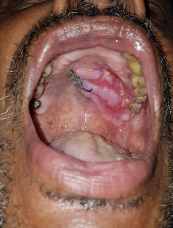
Recurrence of oronasal defect
Fig. 2.

Coronal CT
Fig. 3.

Exposure of auricular cartilage
Fig. 4.
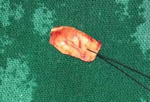
Harvested cartilage graft
Fig. 5.

Placement of graft
Fig. 6.
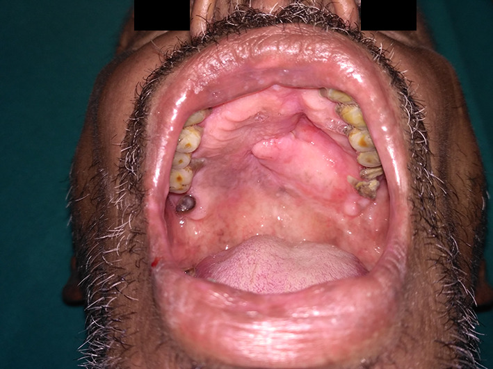
Follow-up photograph
Discussion
When it comes to an oronasal defect, the management protocols differ depending on various factors such as the site of the defect, size and duration span of the defect. The clinical features range from unawareness of the patient about the presence of a communication to nasal regurgitation of liquids and food particles [4] and consequent difficulty of speech, blowing, swallowing etc. In some cases it can even lead to respiratory tract infections. The patient compliance and etiologic factor of a presented oronasal defect plays a major role in deciding the treatment. In this presented case, as the defect was acquired due to trauma and patient reported during the healing phase. First line of treatment was to give a splint appliance to protect the wound and to guide the healing. Old aged patient with poor compliance to wear appliance and inability to maintain oral hygiene led to formation of a fistulous tract which was later opted for closure by a soft tissue pedicled flap. In cases of long standing fistulae, thorough debridement should be done to make sure the fistulous tract is excised inorder to reduce the risk of recurrence. As this case was reported with a recurrence of the oronasal communication, auricular cartilages were taken as option for palatal defect reconstruction. Auricular cartilage grafts are less invasive, provide ease of harvest, and reduced risk of graft failure compared to other cancellous bone graft options and free flaps. Auricular cartilages in this case served as a scaffold to bridge the bony margins of defect, to provide adequate support to guide the healing of nasal as well as the oral cavity with comparatively less donor site morbidity. It is advisable to harvest a graft of size greater in dimension than that of the defect to avoid tension while closure [5, 6] and reducing the risk of failure or resorption leading to recurrence in later stages. Hence, Auricular cartilages grafts can be preferred as an alternative for bridging small to medium sized oronasal defects compared to more invasive options based on a given clinical scenario.
Funding
The author(s) received no financial support for the research, authorship, and/or publication of this article.
Compliance with Ethical Standards
Conflict of interest
The author(s) declared no potential conflicts of interest with respect to the research, authorship, and/or publication of this article.
Informed Consent
Written informed consent was obtained from a legally authorized representative for anonymized patient information to be published in this article.
Footnotes
Publisher's Note
Springer Nature remains neutral with regard to jurisdictional claims in published maps and institutional affiliations.
References
- 1.Sperber GH. The palate. In: Sperber GH, Sperber SM, Guttmann GD, editors. Craniofacial embryogenesis and development. 2. London: BC Deker Inc; 2010. pp. 131–144. [Google Scholar]
- 2.Diah E, Lo LJ, Yun C, et al. Cleft oronasal fistula: a review of treatment results and a surgical management algorithm proposal. Chang Gung Med J. 2007;30:529–537. [PubMed] [Google Scholar]
- 3.Singh V, Agarwal SK, Jajodia N, Garg A. Purse string suture closure: a useful double-layer technique for closure of an oronasal communication. J Maxillofac Oral Surg. 2019;18(2):317–319. doi: 10.1007/s12663-018-1160-x. [DOI] [PMC free article] [PubMed] [Google Scholar]
- 4.Majid OW. Persistent oronasal fistula after primary management of facial gunshot injuries. Br J Oral Maxillofac Surg. 2008;46:50–52. doi: 10.1016/j.bjoms.2006.09.016. [DOI] [PubMed] [Google Scholar]
- 5.Abuabara A, Cortez ALV, Passeri LA, et al. Evaluation of different treatments for oroantral/oronasal communications: experience of 112 cases. Int J Oral Maxillofac Surg. 2006;35:155–158. doi: 10.1016/j.ijom.2005.04.024. [DOI] [PubMed] [Google Scholar]
- 6.Sahoo NK, Desai AP, Roy ID, Kulkarni V. Oro-nasal communication. J Craniofac Surg. 2016;27(6):529–533. doi: 10.1097/SCS.0000000000002815. [DOI] [PubMed] [Google Scholar]