Abstract
Traditional parotidectomy incision was devised by Blair (1912) which was modified by Bailey (1941). Over the years various approaches and techniques have evolved to improve the aesthetic outcome and at the same time for complete disease clearance with reduced complications. In this study, we evaluated the feasibility of our mini-incision parotidectomy technique along with the surgical and quality of life (QOL) outcomes. This prospective case series was conducted at Apollo Hospitals, Bangalore over a period of 2 years (June 2018-August 2020) and includes 20 patients. The surgical outcomes were assessed in terms of feasibility of mini-incision technique with respect to levels of parotid involved and functional outcomes in terms of presence or absence of complications like facial palsy (temporary or permanent), seroma and Frey’s syndrome. Patient related quality of life (QOL) outcomes were assessed in terms of post-operative pain score, patient comfort score and cosmetic score by using numerical rating scale-11 (NRS-11). The mini-incision parotidectomy technique is feasible for lesions in all the parotid levels and conversion or lengthening of incision was not needed in any of the cases. 2(10%) patients had temporary facial palsy (House-Brackman grade III) which was recovered within 3 weeks after surgery. One patient (5%) with adenoid cystic carcinoma had permanent facial palsy. Out of 20 patients 2(10%) had seroma and 1(5%) patient presented with Frey’s syndrome. Mean post-operative pain score at 0, 6 and 24 h were 4.8, 3.4 and 1.8 out of 10 respectively. Mean comfort score was 9 out of 10 and mean cosmetic score was 9.5 out of 10. Mini-incision parotidectomy technique can render improved functional as well as aesthetic outcomes after parotidectomy without compromising the surgical clearance of the disease process.
Keywords: Mini-incision parotidectomy, Parotid level, Aesthetic outcomes, Quality of life outcomes, Cosmetic score
Introduction
Parotidectomy is one of the commonest head and neck surgical procedure performed for both benign and malignant parotid gland lesions. Traditional parotidectomy incision was devised by Blair (1912) which was modified by Bailey (1941). Over the years various approaches and techniques have evolved to improve the aesthetic outcome and at the same time for complete disease clearance with reduced complications. European salivary gland society (ESGS) had proposed various levels in parotid to define, report and compare the resections performed [1]. In this study, we evaluated the feasibility of our mini-incision parotidectomy technique along with the surgical and quality of life (QOL) outcomes.
Material and Methods
This prospective case series was conducted at Apollo Hospitals, Bangalore over a period of 2 years (June 2018–August 2020) after obtaining the Institutional Ethical Committee approval. Our study includes 20 consecutive patients who presented to our outpatient department with benign parotid tumors. Patients underwent contrast enhanced CT scan or MRI of neck along with ultrasound guided fine needle aspiration cytology before the surgery. Informed consent was obtained from all the study participants and they were counselled regarding conversion or lengthening of the incision if the access was inadequate. The level of parotid involvement by the lesion and type of parotidectomy performed was classified as per ESGS 2016 classification (Fig. 1) [1]. The mean operation time was calculated from the time of skin infiltration to the completion of skin sutures.
Fig. 1.
Parotid levels as per ESGS classification
The surgical outcomes were assessed in terms of feasibility of mini-incision technique with respect to levels of parotid involved and functional outcomes in terms of presence or absence of complications like facial palsy (temporary or permanent), seroma and Frey’s syndrome. Patient related quality of life (QOL) outcomes were assessed in terms of post-operative pain score, patient comfort score and cosmetic score by using numerical rating scale-11 (NRS-11). All patients were treated with same analgesics and post-operative pain was assessed at 0, 6 and 24 h after surgery. Patient comfort score was assessed at the time of discharge. All patients were followed up at 1st week, 1st month and 3rd month after surgery. Cosmetic score was evaluated at 3rd month follow-up. Mean hospital stay, day of drain removal and day of return to daily routine was also evaluated.
Surgical Technique
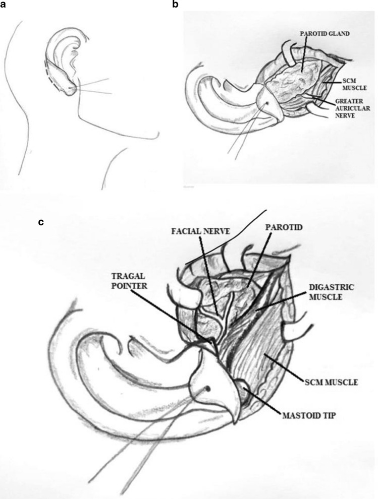
A 3–4 cm vertical skin incision was given in the retro-auricular groove inferiorly reaching till the level of attachment of ear lobule to the face (Fig. 2a). Flaps are elevated deep to the plane of superficial musculo-aponeurotic system (SMAS) and subcutaneous tissue undermined to facilitate retraction of flaps for better access (Fig. 2b). Tragal pointer identified and tail of parotid gland was identified and elevated. Sternocleidomastoid muscle identified and retracted and posterior branch of greater auricular nerve was identified and preserved in all cases. Next step was the identification of posterior belly of digastric muscle. Then the tympano-mastoid suture line was palpated and the main trunk of facial nerve identified (Fig. 2c). Dissection proceeded at the plane of facial nerve trunk and main branches as per standard technique. The tumor was removed in toto along with the cuff of normal parotid tissue. In cases of large tumors the tissue was maneuvered in order to remove through the small incision. After achieving complete hemostasis minivac drain was inserted and secured and wound closed in single layer simple sutures using 3–0 monocryl. The per-operative techniques and post-operative image of the index case is shown in Fig. 3a–f.
Fig. 2.
a Diagrammatic representation of 3–4 cm vertical incision in the post-aural groove. b Diagrammatic representation showing skin undermining and visualization of parotid tissue and sternocleidomastoid muscle. c Diagrammatic representation of identification of facial nerve trunk by the markers like tragal pointer, posterior belly of digastric muscle
Fig. 3.
a Pre-operative image showing swelling in the left parotid region. b Intra-operative image showing main trunk of facial nerve with two main branches. c Complete resection of tumor. d Specimen of tumor along with the normal parotid tissue. e Post-aural wound appearance on post-operative day-1. f Post-operative image of patient at 1st month follow-up without any visible scar
Results
Out of 20 patients included in our study, 8(40%) were males and 12(60%) were females with male to female ratio of 2:3 and the mean age of presentation was 33.5 years (14–68 years). Left side was involved in 13(65%) cases and right side in 7(35%) cases with left to right ratio of 1.8:1. Most common presentation was long standing painless swelling. 2 patients had already been operated for pleomorphic adenoma in the past and presented with recurrent swelling within the period of 3 months from the previous procedure. None of the patients presented with facial palsy or skin involvement at the time of presentation. Mean size of the tumor was 3.9 (± 0.37) × 3.8 (± 0.28) cm. Modification or lengthening of incision was not needed in any case as the access to the tumor and facial nerve was adequate. The levels of parotid levels involved and type of parotidectomy performed is presented in Table 1. Extra capsular dissection (ECD) was performed in one case where only level II was involved and the size of the tumor was less than 2 cm. None of the patient had any intra-operative complications.
Table 1.
Parotid levels involved with respective type of resection performed as per ESGS classification
| Parotid levels involved | Pathology (No. of patients) | Type of resection | Total no. of patients (%) |
|---|---|---|---|
| I and II | Pleomorphic adenoma (8) | Parotidectomy | 8(40) |
| III and IV | Pleomorphic adenoma (3) | Parotidectomy I–IV | 3(15) |
| II and III | Pleomorphic adenoma (1) | Parotidectomy I–IV | 1(5) |
| I and IV | Warthin’s tumor (2) | Parotidectomy I–IV | 2(10) |
| I | Pleomorphic adenoma (1) | Extracapsular Dissection | 2(10) |
| Adenoid cystic carcinoma (1) | Parotidectomy I–IV | ||
| II | Mucoepidermoid carcinoma (2) | Parotidectomy I–IV | 2(10) |
| V | Pleomorphic adenoma (1) | Parotidectomy I, II,V | 2(10) |
| Mucoepidermoid carcinoma (1) | Parotidectomy I–IV |
Pre-operative cytology showed benign tumor pathology in all our patients. However the intra-operative frozen section analysis revealed 4 (20%) cases to be malignant. Since the tumor size in these patients were less than 2 cm (T1) total conservative parotidectomy (Parotidectomy I-IV) was done in these cases along with level II cervical lymph node sampling. As none of the patients with malignant lesion showed lymph nodes metastasis on frozen section, further selective neck dissection was not performed. The resected margins were free of tumor in all these cases. The mean operation time was 118 min (88–148 min). Post-operative histopathological examination was reported as pleomorphic adenoma in 14(70%) patients, Warthin’s tumor in 2(10%) patients, mucoepidermoid carcinoma in 3(15%) patients with high grade in two patients and low grade in 1 patient and adenoid cystic carcinoma- high grade in 1(5%) patient. 2 patients with high grade mucoepidermoid carcinoma and 1 patient with high grade adenoid cystic carcinoma underwent post-operative adjuvant radiation therapy. 2(10%) patients had temporary facial palsy (House-Brackman grade III) which was recovered within 3 weeks after surgery. One patient (5%) with adenoid cystic carcinoma had permanent facial palsy. Out of 20 patients 2(10%) had seroma and 1(5%) patient presented with Frey’s syndrome.
Mean post-operative pain score at 0, 6 and 24 h were 4.8 (4.5–5.1), 3.4 (3.1–3.7) and 1.8 (1.6–2.0) out of 10 respectively. Mean comfort score was 9 (8.6–9.4) out of 10 and mean cosmetic score was 9.5 (9.2–9.8) out of 10. Mean duration of hospital stay was 1.8 days (1.5–2.1) and mean day of drain removal was 1.9 days (1.5–2.3). The mean duration of return to daily activities was 4.9(4.6–5.2) days. Mean follow-up period was 16.5 months (5–27 months) and none of the patients had any recurrent disease till date.
Discussion
Parotid gland neoplasms are one of the most common head and neck masses with 70% of parotid lesion being benign. Parotidectomy is the surgical technique performed for both benign and malignant parotid lesions. European Salivary Gland Society (ESGS) has proposed a classification system for determining the levels of parotid gland that is involved by the pathology and also has defined the type of parotidectomy performed accordingly [1]. The disease clearance has an important goal in the surgical outcome. However, functional and aesthetic outcomes have high impact on patient’s quality of life [2–5].
Over the century, the approaches and incisions have evolved in the field of parotid surgery to improve both the functional as well as aesthetic outcomes. Post-operative scar over the face and neck leads to negative impact on the individual’s quality of life. Modified Blair’s incision by Bailey (1941) is the most common incision used for parotidectomy. Facelift incision, modified facelift incision, intra-auricular modification of facelift incision, retro-auricular hairline incision and small access post aural incision have been experimented to improve the cosmetic outcome after parotid surgeries by various authors [2–12]. The incisions and approaches greatly depend on three factors viz size of the tumor, location of the tumor and the difficulty in resection like deep lobe tumors [12].
In our study we describe a mini-incision parotidectomy technique and have named it as SN’s incision which is a 3–4 cm vertical incision in the post-aural groove. We assessed the feasibility of this incision with respect to various parotid level lesions and the type of resection performed. The post-aural incision usually has limited access to the lesions in anterior and superior levels, tumors greater than 6 cm in size and deep lobe lesions [12]. In our study, we were able to resect the lesions in all five mentioned parotid levels. The conversion or lengthening of incision was not needed in any of our cases as the subcutaneous undermining of the flaps allow for maximal retraction and adequate exposure of the surgical field. However, maneuvering of the tissue through the incision was performed for the tumors greater than 5 cm for removal.
The rate of occurrence of temporary and permanent facial palsy after superficial parotidectomy is 23% and 2% and after total parotidectomy is 54% and 17% respectively [13]. The rate of seroma and Frey’s syndrome in literature is 17% and 43% respectively [13]. In our study, 2(10%) had temporary facial palsy, 1(5%) patient had permanent facial palsy, 2(10%) patients had seroma and 1(5%) patient had Frey’s syndrome which is comparable to the literature. The mean cosmetic score for modified Blair’s incision, Facelift incision and retro-auricular hairline incision was 5.8, 6.8 and 8.1 respectively [6]. The mean cosmetic score in our series was 9.5 out of 10.
We advocate that mini-incision parotidectomy technique can be safely performed in all benign parotid tumors less than 6 cm. We performed 4 cases of parotid malignancy since it was less than 2 cm (T1) and without any nodal metastasis. However, this technique may not be feasible in large malignant lesions with nodal metastasis requiring elective nodal dissection, skin involvement, recurrent tumors, irradiated patients and benign tumors greater than 6 cm. Mini-incision technique requires a learning curve and excellent surgical expertise. Limitations of our study include smaller study population and shorter mean follow-up period.
Conclusion
The mini-incision parotidectomy is feasible for benign parotid tumors involving all the parotid levels. This technique can render improved functional as well as aesthetic outcomes after parotidectomy without compromising the surgical clearance of the disease process.
Author contribution
Satish Nair: Substantial contributions to the conception or design of the work, revising it critically for important intellectual content, final approval of the version to be published. J G Aishwarya: Substantial contributions to the acquisition of data for the work, revising it critically for important intellectual content. Aditya Jain, Pavithra V, Sneha Mohan: Substantial contributions to the acquisition of data for the work and revising it.
Funding
None.
Declarations
Conflict of interest
The authors report no proprietary or commercial interest in any product mentioned or concept discussed in this article.
Footnotes
Publisher's Note
Springer Nature remains neutral with regard to jurisdictional claims in published maps and institutional affiliations.
References
- 1.Quer M, Guntinas-Lichius O, Marchal F, et al. Classification of parotidectomies: a proposal of the European salivary gland society. Eur Arch Otorhinolaryngol. 2016;273(10):3307–3312. doi: 10.1007/s00405-016-3916-6. [DOI] [PubMed] [Google Scholar]
- 2.Dalmia D, Behera SK, Bhatia JS. Anteriorly based partial thickness sternocleidomastoid muscle flap following parotidectomy. Indian J Otolaryngol Head Neck Surg. 2016;68(1):60–64. doi: 10.1007/s12070-015-0906-8. [DOI] [PMC free article] [PubMed] [Google Scholar]
- 3.Panda NK, Kaushal D, Verma R. Do we need to modify the parotidectomy incision? Indian J Otolaryngol Head Neck Surg. 2016;68(4):487–489. doi: 10.1007/s12070-016-0999-8. [DOI] [PMC free article] [PubMed] [Google Scholar]
- 4.Giotakis EI, Giotakis AI. Modified facelift incision and superficial musculoaponeurotic system flap in parotid malignancy: a retrospective study and review of the literature. World J Surg Onc. 2020;18:8. doi: 10.1186/s12957-020-1785-3. [DOI] [PMC free article] [PubMed] [Google Scholar]
- 5.Kim IK, Cho HW, Cho HY, Seo JH, Lee DH, Park SH. Facelift incision and superficial musculoaponeurotic system advancement in parotidectomy: case reports. Maxillofac Plast Reconstr Surg. 2015;37(1):40. doi: 10.1186/s40902-015-0040-2. [DOI] [PMC free article] [PubMed] [Google Scholar]
- 6.Kim DY, Park GC, Cho YW, Choi SH. Partial superficial parotidectomy via retroauricular hairline incision. Clin Exp Otorhinolaryngol. 2014;7(2):119–122. doi: 10.3342/ceo.2014.7.2.119. [DOI] [PMC free article] [PubMed] [Google Scholar]
- 7.Movassaghi K, Lewis M, Shahzad F, May JW., Jr Optimizing the aesthetic result of parotidectomy with a facelift incision and temporoparietal fascia flap. Plast Reconstr Surg Glob Open. 2019;7(2):e2067. doi: 10.1097/GOX.0000000000002067. [DOI] [PMC free article] [PubMed] [Google Scholar]
- 8.Lorenz KJ, Behringer PA, Höcherl D, Wilde F. Improving the quality of life of parotid surgery patients through a modified facelift incision and great auricular nerve preservation. GMS Interdiscip Plast Reconstr Surg DGPW. 2013;2:Doc20. doi: 10.3205/iprs000040. [DOI] [PMC free article] [PubMed] [Google Scholar]
- 9.Wasson J, Karim H, Yeo J, Panesar J. Cervicomastoidfacial versus modified facelift incision for parotid surgery: a patient feedback comparison. Ann R Coll Surg Engl. 2010;92(1):40–43. doi: 10.1308/003588410X12518836440009. [DOI] [PMC free article] [PubMed] [Google Scholar]
- 10.Chen CY, Chen PR, Chou YF. Intra-auricular modification of facelift incision decreased the risk of frey syndrome. Tzu Chi Med J. 2019;31(4):266–269. doi: 10.4103/tcmj.tcmj_117_18. [DOI] [PMC free article] [PubMed] [Google Scholar]
- 11.Yuen AP. Aesthetic consideration of parotidectomy: postaural approach and extended sternomastoid flap: how we do it. Clin Otolaryngol. 2010;35(3):231–234. doi: 10.1111/j.1749-4486.2010.02108.x. [DOI] [PubMed] [Google Scholar]
- 12.Yuen AP. Small access postaural parotidectomy: an analysis of techniques, feasibility and safety. Eur Arch Otorhinolaryngol. 2016;273(7):1879–1883. doi: 10.1007/s00405-015-3691-9. [DOI] [PMC free article] [PubMed] [Google Scholar]
- 13.Thielker J, Grosheva M, Ihrler S, Wittig A, Guntinas-Lichius O. Contemporary management of benign and malignant parotid tumors. Front Surg. 2018;11(5):39. doi: 10.3389/fsurg.2018.00039. [DOI] [PMC free article] [PubMed] [Google Scholar]


