Abstract
Lipomas of the oral cavity are uncommon. Here we report a case of 85 year old female presenting with a progressively increasing large growth in the oropharynx which was diagnosed as lipoma on histopathology. The clinicoradiological and histopathological findings are discussed. To the best of our knowledge; this is one of the largest intraoral lipoma reported in India till date. The present case highlights the need to be aware of intraoral lipomas which can present as large growths at this unusual site so as to avoid any unwarranted aggressive surgery.
Keywords: Intraoral, Lipoma, Large, Pathology, Tumor
Introduction
Lipomas are benign tumors of adipose tissue which occur commonly in soft tissue predominantly of trunk.neck and extremities. Intraoral lipomas are however uncommon accounting for 1–4% [1]. In view of their slow growth, they can achieve large size by the time they seek treatment. Although they mostly present as below 3 cms diameter size, they can rarely achieve large size. Herein we report a large intraoral lipoma in an elderly female which was cured by wide local excision.
Case Report
A 85 year female presented in the E.N.T outpatient department with a growth in oral cavity which was progressively increasing in size for the last six years along with dysphagia and difficulty in chewing since one year. Patient had no co-morbities. Routine investigations were normal. On local examination, the swelling was soft, cystic, globular and forming a pedunculated mass. The mass was in oropharynx and appeared to be arising from right tonsillar fossa and posterior aspect of soft palate.
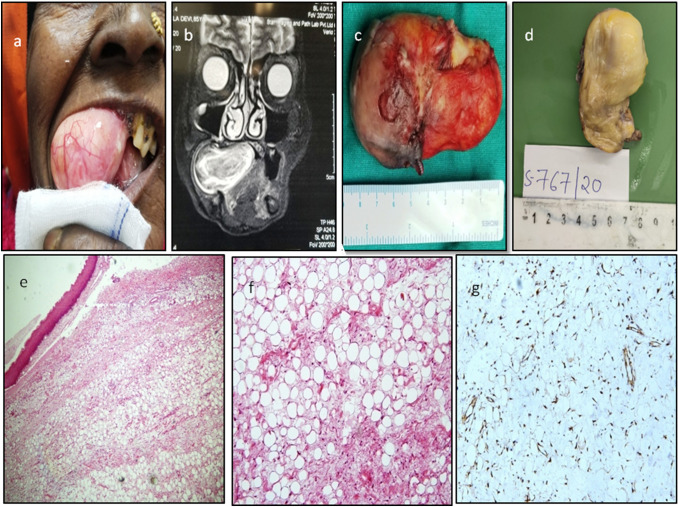
(Fig. 1a) Magnetic resonance imaging (MRI) neck with contrast showed a large, well circumscribed elongated intra-oral soft tissue mass lesion measuring 8 × 3 cms arising from right pharyngeal mucosal space and right posterior soft palate with partial compression of oropharyngeal airway. (Fig. 1b) There was no invasion of adjacent structures. With the clinicoradiological diagnosis of benign lesion, patient underwent complete surgical excision of the mass under general anesthesia. Per operative and gross examination showed a nodular encapsulated lesion measuring 8 × 3.5 × 3 cms. Cut surface was soft, yellow and fatty. (Fig. 1c, d) Microscopic examination showed a lesion composed of lobules of mature adipose tissue. Focal oedema was present. No lipoblasts, atypia, increased mitosis or necrosis was seen. The overlying squamous mucosa showed mild hyperplasia. (Fig. 1e, f) The adipocytes were positive for S100 (Fig. 1g) and negative for MDM2 (Mouse double minute 2 homolog). The features were consistent with lipoma. She had an uneventful postoperative recovery with no recurrences at two year follow up.
Fig. 1.
a Local clinical examination showing a large nodular intraoral growth.Overlying mucosa appears smooth. b Magnetic resonance imaging (MRI) neck with contrast showing a large, well circumscribed elongated intra-oral soft tissue mass lesion measuring 8 × 3 cms arising from right pharyngeal mucosal space and right posterior soft palate with partial compression of oropharyngeal airway. c Per operative examination showing a nodular encapsulated lesion measuring 8 × 3.5 × 3 cm. d Grossly the cut surface is yellow and fatty. e Microscopy showing nodular lesion covered with squamous epithelium (H&Ex20). f The lesion is composed of lobules of mature adipocytes (H&Ex40).G. S100 immunohistochemistry is positive in the adipocytes
Discussion and Review of Literature
Lipomas are common benign mesenchymal tumors composed of mature adipose tissue. The tumors of the oro-pharyngeal space represent 0.5% of all neoplasms of the head and neck. Lipomas of the tonsillar/oropharyngeal space are very rare with only few cases described in literature. Buccal mucosa is the commonest site of intraoral lipomas. Other rare sites include lips, tongue, floor of mouth, buccal sulcus and retromolar area [2]. Intraoral lipomas are usually small with only few large lipomas described in literature [3–5]. Although the exact etiology is unknown, the pathogenesis may be linked to hereditary, fatty degeneration, trauma, infection, infarction and chronic irritation [1]. Clinically, oropharyngeal lipomas are slow growing painless masses which grow slowly over years. Patients may therefore have a large growth at the time of presentation. Depending upon their intraoral location, they may present with dysphagia, hoarseness of voice, dyspnea, foreign body sensation, painless neck mass or obstructive sleep apnea. Uncommonly it may result in compressive symptoms [5, 6]. Although extremely rare, malignant change in larger sized lipomas is described [7]. Rapid sudden increase in size or pain is suggestive of malignancy. Clinically the differential diagnoses include mucoceles, pleomorhic adenoma, lipomas and fibromas [6]. Radioimaging is useful. however histopathology is essential to establish the exact diagnosis. On histopathology, the lesion is composed of lobules of mature adipocytes. No atypical cells or lipoblasts are seen. Benign lesions are positive for S100 and negative for MDM2. Although classical lipomas are the commonest, uncommon variants include pleomorphic, spindle cell, chondroid, myxoid and fibrohistiocytic type [8]. Fine needle aspiration cytology cannot accurately differentiate benign from malignant lipomatous lesions [9]. Treatment includes total surgical excision of the mass with excellent prognosis [1, 2, 5] and these tumors rarely reccur,
Intraoral lipomas are infrequent, and occasionally can attain large size. Rarely can they become life threatening when they compress normal structures. Awareness of these lesions for correct differential diagnosis is important for proper planning and management.
Funding
None.
Declarations
Conflict of Interest
There are no conflicts of interest.
Consent for Publication
Written informed consent for publication of their clinical details and/or clinical images was obtained from the patient. Care has been taken to hide the identity details for confidentiality.
Footnotes
Publisher's Note
Springer Nature remains neutral with regard to jurisdictional claims in published maps and institutional affiliations.
Contributor Information
Sunila Jain, Email: drsunilajain@gmail.com.
Saurabh Garg, Email: gargsaurabh12189@gmail.com.
Nitin Aggarwal, Email: dr.nitinaggarwal@gmail.com.
References
- 1.Kumaraswamy S, Madan N, Keerthi R, Shakti S. Lipomas of oral cavity: case reports with review of literature. J Maxillofac Oral Surg. 2009;8:394–397. doi: 10.1007/s12663-009-0096-6. [DOI] [PMC free article] [PubMed] [Google Scholar]
- 2.Fregnani ER, Pires FR, Falzoni R, Lopes MA, Vargas PA. Lipomas of the oral cavity: clinical findings, histological classification and proliferative activity of 46 cases. Int J Oral Maxillofac Surg. 2003;32:49–53. doi: 10.1054/ijom.2002.0317. [DOI] [PubMed] [Google Scholar]
- 3.Motagi A, Aminzadeh A, Razavi SM. Large oral lipoma: case report and literature review in Iran. Dent Res J (Isfahan) 2012;9(3):350–352. [PMC free article] [PubMed] [Google Scholar]
- 4.Fitzgerald K, Sanchirico PJ, Pfeiffer DC. Large intramuscular lipoma of the tongue. Radiol Case Rep. 2018;13(2):361–364. doi: 10.1016/j.radcr.2018.01.014. [DOI] [PMC free article] [PubMed] [Google Scholar]
- 5.PonceJB, Ferreira GZ, Santos PS, Lara VS (2016) Giant oral lipoma: a rare entity. An Bras Dermatol 91(5 suppl 1): 84 86 10.1590/abd1806-4841.20165008. PMID: 28300904. PMCID: PMC5325003 [DOI] [PMC free article] [PubMed]
- 6.Pires FR, Souza L, Arruda R, Cantisano MH, Picciani BL, Santos Dos TC (2021) Intraoral soft tissue lipomas: clinicopathological features from 91 cases diagnosed in a single Oral Pathology service. Med Oral Patol Oral Cir Bucal 26(1): e90–e96 10.4317/medoral.24023. PMID: 32851988. PMCID: PMC7806349 [DOI] [PMC free article] [PubMed]
- 7.Egido-Moreno S, Lozano-Porras AB, Mishra S, Allegue-Allegue M, Marí- Roig A, López-López J. Intraoral lipomas: review of literature and report of two clinical cases. J Clin Exp Dent. 2016;8(5):e597–e603. doi: 10.4317/jced.52926. [DOI] [PMC free article] [PubMed] [Google Scholar]
- 8.Fanburg-Smith JC, Furlong MA, Childers EL. Liposarcoma of the oral and salivary gland region: a clinicopathologic study of 18 cases with emphasis on specific sites, morphologic subtypes, and clinical outcome. Mod Pathol. 2002;15(10):1020–1031. doi: 10.1097/01.MP.0000027625.79334.F5. [DOI] [PubMed] [Google Scholar]
- 9.Thavikulwat AC, Wu JS, Chen X, Anderson ME, Ward A, Kung J. Image-guided core needle biopsy of adipocytic tumors: diagnostic accuracy and concordance with final surgical pathology. Am J Roentgenol. 2021;216(4):997–1002. doi: 10.2214/AJR.20.23080. [DOI] [PubMed] [Google Scholar]


