

Abstract
Nasal gliomas are congenital masses of dysplastic neuroglial and fibrovascular tissue. However, other congenital nasal masses, including encephaloceles, hemangiomas, and dermoid cysts make clinical diagnosis difficult. Radiological examination is imperative to accurate diagnosis of nasal gliomas. We hereby present the diagnostic imaging features of these lesions, which necessitate inclusion into the differential diagnosis of a congenital nasal mass.
Keywords: Nasal gliomas, Glial heterotopia, Congenital nasal masses, Encephalocele, Hemangioma
Introduction
Nasal gliomas are rare congenital growths composed of dysplastic neuroglial and fibrovascular tissue. These masses are a part of a group of congenital nasal masses, which occur with an estimated annual incidence of one in 20,000–40,000 live births [1]. Although nonneoplastic, nasal gliomas require prompt surgical excision since delayed intervention leads to secondary craniofacial distortions [2]. Therefore, prompt recognition and treatment are paramount. However, the rarity of nasal gliomas and their nonspecific manifestations render clinical diagnosis very difficult.
Radiologic evaluation is consistently regarded as a mainstay in diagnosis of nasal gliomas. However, unaware radiologists are prone to pitfalls in diagnosis. Differentiating between congenital nasal masses is essential as management approaches differ for each. Therefore, an understanding of the characteristic imaging features of such lesions is paramount for radiologists to facilitate proper diagnosis and avoid pre-and postnatal complications. We report a mixed nasal glioma in a neonate and review the appropriate differential diagnosis to establish and radiologic investigations to carry out in such a setting.
Case Report
A 9-day-old boy presented with a right frontonasal mass that was noticed at birth. His delivery was uneventful and his family history was unremarkable. Physical examination revealed a 5 × 5 cm soft, mobile, non-pulsating, pedunculated swelling with telangiectasia at the glabella (Fig. 1).
Fig. 1.
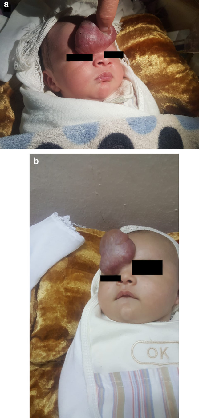
A neonate presented to us a, b with a 5 × 5 cm soft, mobile, non-pulsating, pedunculated swelling with telangiectasia at the glabella
Subsequent Computed tomography (CT) scan revealed a predominantly extranasal midline mass isodense to brain parenchyma with no definite connection to the intracranial vault (Fig. 2). Postnatal magnetic resonance imaging (MRI) revealed a 5 × 5 × 3.5 cm lesion that followed brain parenchymal signal intensity on T1 and T2, thus confirming CT findings. (Fig. 3). Post-contrast MR images showed no appreciable enhancement. A pedicle, measuring 9–10 mm, was observed on the right side of the frontal bone near the midline, causing widening of the frontonasal suture, with insinuation into the right side of the nasal cavity anteriorly and superiorly. No intracranial connection or vascular malformation was seen (Fig. 3). Doppler flow studies showed low resistance and low-velocity arterial flow. On this basis, the diagnosis of an extranasal glioma was made and neurosurgical consultation advised.
Fig. 2.

Coronal oblique CT shows a mass-like lesion in the frontal region anteriorly isodense to brain parenchyma and a slight widening of the right nasofrontal suture. Post-contrast CT images show no enhancement or vascular malformation. No definite enhancing connecting stalk to intracranial structures is observed
Fig. 3.

Axial T2 weighted images a, b revealed a midline mass anterior to the frontal bone and nasal bridge, following brain parenchymal signal intensity. A possible intracranial communication is noted. Axial c Sagittal d T1 brain MRI revealed a mass with isointense signals to brain tissue. A possible intracranial extension is seen in the sagittal image
The patient was subsequently taken to the operating room, where the mass was excised via a midline nasal incision. The procedure was uneventful, no intracranial extension was found, and the patient was discharged in stable condition (Fig. 4a). The follow-up was uneventful (Fig. 4b).
Fig. 4.
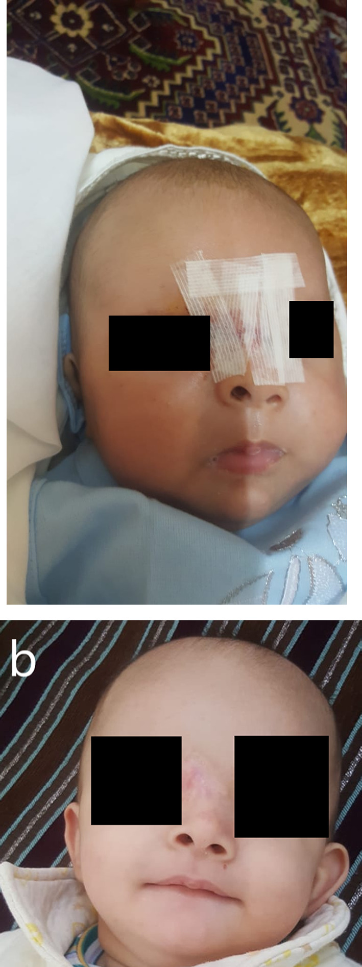
Postoperative image a of the patient—we discharged him in stable condition. The follow-up b was uneventful
Discussion
Nasal gliomas are part of a spectrum of congenital nasal masses that occur once in every 20,000–40,000 live births [3]. Encephaloceles constitute the most common congenital nasal mass, whereas gliomas, dermoid cysts, epidermoid cysts, hemangiomas, and vascular malformations are rare in comparison [2, 4]. Extranasal gliomas account for 60% of cases, with the glabella as the most common site. 30% of patients have intranasal gliomas, which arise most often from the lateral nasal wall or middle turbinate, and occasionally from the nasal septum [5]. Further, intranasal gliomas cause nasal airway obstruction and consequent neonatal respiratory distress. 10% of glioma cases, including ours, are mixed, with both intra- and extra-nasal masses observed.
The pathomechanism behind the development of nasal gliomas remains to be firmly established. The widely accepted “encephalocele hypothesis” implicates aberrant embryogenesis with improper closure of the nasofrontal fontanelle or foramen cecum, resulting in incomplete dural regression, i.e., a sustained connection between neural and surface ectoderms [2, 3]. Encephaloceles retain their intracranial connections, whereas gliomas do not. However, 15–20% of glioma cases exhibit a fibrous connecting stalk into the cranial vault.
Nasal gliomas are detected prenatally via ultrasound. Further characterization is done via MRI, which demonstrates isointense signals to brain parenchyma on T1-weighted images. On T2, hyperintensity is seen due to gliosis; in contrast, dermoid and epidermoid cysts are variable [5]. Enhancement is variable; it may be observed due to compression of the adjacent nasal mucosa. Additionally, MRI can assess for underlying central nervous system (CNS) abnormalities and potential airway obstruction in the setting of intranasal gliomas [2, 6]. As stated above, the principal difference between encephaloceles and gliomas is the lack of an intracranial connection in gliomas. Collectively, this discussion highlights MRI as the imaging modality of choice to diagnose nasal gliomas. Postnatally, MRI constitutes the investigation of choice owing to its ability to visualize a potential intracranial extension. However, the success of MRI in detecting gliomas connected to the CNS via a fibrous stalk is variable. A CT scan is useful to evaluate for bony defects but may create a confusing radiologic appearance due to the ongoing growth and ossification of the craniofacial bones in neonates. Grossly, nasal gliomas appear as firm, incompressible and non-pulsatile masses with a telangiectatic surface [5]. The MRI findings along with these clinical features make hemangioma a potential pitfall in diagnosis. Hemangiomas and gliomas are distinguished by Doppler flow studies: gliomas reveal low-velocity arterial flow, whereas hemangiomas show high-velocity arterial flow [5]. Clinically distinguishing between encephaloceles and gliomas involves increasing intracranial pressure (ICP). Encephaloceles, due to an intact intracranial extension, expand upon increases in ICP, whereas gliomas do not (negative Furstenberg sign).
Although numerous current reports have substantiated our findings [2, 7], other studies have indicated that MRI findings of nasal gliomas are variable depending on the mass contents. For instance, entirely solid masses exhibit hyperintense signals on T2, while others are purely cystic with CSF signal intensity on MRI [8]. Manifestations on post-gadolinium contrast MRI are also variable, with various studies moderate enhancement [9], whereas others show no demonstrable enhancement, as was the case here. Moreover, even though a CT was conducted in our case and others, a recent study demonstrated that MRI alone is sufficient for diagnosis, with no cases of unexpected intracranial extensions and of patients suffering from a subtotal resection after only MRI [10]. Add to this the adverse consequences associated with high-dosages of ionizing radiation delivered by CT to the pediatric age group, CT scans do not seem an appropriate investigations in most cases. However, the authors further state that CT is indicated for cases featuring intracranial connections for assessment of potential calvarial defects [10].
Owing to the benign nature of these lesions, definitive treatment involves surgical excision. Early surgical intervention is imperative to minimize secondary malformations, including nasal septal distortions. The recommended approach in treating extranasal glioma is external rhinoplasty, while intranasal gliomas are excised via minimally invasive endoscopic surgery [11]. For gliomas connected to the CNS via a fibrous stalk, removal of the stalk is imperative to minimize chances of recurrence, and cerebrospinal fluid (CSF) leak with consequent meningitis [2]. Nevertheless, postoperative radiologic follow-ups are essential since recurrence rates are 4–10% [6].
Conclusion
We report a case of a mixed nasal glioma which displayed no enhancement, followed brain parenchymal signal intensity on both T1- and T2-weighted sequences and had a lobulated/gyral marginal contour. No intracranial connection was found, and low-velocity arterial flow was detected on doppler. Since nasal gliomas are extremely rare findings and can easily be misdiagnosed as other congenital midline nasal masses, radiologists must necessarily familiarize themselves with the classic imaging features of nasal gliomas to prevent misdiagnosis and potentially disastrous complications including craniofacial distortions, CSF leak, and meningitis. To optimize patient care, prenatal diagnosis must be emphasized.
Acknowledgements
No acknowledgements and this study had no funding from any source.
Authors Contribution
All authors worked in conceptualizing and drafting the final manuscript. All authors have revised and approved the final version for submission.
Funding
This study had no funding from any source.
Declarations
Conflict of interest
No relationship during the conception of this manuscript can be construed as a conflict of interest.
Consent for Publication
Informed consent was obtained from the patient’s parents for the publication of this manuscript.
Footnotes
Publisher's Note
Springer Nature remains neutral with regard to jurisdictional claims in published maps and institutional affiliations.
References
- 1.Adil E, Robson C, Perez-Atayde A, Heffernan C, Moritz E, Goumnerova L, et al. Congenital nasal neuroglial heterotopia and encephaloceles: an update on current evaluation and management. Laryngoscope. 2016;126:2161–2167. doi: 10.1002/lary.25864. [DOI] [PubMed] [Google Scholar]
- 2.Ajose-Popoola O, Lin H, Silvera V, Teot L, Madsen J, Meara J, et al. Nasal glioma: prenatal diagnosis and multidisciplinary surgical approach. Skull Base Rep. 2011;1:083–88. doi: 10.1055/s-0031-1284210. [DOI] [PMC free article] [PubMed] [Google Scholar]
- 3.Valencia MP, Castillo M. Congenital and acquired lesions of the nasal septum: a practical guide for differential diagnosis. Radiographics. 2008;28:205–23. doi: 10.1148/rg.281075049. [DOI] [PubMed] [Google Scholar]
- 4.Verney Y, Zanolla G, Teixeira R, Oliveira LC. Midline nasal mass in infancy: a nasal glioma case report. Eur J Pediatr Surg. 2001;11:324–327. doi: 10.1055/s-2001-18547. [DOI] [PubMed] [Google Scholar]
- 5.Hedlund G. Congenital frontonasal masses: developmental anatomy, malformations, and MR imaging. Pediatr Radiol. 2006;36:647–662. doi: 10.1007/s00247-005-0100-3. [DOI] [PubMed] [Google Scholar]
- 6.Yan YY, Zhou ZY, Bi J, Fu Y. Nasal glial heterotopia in children: two case reports and literature review. Int J Pediatr Otorhinolaryngol. 2020;129:109728. doi: 10.1016/j.ijporl.2019.109728. [DOI] [PubMed] [Google Scholar]
- 7.Grzegorczyk V, Brasseur-Daudruy M, Labadie G, Cellier C, Verspyck E. Prenatal diagnosis of a nasal glioma. Pediatr Radiol. 2010;40:1706–9. doi: 10.1007/S00247-010-1642-6. [DOI] [PubMed] [Google Scholar]
- 8.Dt G, Cd R. Diagnostic imaging features of congenital nose and nasal cavity lesions. Clin Neuroradiol. 2015;25:3–11. doi: 10.1007/S00062-014-0323-5. [DOI] [PubMed] [Google Scholar]
- 9.Karunakaran P, Duraikannu C, Pulupula VNK. An unusual presentation of neuroglial heterotopia: case report. BJR Case Rep. 2020;6:20190116. doi: 10.1259/BJRCR.20190116. [DOI] [PMC free article] [PubMed] [Google Scholar]
- 10.Adil E, Robson C, Perez-Atayde A, Heffernan C, Moritz E, Goumnerova L, et al. Congenital nasal neuroglial heterotopia and encephaloceles: an update on current evaluation and management. Laryngoscope. 2016;126:2161–7. doi: 10.1002/LARY.25864. [DOI] [PubMed] [Google Scholar]
- 11.Rahbar R, Resto VA, Robson CD, Perez-Atayde AR, Goumnerova LC, McGill TJ, et al. Nasal glioma and encephalocele: diagnosis and management. Laryngoscope. 2003;113:2069–2077. doi: 10.1097/00005537-200312000-00003. [DOI] [PubMed] [Google Scholar]