

Abstract
To present a case of malignant melanoma. A 55-year old female presented with swelling over left upper gingiva which bled on brushing. She was diagnosed with left oral malignant melanoma on histopathology of punch biopsy specimen, which was then followed by C.T. P.N.S and Neck to know the lesion’s extent and Ultrasonography of Abdomen and Pelvis to rule out any metastatic deposits. Patient was managed by left lower partial maxillectomy with left radical neck dissection. No intra-operative or post-operative complications seen. Patient was then sent for chemoradiotherapy. No any complications seen at fifteen months follow-up. Oral malignant melanoma is amenable to surgery and chemoradiotherapy with excellent results if detected at an early stage.
Supplementary Information
The online version contains supplementary material available at 10.1007/s12070-021-02735-1.
Keywords: Oral malignant melanoma, Mucosal melanosis, Hyperpigmented oral lesions, Carcinoma gingiva, Carcinoma palate
Introduction
Weber described the term “mucosal melanoma” for the first time in 1895.
Mucosal melanomas have two patterns of spread:
In situ- Lesion limited to epithelium and epithelial connective tissue interface (junctional)
Invasive- Lesion within supporting connective tissue also.
Greene et al. [1] gave three criteria for confirming the diagnosis of primary malignant melanoma:
Demonstration of malignant melanoma both histologically and clinically
Demonstration of junctional activity
Inability to demonstrate any other primary site.
Lopez et al. [2] and Tanaka et al. [3] described five types of oral malignant melanoma(OMM) based on clinical features:
Pigmented nodular
Non-pigmented nodular
Pigmented macular
Pigmented mixed
Non-pigmented mixed
Lesions are biologically aggressive, being rapidly destructive locally with vertical (nodular) phase [4] but are unnoticed till later stages as they are clinically asymptomatic in the initial stages.
The annual meeting of the Western Society of Teachers of Oral Pathology1995 (WESTOP) concluded that oral lesions have separate classification from cutaneous forms. [5]
In situ OMM
Invasive OMM
Atypical melanocytic proliferation
The clinical and histological criteria should be used for monitoring of pigmented lesions and for recognizing the potentially malignant ones.
The clinical staging for oral malignant melanoma with histopathological microstaging for stage 1 [6]:
Stage I: Only primary tumour present (T any N0M0).
Level I Pure in situ melanoma without evidence of invasion or in situ melanoma with microinvasion.
Level II Invasion up to lamina propria
Level III Deep skeletal tissue invasion into skeletal muscle, bone or cartilage
Stage II: Tumour metastatic to regional lymph nodes (T any N1M0).
Stage III: Tumour metastatic to distant sites (T any N1 M1).
Case
A 55 year old female patient presented to E.N.T. O.P.D. of a tertiary care hospital, Rajkot with primary complaints of left upper gingival swelling associated with bleeding on brushing since last fifteen days.
The patient was relatively asymptomatic before fifteen days. Then, she developedthe above complaints. On taking further history, she did not have any discomfort or pain. She did not have history of smoking or alcohol intake, however, she had a habit of using “Chikani”.
She had no history of trauma or significant medical or surgical co-morbidities.
She had no familial history of carcinoma.
On examination, the hyperpigmented, blackish lesion with irregular, raised, well demarcated margins was present only on maxillary gingiva involving marginal, interdental and attached gingiva from distal aspect of left first pre-molar to left retromolar trigone. Teeth were missing over the extent of the lesion. Palatal aspect of gingiva was also involved upto one third lateral aspect of posterior hard as well as soft palate.
No other patches of hyperpigmentation or induration were observed or felt by digital palpation.
The surface appeared wrinkled, firm, proliferative and bled on touch.
Fundoscopy was normal.
Nasal endoscopy revealed no mucosal pigmentation or mass.
She was advised punch biopsy and the histopathological report was suggestive of Malignant Melanoma.
She also underwent Computed Tomography (CT) of Paranasal Sinuses and Neck which revealed a polypoidal homogenous soft tissue lesion of 21 × 20 × 29 mm in left alveolus projecting in left upper GingivoBuccal sulcus. Sclerosis with minimal erosion of adjacent alveolar arch and floor of left maxillary sinus is noted. An enlarged necrotic left submandibular node of 15 × 10 mm was also noted. CT Thorax and Ultrasonography of Abdomen and Pelvis were undertaken to rule out any metastatic deposits rendering the lesion incurable. However, no evidence of pulmonary or abdominal secondaries were seen.
All routine investigations were performed and were found to be normal. Pre-operative anaesthetic assessment was carried out. She, then, underwent left lower partial maxillectomy and left radical neck dissection, the specimen were sent for histopathological examination which revealed.
Malignant melanoma with involvement of underlying muscle tissue and cortical part of necrotic bony tissue with mild lymphoid reaction at tumour periphery.
Lymphatic permeation was present.
Vascular permeation was absent.
No perineural invasion.
Two lymph nodes out of fifteen, show infiltration with tumour cells with perinodal extension. Rest show reactive non-specific lymphadenitis.
TNM staging- pT4AN1Mx
Post-operatively, she was given betadiene wash for 10 days. Neck drain was removed on 10th post-operative day.
She was advised a removable dental obturator at the site of post-operative defect to aid in eating and speech as well as for regular inspection of the operated site.
She was advised chemotherapy with methotrexate and cisplatin- 6 cycles-each cycle after 21 days and 10 fractions of 20 Gy localized radiotherapy.
No complications were found at the end of fifteen months.
Discussion
Incidence
In the fourth edition of the World Health Organization Classification of Tumours of the Head and Neck, oral and sinonasal mucosal melanomas are recognized as distinct entities [7].
They are seen in the age group of 20–80 years [8] with male predominance [8–10].
Mucosal malignant melanoma is relatively more common in Japanese population, the most frequent site being the oral cavity [9].
Aetiology
Clinical Features
The most common primary lesion is hyperpigmented (blue to black) patch over gingiva which goes unnoticed or un-cared for by the patient. [9, 10] Even if the patient consults the doctor for aesthetic reasons, the doctor draws a relative commoner diagnosis of melanoplakia and reassures the patient regarding the benign nature.
Pain is not so common symptom, only found in the advanced cases [8].
The most common primary sites for oral malignant melanoma are Hard palate and Gingiva [3, 7, 9, 10]
The most common premalignant lesion is oral melanosis [10].
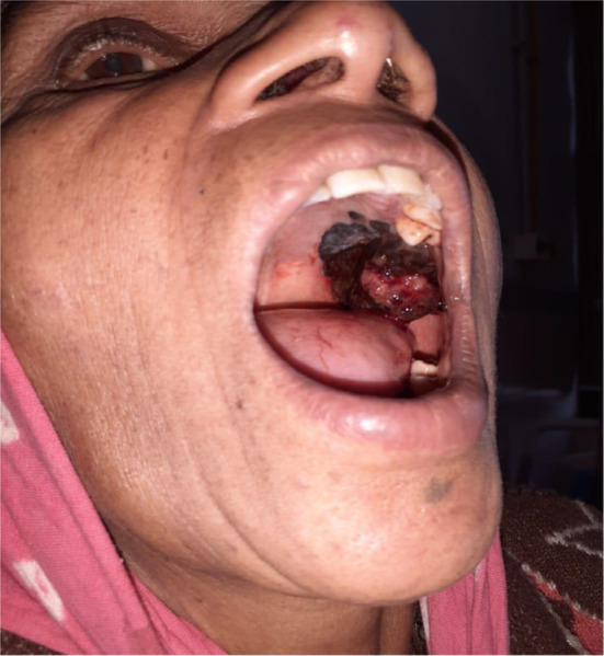
The development of induration is an ominous sign. However, regression of melanoma is a poor prognostic sign probably because of thickness of tumour that shows regression may have been greater than it appears at the time of examination (Fig. 1).
Fig. 1.
Clinical examination
Mucosal melanomas are more aggressive and present in a vertical (nodular) growth phase of the disease. The melanosis turning into the malignant melanoma has great propensity for systemic metastasis to bone marrow, lungs, liver, kidney, stomach, intestine and adrenals leading to systemic symptoms and very poor prognosis [9].
Differential Diagnosis
Physiological pigmentation
Post-inflammatory pigmentation
Acquired melanotic naevus
Amalgam tattoo
Blue naevi
Melanosis associated with smoking and medications
Melanoplakia
Melanoacanthoma
Pituitary based Cushing’s syndrome
Addison’s disease
Peutz-Jegher’s syndrome
Kaposi’s sarcoma
Investigations
Incisional biopsy
All oral pigmentation should be biopsied as clinical diagnosis is precarious and histopathological confirmation is diagnostic (Fig. 2).
The pigmentation shows the following characteristics determining its nature whether benign or malignant.
A-Asymmetry
B-Border irregularity
C-Colour variation
D-Diameter of lesion > 6 mm
E-Evolving
Fig. 2.
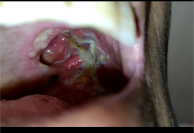
Post-operative clinical examination
If any two out of these five criteria are present in the lesion, it should be biopsied as clinical diagnosis is precarious and histopathological confirmation is diagnostic.
Histologically, atypical melanocytes and clear cells are present in the epithelial and connective tissue junction with hyperpigmentation of basal layers.
However, according to Mohan M. et al [1], incisional biopsy or Fine Needle Aspiration Cytology (F.N.A.C.) causes seeding of tumour cells into adjacent tissue or dissemination via blood or lymph leading to metastasis and decreased five year survival rate who underwent such procedure to 25.9% with respect to those who directly proceeded for definitive surgery (91.7%)
-
2.
Orthopantogram
-
3.
Computed Tomography
-
4.
Magnetic Resonance Imaging
-
5.
Immunohistochemistry is mandatory in cases of amelanotic or poorly pigmented melanomas.
S-100 protein is positive in 97%, HMB-45 in 71% and melan-A in 74% [6, 7].
Diagnosis
According to Takagi et al. [9], histologically, there are three forms of oral malignant melanoma.
Polygonal
Fusiform
Mixed
But the diagnosis becomes difficult because.
Early in situ lesions
Inadequate sampling
Late stage lesions
Treatment
Wide Local Excision with wide margins alongwith neck dissection is the primary treatment along with chemo-radiotherapy as adjuvant due to its grave prognosis. [8] Maxillectomy with a margin of 3–5 cms is recommended for the palate. [1]1 However, the presence of vital structures makes reaching the aim elusive. Hence, chemotherapy is a must due to a high possibility of systemic metastasis and radiotherapy is also needed for bony invasion even with complete excision of the tumour with clear margins.
Post-operative radiotherapy is highly useful [11] in.
Positive surgical margins
Strong likelihood of local metastasis
Possibility of distant spread.
Dacarbazine-DTIC and INF- alpha-2b are considered as chemotherapeutic as well as immunotherapeutic along with BCG and recombinant interleukin-2 [9].
Prognosis
In cases of early diagnosis, the malignant cells are present only in the epithelium with minimal invasion making the melanoma 100% curable by excision with 95% of 5-year survival rate.
Factors with poor prognosis.
> 4 mm at initial presentation
Inadequate resection of margins
Late stage at presentation
Vascular invasion
Absence of melanosis
Metastasis-nodal and/or distant
Follow-Up
All the patients should be closely followed up by either nasal endoscopy or computed tomographic evaluation to check for.
Recurrence
Residual Lesion
For which repeat exploration is warranted.
Points to remember
Proper clinical examination with staging is considered the most important prognosis determining factor [3, 7, 11]. Regular close follow-up of pre-malignant lesions is mandatory for better understanding of the cancerization phenomenon so as to better the current diagnostic and treatment modalities.
Supplementary Information
Below is the link to the electronic supplementary material.
Funding
The authors did not receive any funding for this research work.
Declaration
Conflict of interest
The authors declare no conflicts of interest in this research work.
Ethics Approval
All procedures performed in studies involving human participants were in accordance with the ethical standards of the institutional research committee (Institutional Ethics Committee (Human), P.D.U. Medical College, Rajkot) and with the 1964 Helsinki declaration and its later amendments or comparable ethical standards.
This article does not contain any studies with animals performed by any of the authors.
Consent for Participation and Publication
Informed consent including that for publication was obtained from the participant included in the study.
Footnotes
Publisher's Note
Springer Nature remains neutral with regard to jurisdictional claims in published maps and institutional affiliations.
References
- 1.Greene GW, Haynes JW, Dozier M, Blumberg JM, Bernier JL. Primary malignant melanoma of the oral mucosa. Oral Surg, Oral Med, Oral Pathol. 1953;6(12):1435–1443. doi: 10.1016/0030-4220(53)90242-4. [DOI] [PubMed] [Google Scholar]
- 2.Lopez-Graniel CM, Ochoa-Carrillo FJ, Meneses-García A. Malignant melanoma of the oral cavity: diagnosis and treatment. Oral Oncol. 1999;35(4):425–430. doi: 10.1016/S1368-8375(99)00017-2. [DOI] [PubMed] [Google Scholar]
- 3.Tanaka N, Mimura M, Ogi K, Amagasa T. Primary malignant melanoma of the oral cavity: assessment of outcome from the clinical records of 35 patients. Int J Oral Maxillofac Surg. 2004;33(8):761–765. doi: 10.1016/j.ijom.2004.01.008. [DOI] [PubMed] [Google Scholar]
- 4.Gondivkar SM, Indurkar A, Degwekar S, Bhowate R. Primary oral malignant melanoma–a case report and review of the literature. Quintessence Int. 2009;40(1):41–46. doi: 10.3290/j.qi.a14113. [DOI] [PubMed] [Google Scholar]
- 5.Lourenço SV, Bologna SB, Colucci F, Neto CF, Montenegro FLM, Nico MMS. Oral mucosal melanoma of the mandibular gingiva: a case report. Cutis. 2010;86(2):89–93. [PubMed] [Google Scholar]
- 6.Prasad ML, Jungbluth AA, Iversen K, Huvos AG, Busam KJ. Expression of melanocytic differentiation markers in malignant melanomas of the oral and sinonasal mucosa. Am J Surg Pathol. 2001;25(6):782–787. doi: 10.1097/00000478-200106000-00010. [DOI] [PubMed] [Google Scholar]
- 7.Williams MD (2017) Update from the 4th edition of the world health organization classification of head and neck tumours mucosal melanomas. Head Neck Pathol, 11(1): 110-117, 10.1007/s12105-017-0789-y [DOI] [PMC free article] [PubMed]
- 8.Barker BF, Carpenter WM, Daniels TE, et al. Oral muscosal melanomas. Oral Surg, Oral Med, Oral Pathol, Oral Radiol, Endodontol. 1997;83(6):672–679. doi: 10.1016/S1079-2104(97)90318-8. [DOI] [PubMed] [Google Scholar]
- 9.Takagi M, Ishikawa G, Mori W. Primary malignant melanoma of the oral cavity in Japan with special reference to mucosal melanosis. Cancer. 1974;34(2):358–370. doi: 10.1002/1097-0142(197408). [DOI] [PubMed] [Google Scholar]
- 10.Padhye A, D’souza J. Oral malignant melanoma: a silent killer? J Indian Soc Periodontol. 2011;15(4):425–428. doi: 10.4103/0972-124X.92587. [DOI] [PMC free article] [PubMed] [Google Scholar]
- 11.Mohan M, Sukhadia VY, Pai D, Bhat S. Oral malignant melanoma: systematic review of literature and report of two cases. Oral Surg Oral Med Oral Pathol Oral Radiol. 2013;116(4):e247–254. doi: 10.1016/j.oooo.2011.11.034. [DOI] [PubMed] [Google Scholar]
Associated Data
This section collects any data citations, data availability statements, or supplementary materials included in this article.