

Abstract
We describe our innovative technique of endoscopic cartilage tympanoplasty using cartilage fashioned as umbrella in stapes absent condition using endoscope holder. Tragal cartilage disc of 3 × 3 mm dimensions with a circular slot of 0.8 mm to accommodate the vertical strut measuring 3 mm × 7 mm in cases with absent incus and stapes. Tympanic membrane reconstruction was done, with or without attic reconstruction, using sliced tragal cartilage of less than 0.5 mm thickness.
Supplementary Information
The online version contains supplementary material available at 10.1007/s12070-021-02518-8.
Keywords: Endoscope holder, Two handed technique, Type 3 Endoscopic cartilage Tympanoplasty
Introduction
The earliest attempt at ossiculoplasty was made in 1901. Since then, many techniques and materials have been proposed for ossiculoplasty with varying degree of success. The goal of ossiculoplasty is to repair the conductive hearing loss giving a long term stability. The most commonly used autograft material is the incus body, which is fashioned and placed on footplate of stapes. Various grafts for reconstruction of the ossicular defect include bone cement, cartilage, hydroxyapatite, glass ionomers, alloplastic ossicular prosthesis like total and partial ossicular replacement prosthesis (TORP/PORP) made of teflon, and titanium, gold implants etc. [1]. We have been using tragal cartilage since 2003 for reconstructing the hearing mechanism in cases of absence of stapes superstructure. Till 2013, we performed this technique of ossiculoplasty using microscope. In 2013, after development of endoscope holders, we have been performing these techniques in middle ear surgery with the additional advantages of endoscope and endoscope holders [2–14]. We report the use of the table model of endoscope holder having gas spring action and rack and pinion mechanism for two handed endoscopic earsurgery. We fashion the tragal cartilage graft as an umbrella as total ossicular replacement to be placed over the stapes footplate in cases of absence of incus and stapes superstructure.
Methods
Operating Theatre Requirements
Zero degree 3 mm Karl Storz endoscope, triple charge coupled device Camera (Karl Storz, Germany), and table model of endoscope holder, Endohold GS 201 B (Dr. Khan’s Creations, India).
The table model of the Endoscope holder (Fig. 1) is based on the concept of gas spring action with rack and pinion mechanism [2, 3]. A gas spring uses compressed gas contained within an enclosed cylinder sealed by a sliding piston to pneumatically store potential energy and withstand external force applied parallel to the direction of the piston shaft. The endoscope holder is dynamic and non-static. It allows the endoscope to be tilted in any direction (rotated forwards, rotated backwards, upwards, downwards and sidewise angular rotation). The peculiarity of the endoscope holder is that it is provided with rack and pinion mechanism which allows the additional few centimetres closer motion of the endoscope into the ear canal and middle ear cavity.
Fig. 1.

Endoscope holder, Endohold GS 201 B in endoscopic ear surgery
The parts of the endoscope holder:
O.T. table gripping attachment
Horizontal metallic rod
Gas Spring arm
Metallic connecting rod
Rack and Pinion system attached
Attachment with slot for holding the endoscope
Position of Endoscope Holder
The endoscope holder is fixed to the operation table on the opposite side of the ear to be operated. The endoscope is mounted onto the endoscope holder with the camera (Fig. 1). The endoscope is directed into the ear canal. The operating surgeon sits on the same side as the ear with the monitor facing the surgeon. The surgeon operates by seeing onto the monitor thus requiring hand-eye-monitor coordination.
Preparation of the Patient
The patient is prepared and draped in the usual fashion with the ipsilateral shoulder pulled down. The ear canal is infiltrated with 2 percent lidocaine with 1 in 2,00,000 adrenaline.
Harvest of the Tragal Cartilage
We prefer tragal cartilage as the graft of choice for tympanic membrane and ossicular reconstruction. It is harvested via the horizontal incision on the tragus. Approximately 15 × 15 mm cartilage is harvested.
Preparation of the Graft
A 4 × 4 mm full thickness cartilage is taken. Perichondrium is removed from both sides. A central circular slot is made into it with 1 mm punch rod (Fig. 2). The vertical strut of cartilage (with intact perichondrium on both sides) measuring 6–7 mm × 2 mm is taken (Fig. 3). The vertical strut is passed through the circular cartilage disc of 4 mm x 4 mm so as to form an umbrella cartilage (Fig. 4). This stable assembly is then placed over the stapes footplate. The remaining tragal cartilage graft is sliced [2, 4] with metallic thickness plate of 0.3 mm with cartilage splitter to obtain 2 sliced tragal grafts (approximately 0.3 mm and 0.5 mm thickness (assuming the tragal cartilage is 1 mm in thickness, each perichondrium 0.1 mm and after slicing with 0.3 mm we get 0.3 mm and 0.5 mm).
Fig. 2.
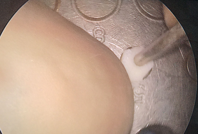
Creation of Cartilage disc with a central hole
Fig. 3.
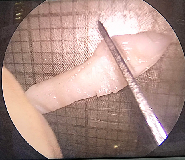
Creation of Vertical strut
Fig. 4.
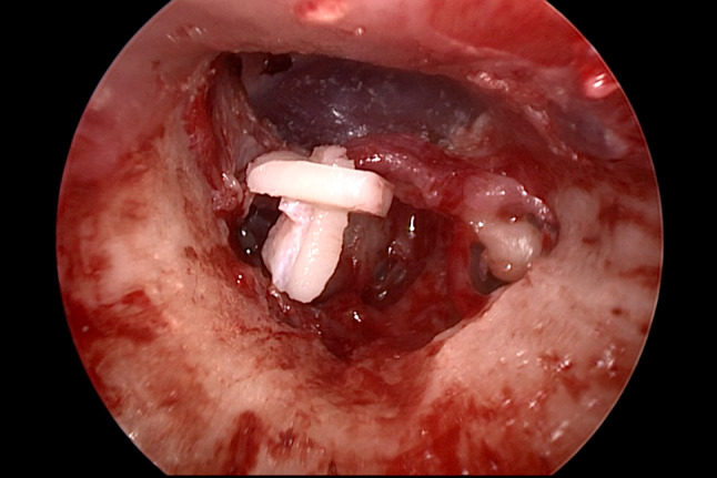
View before Placement of Cartilage umbrella on stapes footplate
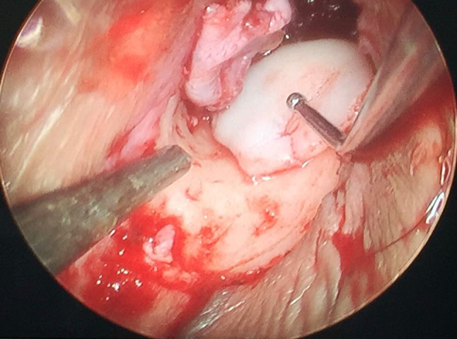
Procedure of Endoscopic Cartilage Umbrella Ossiculoplasty (Figs. 5, 6, video)
Fig. 5.

Placement of Cartilage umbrella on stapes footplate
Fig. 6.
Reinforcement with Sliced tragal graft. VIDEO: Endoscopic cartilage umbrella ossiculoplasty
Instruments are similar to those used in microscopic ear surgery. Pinna stay suture is taken to retract the ear canal and to provide additional few millimetres for instrumental manipulation [15]. Transcanal/endaural incision is taken. Tympanomeatal flap is elevated. Middle ear inspection is done to detect ossicular erosion. The ventilation block due to granulations or cholesteatoma is cleared depending on the type of pathology (squamosal or mucosal otitis media) by inside-out atticotomy and antrostomy. In the absence of all ossicles, the prepared cartilage umbrella graft placed onto the footplate of the stapes (Fig. 5). This graft assembly is stable. Sliced cartilage of 0.5 mm thickness kept as underlay shield graft for tympanic membrane reconstruction (Fig. 6). Tympanomeatal flap reposed back. Throughout the procedure, the suction is held in left hand to avoid fogging and to achieve cooling of endoscope generated heat [16]. Intermittent irrigation is done for cooling of endoscope as well as to achieve cleaning of endoscope lens [16]. The 3 mm 14 cm Karl Storz zero degree endoscope gives a panoramic view even in narrow canals, minimising the need for canalplasty. All procedures performed are recorded for documentation. The advantage of endoscope holder is that both the hands are free for intervention with the left hand holding the suction and right hand holding the microear instruments.
Discussion
The reconstruction of the ossicular chain as a surgical treatment of chronic otitis media is challenging to the otologist. Ossiculoplasty involves reconstruction of sound-conducting mechanism between the graft and oval window. We use tragal cartilage fashioned as cartilage umbrella as an ossiculoplasty option when all ossicles are absent.
Advantages of two handed endoscopic cartilage umbrella ossiculoplasty:
Uses of endoscope holders
-
A.
Stable image on the monitor
-
B.
No fatigue as the endoscope is mounted on endoscope holder
-
C.
Simultaneous use of both hands similar to microscopic ear surgery
-
D.
Simultaneous use of drill and the suction possible similar to microscopic surgery
-
E.
No repeated fogging due to the suction in the non-dominant hand unlike single handed endoscopic ear surgery
-
F.
Comfort during handling and manipulation during ossiculoplasty
-
G.
Two handed technique allows control of bleeding through continuous suction held in the non-dominant hand
-
H.
Precision during cartilage graft placement due to the two handed technique
-
2.
Advantages of our Cartilage Umbrella placed on stapes footplate
Gives better graft stability
No displacement or extrusion
Good hearing with post-operative airbone gap closure upto 20 dB.
It is reinforced by a sliced tragal cartilage for TM reconstruction.
-
3.Advantages of Endoscopes
- Panoramic image
- Complete view of tympanic membrane and the canal wall
- Visualisation of the middle ear spaces without much bone removal
- Better understanding of the mucosal folds and the ventilation pathways
Limitations
Lack of depth perception.
Conclusion
Cartilage umbrella is a good option in cases of absence of stapes superstructure. Using endoscope and endoscope holders gives additional advantage of panoramic image along with benefits of two hands in ear surgery.
Supplementary Information
Below is the link to the electronic supplementary material.
Acknowledgements
We express our sincere gratitude to Dr.Mrs. Shirin Mubarak Khan for all the technical support provided during the research study and manuscript preparation.
Author Contribution
Dr SRP: conceptualisation, study design, manuscript drafting. Dr MMK: conceptualisation, study design, and manuscript drafting.
Funding
This study was not financially supported from external sources.
Declarations
Conflict of interest
Endohold GS 201 B is registered and trademarked under the name of Dr. Mubarak M. Khan and applied for patent under the name of Dr. Mubarak Khan under Patent Act, India.
Footnotes
Publisher's Note
Springer Nature remains neutral with regard to jurisdictional claims in published maps and institutional affiliations.
References
- 1.Galy-Bernadoy C, Akkari M, Mathiolon C, Mondain M, Uziel A, Venail F. Comparison of early hearing outcomes of type 2 ossiculoplasty using hydroxyapatite bone cement versus other materials. Euro Ann Otorhinolaryngol Head Neck Dis. 2014;131(5):289–292. doi: 10.1016/j.anorl.2013.03.009. [DOI] [PubMed] [Google Scholar]
- 2.Khan MM, Parab SR. Endoscopic cartilage tympanoplasty: a two-handed technique using an endoscope holder. Laryngoscope. 2016;126:1893–1898. doi: 10.1002/lary.25760. [DOI] [PubMed] [Google Scholar]
- 3.Khan MM, Parab SR. Concept, design and development of innovative endoscope holder system for endoscopic otolaryngological surgeries. Ind J Otolaryngol Head Neck Surg. 2015;67(2):113–119. doi: 10.1007/s12070-014-0738-y. [DOI] [PMC free article] [PubMed] [Google Scholar]
- 4.Parab SR, Khan MM. New cartilage slicer for slicing techniques in tympanoplasty: design and applications. Ind J Otolaryngol Head Neck Surg. 2018;70(4):515–520. doi: 10.1007/s12070-018-1467-4. [DOI] [PMC free article] [PubMed] [Google Scholar]
- 5.Khan Mubarak M., Parab Sapna R. Primary cartilage tympanoplasty: our technique and results. American Journal of Otolaryngology. 2011;32:381–387. doi: 10.1016/j.amjoto.2010.07.010. [DOI] [PubMed] [Google Scholar]
- 6.Khan MM, Parab SR. Day care ear surgery: our experience of 4 years. Indian Journal of Otolaryngology and Head & Neck Surgery. 2012;64(3):280–284. doi: 10.1007/s12070-011-0303-x. [DOI] [PMC free article] [PubMed] [Google Scholar]
- 7.Khan MM, Parab SR. Comparative study of sliced tragal cartilage and temporalis fascia in type I tympanoplasty. J Laryngol Otol. 2015;129(1):16–22. doi: 10.1017/S0022215114003132. [DOI] [PubMed] [Google Scholar]
- 8.Khan MM, Parab SR. Average thickness of tragal cartilage for slicing techniques in tympanoplasty. J Laryngol Otol. 2015;129(5):435–439. doi: 10.1017/S0022215115000055. [DOI] [PubMed] [Google Scholar]
- 9.Khan MM, Parab SR. Endoscopic Septoplasty—Two Handed Technique with Endoscope Holder: A Novel Approach. Indian J Otolaryngol Head Neck Surg. 2016;68(4):475–80. doi: 10.1007/s12070-016-0997-x. [DOI] [PMC free article] [PubMed] [Google Scholar]
- 10.Parab SR, Khan MM. Minimal invasive endoscopic ear surgery: a two handed technique. Indian J Otolaryngol Head Neck Surg. 2019;71(2):1334–42. doi: 10.1007/s12070-018-1411-7. [DOI] [PMC free article] [PubMed] [Google Scholar]
- 11.Parab SR, Khan MM. Endoscopic Management of Tympanic Membrane Retraction Pockets: A Two Handed Technique with Endoscope Holder. Indian J Otolaryngol Head Neck Surg. 2019;71(4):504–11. doi: 10.1007/s12070-019-01682-2. [DOI] [PMC free article] [PubMed] [Google Scholar]
- 12.Parab Sapna R, Khan Mubarak M. Modified Endoscope Holder for Two Handed Endoscopic Ear Surgery. Indian J Otolaryngol Head Neck Surg. 2020;72(3):335–341. doi: 10.1007/s12070-020-01841-w. [DOI] [PMC free article] [PubMed] [Google Scholar]
- 13.Khan MM, Parab SR. Novel concept of attaching endoscope holder to microscope for two handed endoscopic tympanoplasty. Indian J Otolaryngol Head Neck Surg. 2016;68(2):230–40. doi: 10.1007/s12070-015-0916-6. [DOI] [PMC free article] [PubMed] [Google Scholar]
- 14.Parab, S.R., Khan, M.M. Zaidi A. 2020 Endoscopic cartilage butterfly tympanoplasty: a two-handed technique with endoscope holder. Indian J Otolaryngol Head Neck Surg. 10.1007/s12070-020-01875-0 [DOI] [PMC free article] [PubMed]
- 15.Sapna Ramkrishna Parab, Mubarak Muhamed Khan, (2020) Pinna stay suture in two handed endoscopic ear surgery: Our experience. American Journal of Otolaryngology 41 (5):102582 [DOI] [PubMed]
- 16.Elliott D. Kozin, Ashton Lehmann, Margaret Carter, Ed Hight, Michael Cohen, Hideko H. Nakajima, Daniel J. Lee, (2014) Thermal effects of endoscopy in a human temporal bone model: Implications for endoscopic ear surgery. The Laryngoscope 124 (8):E332-E339 [DOI] [PMC free article] [PubMed]
Associated Data
This section collects any data citations, data availability statements, or supplementary materials included in this article.