Abstract
Aims:
Prospective measures of high knee abduction moment (KAM) during landing identify female athletes at increased risk for anterior cruciate ligament (ACL) injury. Laboratory-driven measurements predict high KAM with 90% accuracy. This study aimed to validate the clinic-based variables against 3-dimensional motion analysis measurements.
Methods:
Twenty female basketball, soccer, and volleyball players (age, 15.9 ± 1.3 years; height, 163.6 ± 9.9 cm; body mass, 57.0 ± 12.1 kg) were tested using 3-dimensional motion analysis and clinic-based techniques simultaneously. Multiple logistic regression models have been developed to predict high KAM (a surrogate for ACL injury risk) using both measurement techniques. Clinic-based measurements were validated against 3-dimensional motion analysis measures, which were recorded simultaneously, using within- and between-method reliability as well as sensitivity and specificity comparisons.
Results:
The within-variable analysis showed excellent inter-rater reliability for all variables using both 3-dimensional motion analysis and clinic-based methods, with intraclass correlation coefficients (ICCs) that ranged from moderate to high (0.60–0.97). In addition, moderate-to-high agreement was observed between 3-dimensional motion analysis and clinic-based measures, with ICCs ranging from 0.66 to 0.99. Bland-Altman plots confirmed that each variable provided no systematic shift between 3-dimensional motion analysis and clinic-based methods, and there was no association between difference and average. A developed regression equation also supported model validity with > 75% prediction accuracy of high KAM using both the 3-dimensional motion analysis and clinic-based techniques.
Conclusion:
The current validation provides the critical next step to merge the gap between laboratory identification of injury risk factors and clinical practice. Implementation of the developed prediction tool to identify female athletes with high KAM may facilitate the entry of female athletes with high ACL injury risk into appropriate injury-prevention programs.
Keywords: anterior cruciate ligament injury, knee, drop-vertical jump landing, young athletes, high-risk biomechanics, injury prevention
Introduction
Prospective measures of high knee abduction moment (KAM) during landing have been demonstrated to predict anterior cruciate ligament (ACL) injury risk in young female soccer and basketball players.1 In addition, a large-scale prospective study found that military cadets who sustained ACL injury demonstrated knee landing mechanics related to high KAM.2 In validation of these findings, retrospective observations of ACL injury mechanics in female athletes report knee landing and cutting alignments associated with high KAM at the time of injury.3-5
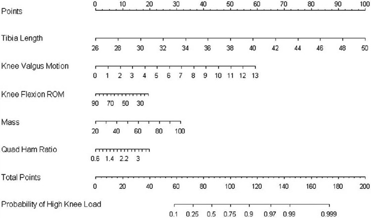
In the development of a KAM-predictive nomogram, reports have isolated 5 laboratory-captured measurements: 1) increased knee abduction angle, 2) increased relative quadriceps recruitment, 3) decreased knee flexion range of motion (ROM), 4) increased tibia length, and 5) mass normalized to body height that accompanies growth, all of which contribute to approximately 80% of the measured variance in KAM during landing.6 However, expensive biomechanical laboratories, which require costly measurement tools (eg, motion analysis systems) and labor-intensive data collection sessions, are necessary to ascertain these measurements. These factors limit the potential to perform athlete risk assessments on a large scale, precluding the opportunity to target athletes at high risk of injury with the appropriate intervention strategies. If simpler assessment tools are developed that can be administered in a clinic or field testing environment (clinic-based) that are validated by the highly accurate 3-dimensional motion analysis assessment, screening for ACL injury risk can be performed on a more widespread basis. Therefore, a clinic-based assessment algorithm was systematically developed and validated to improve the potential to identify and target injury-prevention training to female athletes with increased KAM.7 The validated clinic-based assessment algorithm delineated 5 clinically measurable factors, which, when combined, accurately identify high KAM during landing (Figure 1).6,7 Preliminary results indicate that this clinic-based assessment tool, when used as part of an algorithmic methodology, can precisely replicate 3-dimensional motion analysis kinematics and accurately predict the critical outcome of high KAM, which determines the risk of sustaining an ACL injury.1,8
Figure 1.
Nomogram that was developed from the regression analysis, which can be used to predict outcome based on tibia length, knee valgus motion, knee flexion range of motion, body mass, and quadriceps-to-hamstrings ratio.
To use the nomogram, one should place a straight edge vertically so that it touches the designated variable on the axis for each predictor, and record the value that each of the 5 predictors provides on the “points” axis at the top of the diagram. All of the recorded points are then summed and this value is located on the total points line with a straight edge. A vertical line drawn down from the total points line to the probability line will identify the probability that the athlete will demonstrate high knee abduction moment during the drop-vertical jump based on the utilized predictive variables.
Observational screenings, based on observed valgus alignment during landing, provide a promising low-cost assessment tool for athletes who are at high risk for injury. However, simplified identification of KAM may yield the most accurate classification of high risk for ACL injury.1,9 The purpose of this study is to validate the clinic-based variables against 3-dimensional motion analysis measurements to determine their viability for use in an ACL injury risk-prediction algorithm. The hypothesis was that clinically obtainable correlates derived from highly predictive 3-dimensaional motion analysis models would demonstrate high accuracy for determination of high KAM status.
Materials and Methods
Subjects
Twenty female basketball, soccer, and volleyball players (age, 15.9 ± 1.3 years; height, 163.6 ± 9.9 cm; body mass, 57.0 ± 12.1 kg) were tested using 3-dimensional motion analysis and clinic-based techniques simultaneously. All athletes were reported to be injury free at the time of testing.
Procedures
The Cincinnati Children’s Hospital Medical Center and Rocky Mountain University of Health Professions’ institutional review boards approved the data collection procedures and consent forms. Parental and athlete consent were received prior to data collection. Subjects were tested prior to the start of their competitive basketball, soccer, and volleyball seasons. Testing consisted of a knee examination, medical history, and dynamic strength and biomechanical landing analysis. Testing was performed by a research team, which included athletic trainers and research biomechanists.
Anthropometrics and Dynamic Strength
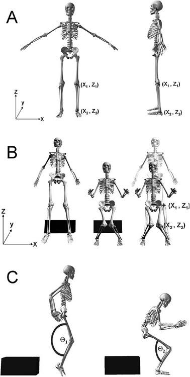
Body mass was measured on a calibrated physician scale. Clinic-based tibia length was measured with a standard measuring tape and was equal to the distance between the lateral knee joint line and the lateral malleolus (Figure 2A). Isokinetic knee extension/flexion (concentric/concentric muscle action) strength was measured on a Biodex System 2 (Biodex Medical Systems, Shirley, NY) and consisted of 10 knee flexion/extension repetitions for each leg at 300° per second.10 The QuadHam ratio was calculated as the ratio of quadriceps-to-hamstrings peak isokinetic torque.
Figure 2.
A) Tibia length was calculated ([3-dimensional motion analysis/measured [clinic-based]) as the distance between knee joint center and ankle joint center (Z2− Z1). B) Example of initial measure of frontal plane knee alignment and frontal plane knee alignment taken at maximum knee flexion during landing. The calibrated displacement measure between the 2 marked knee alignments (X2− X1) is representative of knee valgus motion during the drop-vertical jump. C) Example of initial measure of sagittal plane knee flexion (Θ1) and knee flexion angle taken at maximum knee flexion (Θ2) during landing. The displacement of knee flexion is found by calculating the differences in knee flexion position (Θ1−Θ2).
Laboratory-Based Landing Biomechanics
Three-dimensional hip, knee, and ankle kinematic and kinetic data were quantified for the contact phase of 3 drop-vertical jump tasks. One investigator instrumented each subject, with 37 retroreflective markers placed on the sacrum, left posterior superior iliac spine (PSIS), sternum, and bilaterally on the shoulder, elbow, wrist, anterior superior iliac spine (ASIS), greater trochanter, mid-thigh, medial and lateral knee, tibial tubercle, mid-shank, distal shank, medial and lateral ankle, heel, dorsal surface of the midfoot, lateral foot (fifth metatarsal), and toe (between second and third metatarsals). First, a static trial was conducted, in which the subject was instructed to stand still with foot placement standardized to the laboratory coordinate system. This static measurement was used as each subject’s neutral (zero) alignment; subsequent kinematic measures were referenced in relation to this position.11 The static standing trial was also used to calculate segment lengths as the estimated distance between the proximal and distal joint center (eg, tibia segment distance was equal to the distance between the knee joint center and ankle joint center) (Figure 2A). The drop-vertical jump involved the subject standing on top of a box (31 cm high) with her feet positioned 35 cm apart. The subjects were instructed to drop directly down off the box and immediately perform a maximum vertical jump, raising both arms while jumping for a basketball rebound.12
Joint kinematics and kinetics were measured, as described previously, and knee joint angles were calculated via 3-dimensional motion analysis according to the cardan/euler rotation sequence.11 Ground reaction force and kinematic data were filtered through a low-pass fourth order Butterworth filter at a cutoff frequency of 12 Hz to minimize possible impact peak errors. Kinematic data were combined with force data to calculate knee joint moments using inverse dynamics. Net external knee moments are reported, and KAM measures represent the external load on the joint. The kinematic and kinetic data were exported to MATTAB (MathWorks, Natick, MA), and the peak knee abduction angle and moment (negative) were identified during the deceleration phase of the initial stance phase of the drop-vertical jump. The deceleration phase was operationally defined from initial contact (vertical ground reaction force [VGRF] first exceeded 10 N) to the lowest vertical position of the body center of mass. Knee valgus motion was calculated as the frontal plane displacement of the knee from initial contact to the end of the deceleration phase of the drop-jump landing task (Figure 2B). Knee flexion ROM was calculated as the angular displacement of the knee during the stance phase of the drop-vertical jump (Figure 2C). The left side data were used for statistical analysis. The cut-point (threshold) used to classify the dependent variable status (high KAM) was > 21.74 Nm of KAM, which was based on published prediction modeling of ACL injury risk prediction and the goal to achieve sensitive prediction of high KAM. Using this classification, subjects were categorized into a dichotomous value (high KAM; “yes” or “no”) and used as the dependent variable.
Clinic-Based Landing Biomechanics
Two-dimensional frontal and sagittal plane knee kinematic data were collected using standard video cameras during the 5 recorded trials of drop-vertical jump. Two- and 3-dimensional techniques were collected simultaneously during the 5 trials, handing sequence images were captured with VirtualDub, and kinematic coordinate data were compiled using ImageJ (National Institutes of Health). These kinematic data were exported to MATTAB, and the frontal plane camera provided coordinate position (x axis; Figure 2B) captured from the video frame just before initial contact and the coordinate of maximum medial displacement. Video correction factor was averaged from 2 known distances (force plate width at floor level and box width at 31 cm height). Coordinate data were calibrated with video correction factor, and medial knee displacement was calculated (Figure 2B). The sagittal plane video camcorder was used to capture knee flexion angles, which were calculated in the video frame just prior to initial contact and maximum knee flexion. Knee flexion ROM was calculated as the difference in knee flexion between 2 positions, specifically between positions (Θ1 − Θ2; Figure 2C).13
Statistical Analyses
Data were exported to SPSS version 16 (SPSS, Inc., Chicago, IL) and SAS version 9.1 (SAS Institute, Cary, NC) for statistical analysis. A multiple logistic regression model previously developed7,8 was used to examine the prediction of high KAM (a surrogate for ACL injury risk) using both measurement techniques. Within-methods reliability was assessed using intraclass correlation coefficients (ICC), and measurements from 5 reviewers were used. Clinic-based methods were also validated against 3-dimensional motion analysis measures using between-method reliability (ICC and Bland-Altman plots). The 3-dimensional motion analysis and clinic-based measurements were validated by solving the prediction equation for each subject in the current data set. The resulting logit was converted to probability to determine which group (high KAM vs low KAM) the athletes would be classified into based on a 0.50 probability cut-score. The sensitivity, specificity, and percent correctly classified were calculated for the resulting 2 × 2 table of actual versus model-predicted classifications.
Results
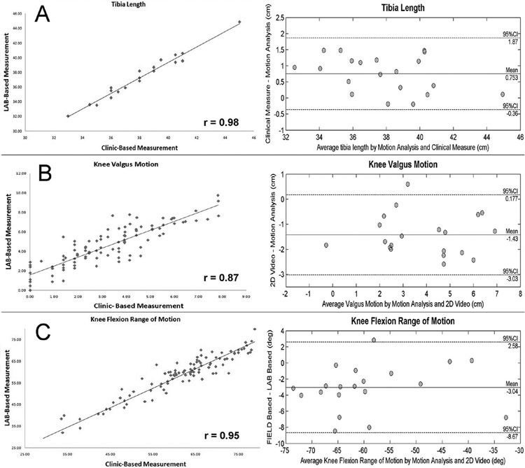
In the current validation data set, the within-variable analysis showed good reliability for all variables using both 3-dimensional motion and clinic-based methods with ICCs that ranged from moderate to high (0.60–0.78). In addition, moderate-to-high agreement was observed between 3-dimensional motion analysis measures and its clinic-based surrogate. Specifically, tibia length provided a between-method ICC of 0.95 and a mean difference (3-dimensional motion analysis clinic-based variables) of 0.75 cm (95% confidence interval [CI], −0.35 to 1.86). Knee valgus motion provided a between-method ICC of 0.66 and a mean difference of −1.42 cm (95% CI, −3.02 to 0.18). Knee valgus ROM provided a between-method ICC of 0.99 and a mean difference of −3.04 (95% CI, −8.67 to 2.58). Bland-Altman plots confirmed that no variable showed any systematic shift between 3-dimensional motion analysis and clinic-based methods, or association between difference and average (Figure 3). The Pearson correlation of average versus between-method differences (r) were −0.35, −0.04, and 0.16 for the tibia length, knee valgus motion, and knee flexion ROM measures, respectively.
Figure 3.
Bland-Altman plots for tibia length, knee valgus motion, and knee flexion range of motion confirmed that no variable showed any systematic shift between 3-dimensional motion analysis and clinic-based methods, or association between difference and average.
The defined variables from the published models for high KAM prediction were employed in the current sample and were measured with both 3-dimensional motion analysis and clinic-based techniques.7,8 The applied prediction equation coefficients (β) with y-intercept at −14.78 included knee valgus motion (cm); 0.358, knee flexion ROM (degree); −0.016, body mass (kg); 0.038, tibia length (cm); 0.320, and QuadHam (ratio) 0.528. Three-dimensional motion analysis measurements demonstrated a combined classification accuracy of 80% to correctly classify high KAM status using the prediction equation. The sensitivity, specificity, and percent correctly classified were calculated for the resulting 2 × 2 table of actual versus model-predicted classifications, indicating that the 3-dimensional motion analysis measures provided 79% sensitivity and 83% specificity for correct classification of high KAM status. Similar to tested model measured using 3-dimensional motion analysis-measured variables, the clinic-based measurements demonstrated a combined classification accuracy of 75% to correctly classify high KAM status. The resulting 2 × 2 table of actual versus model-predicted classification demonstrated a 77% sensitivity and 71% specificity for correct KAM classification using clinic-based measurements.
Discussion
In female athletes, KAM during landing is a measure that can be quantified using highly responsive 3-dimensional motion analysis techniques and can predict ACL injury risk with high sensitivity and specificity.1 A recent report has isolated biomechanical components that combine to nearly 80% of the measured variance in KAM during landing.6 Subsequently, clinically obtainable correlates to measures used in the highly predictive laboratory-based models were defined, which also demonstrated high accuracy in determination of high KAM status.7,8 These combined correlates, which include increased knee valgus motion, knee flexion ROM, body mass, tibia length, and quadriceps-to-hamstrings ratio, have all been related to increased risk of ACL injury in previous prospective and retrospective epidemiological reports.1,2,5,14
The purpose of the current study was to validate the combination of clinic-based variables against 3-dimensional motion analysis measurements to confirm their viability for use in a ACL injury risk-prediction algorithm. The results indicate that the individual variables related to increased ACL injury risk may combine to accurately identify female athletes at increased risk of injury. In addition, the accuracy of the simplistic approach to ACL injury risk prediction could facilitate widespread use and help implement focused interventions to prevent ACL injuries in young female athletes.
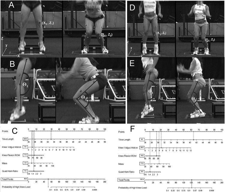
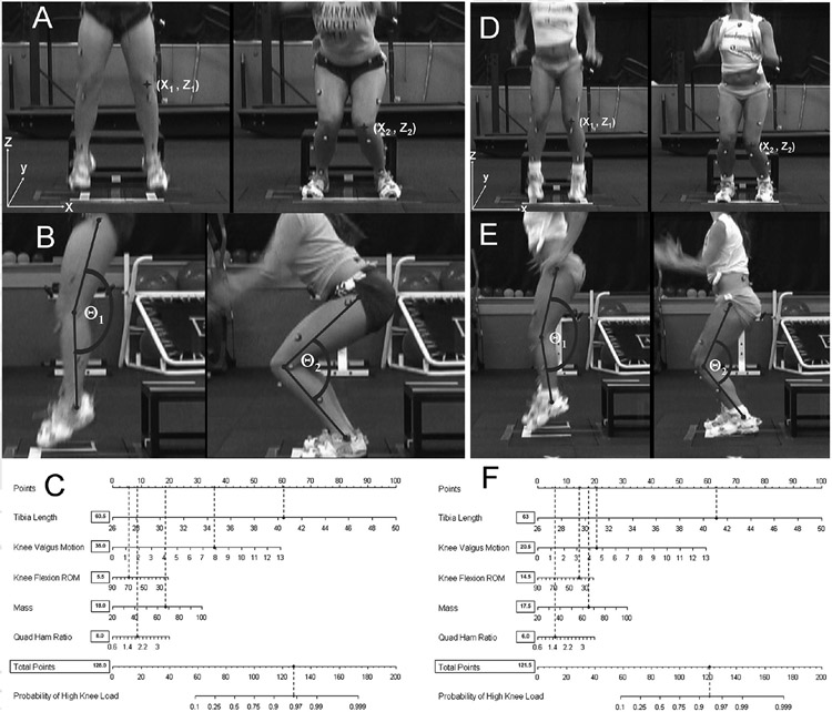
A longitudinal study of children aged 5 to 12 years in youth soccer demonstrated that there is no gender difference in knee injury risk in prepubescent athletes. However, being aged > 11 years was a significant risk factor for knee injury in girls.15 Figure 4A-C presents a subject in early puberty whose combination of decreased tibia length and mass prior to her rapid growth spurt contributes to her very small risk to demonstrate high KAM landing mechanics. As presented in the completed nomogram (Figure 4C), this subject would indicate a 12% probability of high KAM during landing. The accuracy of the clinic-based algorithm, which was confirmed as simultaneous 3-dimensional motion analysis quantification of actual KAM load during this drop-vertical jump trial, was 13.4 Nm. Following the growth spurt associated with maturation, female athletes are reported to be 4 to 6 times more likely sustain a sports-related noncontact ACL injury compared with males.16 Together, these factors suggest that following the onset of puberty, rapid increases in bone length and body mass (in the absence of matched increases in strength and recruitment of the musculature of the lower extremity posterior chain) underlie the tendency for increased KAM during landing tasks in female athletes.17-19
Figure 4.
A) Subject whose combination of decreased tibia length and mass prior to her rapid growth spurt diminish risk to demonstrate high knee abduction moment (KAM) landing mechanics. Knee valgus motion during the drop-vertical jump is calculated as the displacement measure between the 2 marked knee alignments in the X plane measured at the frame prior to initial contact and the frame with maximum knee flexion (X2−X1). B) Knee flexion range of motion (ROM) during the drop-vertical jump is calculated as the difference in knee flexion angle between initial contact and maximum knee flexion positions (Θ1−Θ2). C) Completed nomogram for the representative subject (tibia length, 32.5 cm; knee valgus motion, 4.2 cm; knee flexion ROM, 74.1°; mass, 30.7 kg; QuadHam, 1.64). Based on her demonstrated measurements, this subject would have a 12% (62.5 points) chance to demonstrate high KAM during the drop-vertical jump. Her actual KAM measure for the presented drop-vertical jump that was quantified simultaneously with 3-dimensional motion analysis was 13.4 Nm of knee abduction load. D) Subject whose combination of increased tibia length and mass, associated with her rapid growth, contribute to her increased risk to demonstrate high KAM landing mechanics when using the clinic-based ACL injury risk prediction algorithm. Knee valgus motion during the drop-vertical jump is calculated (X2−X1). E) Knee flexion ROM during the drop-vertical jump is calculated (Θ1−Θ2). F) Completed nomogram for the representative subject (tibia length, 45 cm; knee valgus motion, 3.0 cm; knee flexion ROM, 60.0°; mass, 71 kg; QuadHam, 1.19). Based on her demonstrated measurements, this subject would have a 91% (116.5 points) chance to demonstrate high KAM during the drop-vertical jump. Her actual KAM measure for the presented drop-vertical jump that was quantified simultaneously with 3-dimensional motion analysis was 48.5 Nm of knee abduction load.
The prevalence of ACL injuries incrementally increases each year until age 16 years in females, when the prevalence reaches its peak.20 Increased thigh length is an additional injury risk factor in female athletes.21 Cross-sectional data indicate that female athletes demonstrate incremental increases in KAM as they increase in age. Interestingly, the KAM loads that are related to increased knee injury risk also peak at 16 years of age, concomitant with peak ACL injury incidence in female athletes.20,22 Figure 4D-F depicts a 16-year-old girl whose combination of increased tibia length and body mass, associated with her rapid growth, contributes to her increased risk of high KAM landing mechanics. As demonstrated in the clinic-based ACL injury risk-prediction algorithm (Figure 4F), the combined anthropometric measures of increased tibia length and body mass associated with moderate knee valgus motion indicate nearly a 100% risk to demonstrate high KAM landing mechanics. If female athletes reach maturity with anthropometrics similar to those presented in Figure 4D-F (and in the absence of adaptation in core power and control to match whole body increases in inertial load), their tendency to demonstrate increased ground reaction forces and KAM during dynamic tasks may be increased.19 The current results indicate that tibia length can be reliably obtained through clinical measures that are highly related to 3-dimensional motion analysis measures, which have demonstrated a strong predictive relationship with high KAM.6
In addition to the effects of increased body mass with longer joint levers (ie, tibia length) to initiate greater joint forces that are more difficult to balance and dampen at the lower-extremity joints during high-velocity maneuvers, maturing female athletes do not naturally demonstrate an associated maturational “neuromuscular spurt” (ie, increased strength and power during maturational growth and development).18,23 During the maturation developmental period, male athletes naturally demonstrate this neuromuscular spurt to match the increased demands of growth and development. However, these same increases are not demonstrated in KAM in females.18,23-26 Female athletes do not demonstrate similar neuromuscular adaptations to match the increase demands created from structural and inertial changes during pubertal development.18,23-26
The knee joint, which is a hinge articulation that connects the body’s 2 longest bones, is equipped with strong active muscular restraints to adequately dampen knee joint loads in motions aligned in the sagittal plane.27 In the absence of active neuromuscular control, as evidenced by increased knee abduction motion,12,18 female athletes demonstrate a puberty-related divergence in KAM relative to age- and sport-matched male athletes.28 The decreased active control of knee alignments and loads may destabilize the knee, increase KAM, and increase the risk of ACL injury in maturing female athletes.1,29 Figure 5A-C provides an example of a subject from the current study whose excessive knee valgus motion during the drop-vertical jump provided a strong contribution to her 95% chance of having high KAM landing mechanics. Confirming the utility of the proposed clinic-based measurement of knee valgus motion and its use in the ACL injury risk algorithm, 3-dimensional motion analysis indicated an actual KAM measure of 44.1 Nm for the presented drop-vertical jump trial (Figure 5C).
Figure 5.
A) Subject with excessive knee valgus during the drop-vertical jump that contributes to her increased risk to demonstrate high knee abduction moment (KAM) landing mechanics. Knee valgus motion during the drop-vertical jump is calculated (X2−X1). B) Knee flexion range of motion (ROM) during the drop-vertical jump is calculated (Θ1−Θ2). C) Completed nomogram for the representative subject (tibia length, 40.5 cm; knee valgus motion, 7.8 cm; knee flexion ROM, 69.8°; mass, 67.5 kg; QuadHam, 1.90). Based on her demonstrated measurements, this subject would have a 95% (128 points) chance to demonstrate high KAM during the drop-vertical jump. Her actual KAM measure for the presented drop-vertical jump that was quantified simultaneously with 3-dimensional motion analysis was 44.1 Nm of knee abduction load. D) Example of representative subject with a combination of excessive knee valgus motion and small knee flexion ROM during the drop-vertical jump contribute to her increased risk to demonstrate high KAM landing mechanics. Knee valgus motion during the drop-vertical jump is calculated (X2−X1). E) Knee flexion range of ROM during the drop-vertical jump is calculated (Θ1−Θ2). F) Completed nomogram for the representative subject (tibia length, 41 cm; knee valgus motion, 4.6 cm; knee flexion ROM, 37.9°; mass, 65.3 kg; QuadHam, 1.51). Based on her demonstrated measurements, this subject would have a 93% (121.5 points) chance to demonstrate high KAM during the drop-vertical jump. Her actual KAM measure for the presented drop-vertical jump that was quantified simultaneously with 3-dimensional motion analysis was 77.6 Nm of knee abduction load.
The active restraints about the knee have poor mechanical advantage to adequately control excessive frontal plane knee abduction motion, common in young female athletes. Accordingly, active neuromuscular control from the sagittal plane musculature is required to prevent potential high KAM and is needed to maintain dynamic knee stability during landing and pivoting.30-32 Small knee flexion motions, exacerbated by early quadriceps or delayed/decreased hamstrings recruitment, limit the potential for active control of frontal plane alignments and contribute to increased KAM.6,33 Griffin et al34 postulated that increased body mass index would result in a more extended lower extremity position, which would influence decreased knee flexion on landing. Figure 5D-F presents an example of the consequences of limited sagittal plane mechanics combined with excessive knee valgus motions, which provides a 93% risk of high KAM according to the prediction algorithm. Measurements from simultaneously recorded 3-dimensional motion analysis confirm predicted outcome, as the actual KAM calculated during the presented drop-vertical jump trial was 77.6 Nm. A sagittal plane position of the knee near full extension when landing or cutting is commonly observed in video analysis of ACL injuries in female athletes.35 In addition, a prospective study indicated that female athletes who subsequently sustained ACL injuries demonstrated significantly less (10.5°) knee flexion during a drop-vertical jump than those who did not sustain injury.1 Thus, the reliable measurement demonstrated by clinic-based sagittal plane measurements and accuracy to achieve high KAM status support the inclusion of knee flexion ROM in the proposed ACL injury risk-prediction algorithm.
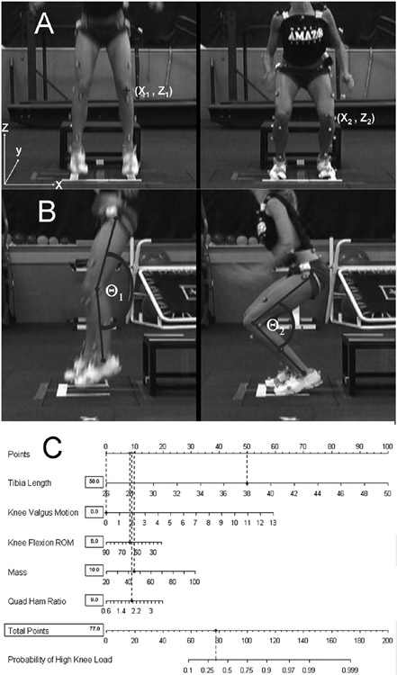
Recent studies demonstrate that neuromuscular training reduces the high KAM risk factor for ACL injury and decreases knee and ACL injury incidence in female athletes.35-38 However, re-evaluation of ACL injury rates in female athletes indicate that this important health issue has yet to be resolved, as increased knowledge and application of injury-prevention techniques have not led to measureable reductions in ACL injury incidence in female athletes.16 A recent investigation by Grindstaff et al39 indicates that standard, nontargeted neuromuscular training programs may require application to 89 female athletes to prevent a single ACL injury. It is possible that the identification of female athletes who demonstrate risk factors for ACL injury, such as high KAM using the presented algorithmic approach, could improve the efficiency of neuromuscular training targeted to these individuals. Pilot work testing this theory indicates that female athletes who are categorized as high risk for ACL injury, due to high KAM, are more responsive to neuromuscular training aimed at reducing this risk factor.1,36 Figure 6 presents optimal landing biomechanics during the drop-vertical jump, in which the athlete demonstrated desirable knee flexion ROM without any knee valgus motion, which ultimately limits her potential to demonstrate high KAM risk factor. Based on her recorded clinic-based measurement, the subject demonstrated an 18% chance for high KAM using the proposed prediction algorithm. Simultaneous 3-dimensional motion analysis confirmed the accuracy of the proposed algorithm as the subject demonstrated an actual KAM measurement of 7.6 Nm.
Figure 6.
Example of representative subject with optimal neuromuscular control during the drop-vertical jump that limits her potential to demonstrate high KAM risk factor. A) Knee valgus motion during the drop-vertical jump is calculated (X2 − X1). B) Knee flexion range of ROM during the drop-vertical jump is calculated (Θ1 − Θ2). C) Completed nomogram for the representative subject (tibia length, 38 cm; knee valgus motion, 0.0 cm; knee flexion ROM, 66.5°; mass, 44.9 kg; QuadHam, 1.89). Based on her demonstrated measurements this subject would have an 30% (77 points) percent chance to demonstrate high KAM during the drop vertical jump. Her actual KAM measure for the presented drop-vertical jump that was quantified simultaneously with 3-dimensional motion analysis was 7.6 Nm of knee abduction load.
Conclusion
The current validation indicates that we can successfully apply laboratory-based identification of injury risk factors1,2 into clinical practice.7,8 The simplicity, and decreased equipment and labor costs (relative to laboratory-based assessments that may be > $1000 per athlete) associated with use of the provided tool, may facilitate the identification of athletes at high risk of ACL injury in clinical and field settings. Prior research has used laboratory-based measures to determine the potential differing effects of neuromuscular training in female athletes who demonstrate high KAM landing strategies relative to those who do not.36 Implementation of the developed prediction tool will likely increase both the efficacy and efficiency of prevention strategies for noncontact ACL injury, and its widespread use may impact injury prevention in female athletes.
Acknowledgments
The authors would like to acknowledge funding support from National Institutes of Health/NIAMS Grants R01-AR049735, R01-AR05563, and R01-AR056259. The authors would like to thank Boone County School District, KY, particularly School Superintendent Randy Poe, for participation in this study. The authors would also like to thank Mike Blevins, Ed Massey, and the athletes and coaches of Boone County public school district for their participation in this study. The authors would like to acknowledge the Sports Medicine Biodynamics Team, who worked together to make large data collection sessions. Finally, the authors would like to acknowledge Sam Wordeman for his assistance with the use of R-project software and Dr. Mitch Rauh for his helpful advice throughout this investigation. Results of the present study do not constitute endorsement by American College of Sports Medicine.
Footnotes
Conflict of Interest Statement
Gregory D. Myer, PhD, CSCS, Kevin R. Ford, PhD, FACSM, Jane Khoury, PhD, and Timothy E. Hewett, PhD, FACSM disclose no conflicts of interest.
References
- 1.Hewett TE, Myer GD, Ford KR, et al. Biomechanical measures of neuromuscular control and valgus loading of the knee predict anterior cruciate ligament injury risk in female athletes: a prospective study. Am J Sports Med. 2005;33(4):492–501. [DOI] [PubMed] [Google Scholar]
- 2.Padua DA, Marshall SW, Beutler AI, Garrett WE. Prospective cohort study of biomechanical risk factors of ACL injury: the JUMP-ACL Study. In: Book Prospective Cohort Study of Biomechanical Risk Factors of ACL Injury: The JUMP-ACL Study. 2009:393–395. [Google Scholar]
- 3.Olsen OE, Myklebust G, Engebretsen L, Bahr R. Injury mechanisms for anterior cruciate ligament injuries in team handball: a systematic video analysis. Am J Sports Med. 2004;32(4):1002–1012. [DOI] [PubMed] [Google Scholar]
- 4.Krosshaug T, Nakamae A, Boden BP, et al. Mechanisms of anterior cruciate ligament injury in basketball: video analysis of 39 cases. Am J Sports Med. 2007;35(3):359–367. [DOI] [PubMed] [Google Scholar]
- 5.Boden BP, Dean GS, Feagin JA, Garrett WE. Mechanisms of anterior cruciate ligament injury. Orthopedics. 2000;23(6):573–578. [DOI] [PubMed] [Google Scholar]
- 6.Myer GD, Ford KR, Khoury J, Succop P, Hewett TE. Biomechanics laboratory-based prediction algorithm to identify female athletes with high knee loads that increase risk of ACL injury. Br J Sports Med. In press. [DOI] [PMC free article] [PubMed] [Google Scholar]
- 7.Myer GD, Ford KR, Khoury J, Succop P, Hewett TE. Clinical correlates to laboratory measures for use in non-contact anterior cruciate ligament injury risk prediction algorithm. Clin Biomech. 2010;25(7):693–699. [DOI] [PMC free article] [PubMed] [Google Scholar]
- 8.Myer GD, Ford KR, Khoury J, Succop P, Hewett TE. Development and validation of a clinic-based prediction tool to identify female athletes at high risk for anterior cruciate ligament injury. Am J Sports Med. In press. [DOI] [PMC free article] [PubMed] [Google Scholar]
- 9.Ekegren CL, Miller WC, Celebrini RG, Eng JJ, Macintyre DL. Reliability and validity of observational risk screening in evaluating dynamic knee valgus. J Orthop Sports Phys Ther. 2009;39(9):665–674. [DOI] [PMC free article] [PubMed] [Google Scholar]
- 10.Myer GD, Ford KR, Barber Foss KD, Liu C, Nick TG, Hewett TE. The relationship of hamstrings and quadriceps strength to anterior cruciate ligament injury in female athletes. Clin J Sport Med. 2009;19(1):3–8. [DOI] [PMC free article] [PubMed] [Google Scholar]
- 11.Ford KR, Myer GD, Hewett TE. Reliability of landing 3D motion analysis: implications for longitudinal analyses. Med Sci Sports Exerc. 2007;39(11):2021–2028. [DOI] [PubMed] [Google Scholar]
- 12.Ford KR, Myer GD, Hewett TE. Valgus knee motion during landing in high school female and male basketball players. Med Sci Sports Exerc. 2003;35(10):1745–1750. [DOI] [PubMed] [Google Scholar]
- 13.Myer GD, Ford KR, Hewett TE. New method to identify athletes at high risk of ACL injury using clinic-based measurements and freeware computer analysis. Br J Sports Med. In press. [DOI] [PMC free article] [PubMed] [Google Scholar]
- 14.Uhorchak JM, Scoville CR, Williams GN, Arciero RA, St Pierre P, Taylor DC. Risk factors associated with noncontact injury of the anterior cruciate ligament: a prospective four-year evaluation of 859 West Point cadets. Am J Sports Med. 2003;31(6):831–842. [DOI] [PubMed] [Google Scholar]
- 15.Buehler-Yund C A longitudinal study of injury rates and risk factors in 5 to 12 year old soccer players. Environ Health. 1999;161. [Google Scholar]
- 16.Agel J, Arendt EA, Bershadsky B. Anterior cruciate ligament injury in national collegiate athletic association basketball and soccer: a 13-year review. Am J Sports Med. 2005;33(4):524–530. [DOI] [PubMed] [Google Scholar]
- 17.Ford KR, Myer GD, Hewett TE. Increased trunk motion in female athletes compared to males during single leg landing. Med Sci Sports Exerc. 2007;39(5):S70. [Google Scholar]
- 18.Hewett TE, Myer GD, Ford KR. Decrease in neuromuscular control about the knee with maturation in female athletes. J Bone Joint Surg Am. 2004;86-A(8):1601–1608. [DOI] [PubMed] [Google Scholar]
- 19.Myer GD, Chu DA, Brent JL, Hewett TE. Trunk and hip control neuromuscular training for the prevention of knee joint injury. Clin Sports Med. 2008;27(3):425–448, ix. [DOI] [PMC free article] [PubMed] [Google Scholar]
- 20.Shea KG, Pfeiffer R, Wang JH, Curtin M, Apel PJ. Anterior cruciate ligament injury in pediatric and adolescent soccer players: an analysis of insurance data. J Pediatr Orthop. 2004;24(6):623–628. [DOI] [PubMed] [Google Scholar]
- 21.Beynnon B, Slauterbeck J, Padua D, Hewett TE. Update on ACL risk factors and prevention strategies in the female athlete. Proceedings of the National Athletic Trainers’ Association 52nd Annual Meeting and Clinical Symposia. Champaign, IL: Human Kinetics Publishers; 2001:15–18. [Google Scholar]
- 22.Ford KR, Myer GD, Divine JG, Hewett TE. Landing differences in high school female soccer players grouped by age. Med Sci Sports Exerc. 2004;36(5):S293. [Google Scholar]
- 23.Hewett TE, Myer GD, Ford KR, Slauterbeck JR. Preparticipation physical examination using a box drop vertical jump test in young athletes: the effects of puberty and sex. Clin J Sport Med. 2006;16(4):298–304. [DOI] [PubMed] [Google Scholar]
- 24.Quatman CE, Ford KR, Myer GD, Hewett TE. Maturation leads to gender differences in landing force and vertical jump performance: a longitudinal study. Am J Sports Med. 2006;34(5):806–813. [DOI] [PubMed] [Google Scholar]
- 25.Kellis SE, Tsitskaris GK, Nikopoulou MD, Mousikou KC. The evaluation of jumping ability of male and female basketball players according to their chronological age and major leagues. J Strength Cond Res. 1999;13(1):40–46. [Google Scholar]
- 26.Malina RM, Bouchard C. Timing and sequence of changes in growth, maturation, and performance during adolescence. In: Growth, Maturation, and Physical Activity. Champaign, IL: Human Kinetics; 1991:267–272. [Google Scholar]
- 27.Beynnon BD, Fleming BC. Anterior cruciate ligament strain in-vivo: a review of previous work. J Biomech. 1998;31(6):519–525. [DOI] [PubMed] [Google Scholar]
- 28.Ford KR, Shapiro R, Myer GD, van den Bogert AJ, Hewett TE. Longitudinal sex differences during landing in knee abduction in young athletes. Med Sci Sports Exerc. 2010;42(10):1923–1931. [DOI] [PMC free article] [PubMed] [Google Scholar]
- 29.Myer GD, Ford KR, Divine JG, Wall EJ, Kahanov L, Hewett TE. Longitudinal assessment of noncontact anterior cruciate ligament injury risk factors during maturation in a female athlete: a case report. J Athl Train. 2009;44(1):101–109. [DOI] [PMC free article] [PubMed] [Google Scholar]
- 30.Besier TF, Lloyd DG, Cochrane JL, Ackland TR. External loading of the knee joint during running and cutting maneuvers. Med Sci Sports Exerc. 2001;33(7):1168–1175. [DOI] [PubMed] [Google Scholar]
- 31.Li G, Rudy TW, Sakane M, Kanamori A, Ma CB, Woo SL. The importance of quadriceps and hamstring muscle loading on knee kinematics and in-situ forces in the ACL. J Biomech. 1999;32(4):395–400. [DOI] [PubMed] [Google Scholar]
- 32.Ford KR, van den Bogert AJ, Myer GD, Shapiro R, Hewett TE. The effects of age and skill level on knee musculature co-contraction during functional activities: a systematic review. Br J Sports Med. 2008;42(7):561–566. [DOI] [PMC free article] [PubMed] [Google Scholar]
- 33.Shultz SJ, Perrin DH, Adams MJ, Arnold BL, Gansneder BM, Granata KP. Neuromuscular response characteristics in men and women after knee perturbation in a single-leg, weight-bearing stance. J Athl Train. 2001;36(1):37–43. [PMC free article] [PubMed] [Google Scholar]
- 34.Griffin LY, Albohm MJ, Arendt EA, et al. Understanding and preventing noncontact anterior cruciate ligament injuries: a review of the Hunt Valley II meeting, January 2005. Am J Sports Med. 2006;34(9):1512–1532. [DOI] [PubMed] [Google Scholar]
- 35.Hewett TE, Stroupe AL, Nance TA, Noyes FR. Plyometric training in female athletes. Decreased impact forces and increased hamstring torques. Am J Sports Med. 1996;24(6):765–773. [DOI] [PubMed] [Google Scholar]
- 36.Myer GD, Ford KR, Brent JL, Hewett TE. Differential neuromuscular training effects on ACL injury risk factors in “high-risk” versus “low-risk” athletes. BMC Musculoskelet Disord. 2007;8:39. [DOI] [PMC free article] [PubMed] [Google Scholar]
- 37.Myer GD, Ford KR, McLean SG, Hewett TE. The effects of plyometric versus dynamic stabilization and balance training on lower extremity biomechanics. Am J Sports Med. 2006;34(3):445–455. [DOI] [PubMed] [Google Scholar]
- 38.Myer GD, Ford KR, Palumbo JP, Hewett TE. Neuromuscular training improves performance and lower-extremity biomechanics in female athletes. J Strength Cond Res. 2005;19(1):51–60. [DOI] [PubMed] [Google Scholar]
- 39.Grindstaff TL, Hammill RR, Tuzson AE, Hertel J. Neuromuscular control training programs and noncontact anterior cruciate ligament injury rates in female athletes: a numbers-needed-to-treat analysis. J Athl Train. 2006;41(4):450–456. [PMC free article] [PubMed] [Google Scholar]


