

Abstract
Background
Intracranial artery stenosis (ICAS) is an arterial narrowing in the brain that can cause stroke. Endovascular therapy (ET) and conventional medical treatment (CMT) may prevent recurrent ischaemic stroke caused by ICAS. However, there is no consensus on the best treatment for people with ICAS.
Objectives
To evaluate the safety and efficacy of endovascular therapy plus conventional medical treatment compared with conventional medical treatment alone for the management of symptomatic intracranial artery stenosis.
Search methods
We searched the Cochrane Stroke Group Trials Register, CENTRAL, MEDLINE, Embase, four other databases, and three trials registries on 16 August 2022. We contacted study authors and researchers when we required additional information.
Selection criteria
We included randomised controlled trials (RCTs) comparing ET plus CMT with CMT alone for the treatment of symptomatic ICAS. ET modalities included angioplasty alone, balloon‐mounted stent, and angioplasty followed by placement of a self‐expanding stent. CMT included antiplatelet therapy in addition to control of risk factors such as hypertension, hyperlipidaemia, and diabetes.
Data collection and analysis
Two review authors independently screened the records to select eligible RCTs, then extracted data from them. We resolved any disagreements through discussion, reaching consensus decisions among the full team. We assessed risk of bias and applied the GRADE approach to assess the certainty of the evidence. The primary outcome was death by any cause or non‐fatal stroke of any type within three months of randomisation. Secondary outcomes included all‐cause death or non‐fatal stroke of any type occurring more than three months after randomisation, ipsilateral stroke, transient ischaemic attack, ischaemic stroke, haemorrhagic stroke, death, restenosis, dependency, and health‐related quality of life.
Main results
We included four RCTs with 989 participants who had symptomatic ICAS, with an age range of 18 to 85 years. We identified two ongoing RTCs. All trials had high risk of performance bias, as it was impossible to blind participants and personnel to the intervention. Three trials were terminated early. One trial was at high risk of attrition bias because of substantial loss to follow‐up after one year and a high proportion of participants transferring from ET to CMT. The certainty of evidence ranged from low to moderate; we downgraded for imprecision.
Compared to CMT alone, ET plus CMT probably increases the risk of short‐term death or stroke (risk ratio (RR) 2.93, 95% confidence interval (CI) 1.81 to 4.75; 4 RCTs, 989 participants; moderate certainty), short‐term ipsilateral stroke (RR 3.26, 95% CI 1.94 to 5.48; 4 RCTs, 989 participants; moderate certainty), short‐term ischaemic stroke (RR 2.24, 95% CI 1.30 to 3.87; 4 RCTs, 989 participants; moderate certainty), and long‐term death or stroke (RR 1.49, 95% CI 1.12 to 1.99; 4 RCTs, 970 participants; moderate certainty). Compared to CMT alone, ET plus CMT may increase the risk of short‐term haemorrhagic stroke (RR 13.49, 95% CI 2.59 to 70.15; 4 RCTs, 989 participants; low certainty), short‐term death (RR 5.43, 95% CI 1.21 to 24.40; 4 RCTs, 989 participants; low certainty), and long‐term haemorrhagic stroke (RR 7.81, 95% CI 1.43 to 42.59; 3 RCTs, 879 participants; low certainty). It is unclear if ET plus CMT compared with CMT alone has an effect on the risk of short‐term transient ischaemic attack (RR 0.79, 95% CI 0.30 to 2.07; 3 RCTs, 344 participants; moderate certainty), long‐term transient ischaemic attack (RR 1.05, 95% CI 0.50 to 2.19; 3 RCTs, 335 participants; moderate certainty), long‐term ipsilateral stroke (RR 1.78, 95% CI 1.00 to 3.17; 4 RCTs, 970 participants; moderate certainty), long‐term ischaemic stroke (RR 1.56, 95% CI 0.77 to 3.16; 4 RCTs, 970 participants; moderate certainty), long‐term death (RR 1.61, 95% CI 0.77 to 3.38; 4 RCTs, 951 participants; moderate certainty), and long‐term dependency (RR 1.51, 95% CI 0.93 to 2.45; 4 RCTs, 947 participants; moderate certainty). No subgroup analyses significantly modified the effect of ET plus CMT versus CMT alone. The trials included no data on restenosis or health‐related quality of life.
Authors' conclusions
This review provides moderate‐certainty evidence that ET plus CMT compared with CMT alone increases the risk of short‐term stroke and death in people with recent symptomatic severe ICAS. This effect was still apparent at long‐term follow‐up but appeared to be due to the early risks of ET; therefore, there may be no clear difference between the interventions in terms of their effects on long‐term stroke and death. The impact of delayed ET intervention (more than three weeks after a qualifying event) warrants further study.
Keywords: Adolescent; Adult; Aged; Aged, 80 and over; Humans; Middle Aged; Young Adult; Angioplasty; Angioplasty/adverse effects; Arteries; Constriction, Pathologic; Constriction, Pathologic/therapy; Hemorrhagic Stroke; Hemorrhagic Stroke/complications; Ischemic Attack, Transient; Ischemic Attack, Transient/complications; Ischemic Stroke; Ischemic Stroke/complications; Stroke; Stroke/etiology; Stroke/prevention & control
Plain language summary
Endovascular therapy versus conventional medical treatment for symptomatic intracranial artery stenosis
What is intracranial artery stenosis?
Intracranial artery stenosis (ICAS) is the narrowing of the blood vessels in the brain caused by buildup of plaque (fatty deposits). It is a common cause of stroke worldwide. When this condition causes stroke symptoms, it is known as symptomatic ICAS.
How can intracranial artery stenosis be treated?
ICAS can be treated with endovascular therapy or conventional medical treatment, but it is unclear which approach works best. In endovascular therapy, the surgeon inserts a thin tube called a catheter into an artery in the arm or groin of the person being treated and guides it to the affected artery in the brain. The surgeon can then expand the narrowed artery with a small balloon or place a small mesh tube inside the narrowed artery to hold it open, or perform both techniques in the same procedure. Conventional medical treatment includes taking antiplatelets (medicines that stop platelets in the blood from sticking together) and trying to control factors that increase the risk of stroke (high blood pressure, high cholesterol, and diabetes) with medicines and lifestyle changes.
What did we want to find out?
We wanted to find out whether endovascular therapy in addition to conventional medical treatment was more effective than conventional medical treatment alone for preventing stroke and death in people with symptomatic ICAS.
What did we do?
We searched for randomised controlled trials (studies that assign participants to one of two or more treatment groups using a random method) that compared the two treatments in people with symptomatic ICAS. We compared and summarised the results of the studies and rated our confidence in the evidence, based on factors such as study methods and sizes.
What did we find?
We included four trials with a total of 989 participants who had recent symptoms of ICAS. Three trials were carried out across multiple centres and compared endovascular therapy involving stents with conventional medical treatment. Two trials took place in Chinese centres and compared different types of endovascular therapy with conventional medical treatment in Chinese participants.
Main results
People who received endovascular therapy as well as conventional medical treatment were more likely to die or have a stroke in the short term and in the long term. There were no major differences in the rates of ischaemic stroke and death or dependency in the long term.
What are the limitations of the evidence?
We are moderately confident in some results and have little confidence in others, because the studies enroled few people.
How up to date is the evidence?
The evidence is up to date to 16 August 2022.
Summary of findings
Summary of findings 1. Endovascular therapy plus conventional medical treatment compared to conventional medical treatment alone for symptomatic intracranial artery stenosis.
| Endovascular therapy plus conventional medical treatment compared to conventional medical treatment alone for symptomatic intracranial artery stenosis | ||||||||
| Patient or population: people with symptomatic intracranial artery stenosis Setting: hospital Intervention: endovascular therapy Comparison: conventional medical treatment | ||||||||
| Outcomes | Anticipated absolute effects* (95% CI) | Relative effect (95% CI) | No. of participants (studies) | Certainty of the evidence (GRADE) | Comments | |||
| Risk with CMT alone | Risk with ET plus CMT | |||||||
| Death or stroke | Short‐term follow‐up: mean 30 days (primary outcome) |
Study population |
RR 2.93 (1.81 to 4.75) |
989 (4 RCTs) | ⊕⊕⊕⊝ Moderatea | — | ||
| 40 per 1000 | 114 per 1000 (72 to 178) | |||||||
| Long‐term follow‐up: mean 12 months | Study population |
RR 1.49 (1.12 to 1.99) |
970 (4 RCTs) | ⊕⊕⊕⊝ Moderatea | — | |||
| 132 per 1000 | 190 per 1000 (147 to 246) | |||||||
| Ipsilateral stroke | Short‐term follow‐up: mean 30 days | Study population |
RR 3.26 (1.94 to 5.48) |
989 (4 RCTs) | ⊕⊕⊕⊝ Moderatea | — | ||
| 34 per 1000 | 108 per 1000 (66 to 174) | |||||||
| Long‐term follow‐up: mean 12 months |
Study population |
RR 1.78 (1.00 to 3.17) |
970 (4 RCTs) | ⊕⊕⊕⊝ Moderatea | — | |||
| 99 per 1000 | 170 per 1000 (125 to 228) | |||||||
| Type of recurrent event | Transient ischaemic attack | Short‐term follow‐up: mean 30 days | Study population |
RR 0.79 (0.30 to 2.07) |
344 (3 RCTs) |
⊕⊕⊕⊝ Moderatea | — | |
| 49 per 1000 | 39 1000 (15 to 99) |
|||||||
| Long‐term follow‐up: mean 12 months |
Study population |
RR 1.05 (0.50 to 2.19) |
335 (3 RCTs) |
⊕⊕⊕⊝ Moderatea | — | |||
| 76 per 1000 | 80 per 1000 (39 to 159) |
|||||||
| Ischaemic stroke | Short‐term follow‐up: mean 30 days | Study population |
RR 2.24 (1.30 to 3.87) |
989 (4 RCTs) | ⊕⊕⊕⊝ Moderatea | — | ||
| 34 per 1000 | 75 per 1000 (44 to 126) |
|||||||
| Long‐term follow‐up: mean 12 months |
Study population |
RR 1.56 (0.77 to 3.16) |
978 (4 RCTs) | ⊕⊕⊕⊝ Moderatea | — | |||
| 99 per 1000 | 149 per 1000 (109 to 204) |
|||||||
| Haemorrhagic stroke | Short‐term follow‐up: mean 30 days | Study population |
RR 13.49 (2.59 to 70.15) |
989 (4 RCTs) | ⊕⊕⊝⊝ Lowa,b | — | ||
| 0 per 1000 | 0 per 1000 (0 to 0) |
|||||||
| Long‐term follow‐up: mean 12 months |
Study population |
RR 7.81 (1.43 to 42.59) |
879 (3 RCTs) |
⊕⊕⊝⊝ Lowa,b | — | |||
| 2 per 1000 | 17 per 1000 (3 to 92) |
|||||||
| Death | Short‐term follow‐up: mean 30 days | Study population |
RR 5.43 (1.21 to 24.40) |
989 (4 RCTs) | ⊕⊕⊝⊝ Lowa,b | — | ||
| 2 per 1000 | 11 per 1000 (2 to 48) | |||||||
| Long‐term follow‐up: mean 12 months |
Study population |
RR 1.61 (0.77 to 3.38) |
951 (4 RCTs) | ⊕⊕⊕⊝ Moderatea | — | |||
| 23 per 1000 | 37 per 1000 (18 to 76) | |||||||
| Dependency | Long‐term follow‐up: mean 12 months |
Study population |
RR 1.51 (0.93 to 2.45) |
947 (4 RCTs) | ⊕⊕⊕⊝ Moderatea | — | ||
| 53 per 1000 | 79 per 1000 (50 to 126) | |||||||
| Restenosis (≥ 50%) | — | — | — | — | — | No data for analysis | ||
| Health‐related quality of life | — | — | — | — | — | No data for analysis | ||
| *The risk in the intervention group (and its 95% confidence interval) is based on the assumed risk in the comparison group and the relative effect of the intervention (and its 95% CI). CI: confidence interval; CMT: conventional medical treatment; ET: endovascular therapy; RCT: randomised controlled trial; RR: risk ratio. | ||||||||
| GRADE Working Group grades of evidence High certainty: we are very confident that the true effect lies close to that of the estimate of the effect. Moderate certainty: we are moderately confident in the effect estimate; the true effect is likely to be close to the estimate of the effect, but there is a possibility that it is substantially different. Low certainty: our confidence in the effect estimate is limited; the true effect may be substantially different from the estimate of the effect. Very low certainty: we have very little confidence in the effect estimate; the true effect is likely to be substantially different from the estimate of effect. | ||||||||
a Downgraded one level for imprecision (few events and small sample size). b Downgraded one level for imprecision (wide confidence interval).
Background
Description of the condition
Stroke, encompassing ischaemic and haemorrhagic stroke, is the second leading cause of disability and death worldwide. It places a considerable economic burden on families and healthcare systems (Banerjee 2017; Benjamin 2017; Saini 2021; WHO 2017). Intracranial artery stenosis (ICAS) represents an advanced stage of intracranial atherosclerotic disease (ICAD), with narrowing of vessel lumen by 50% to 90% (Qureshi 2009). ICAS is thought to be the most common cause of stroke worldwide (Arenillas 2011; Bang 2014; Chatterjee 2015; Gorelick 2008; Holmstedt 2013; Qureshi 2014). The most common site for ICAS is the middle cerebral arteries, followed by the basilar artery and the internal carotid arteries. The vertebral arteries, posterior cerebral arteries, and anterior cerebral arteries are less commonly affected, while the cerebellar and communicating arteries are rarely involved (Resch 1970; Van der Kolk 2015; Wityk 1996). ICAD often occurs concomitantly with systemic atherosclerosis, involving extracranial, coronary, or peripheral arteries (Manzano 2012).
ICAS is more prevalent in Asia, Africa, South America, and the Middle East than in Europe or North America (Gorelick 2008; Liu 1996; Sacco 1995; Wang 2014; White 2005). It causes 30% to 50% of ischaemic strokes in Asia, compared with 8% to 10% of ischaemic strokes in North America (Feldmann 1990; Leung 1993; Solberg 1972; White 2005; Wong 2006; Wong 2007). The precise reason for this is unknown; however, genetic susceptibility and environmental factors are thought to play an important role. The high prevalence of ICAS among African Americans may be partly attributable to the disproportionately high rate of specific risk factors for atherosclerotic disease in this population, including hypertension, diabetes mellitus, and hyperlipidaemia (Carson 2011; Ritz 2014; Waddy 2009). On the other hand, the white population in the USA and Europe has a higher rate of extracranial atherosclerotic disease (Stevens 2008). Based on clinical manifestation, ICAS can be classified into asymptomatic and symptomatic types; symptomatic ICAS correlates with a high clinical recurrence rate of stroke (Chimowitz 2005).
Physicians can diagnosis ICAS using invasive neuroimaging, namely catheter angiography, which is the gold standard for accurately measuring the degree of stenosis, differentiating occlusion from severe stenosis, and evaluating collateral flow. Non‐invasive modalities include transcranial Doppler (TCD), magnetic resonance angiography (MRA), and computed tomographic angiography (CTA). These offer safer, accessible, and less expensive methods of evaluating intracranial circulation (Feldmann 2007; Hirai 2002; Hou 2009; Sloan 2004). Moreover, high‐resolution magnetic resonance imaging (MRI), intravascular ultrasonography, and optical coherence tomography (OCT) have enabled visualisation of the submillimetric structure of the intracranial arterial wall. These modalities help physicians to characterise plaque morphology and identify high‐risk plaque components, such as intraplaque haemorrhage, thin or ruptured fibrous cap, and high lipid core scores (Aoki 1995; Chu 2004; Hatsukami 2000; Patel 2013; Saam 2005; Turan 2015).
Description of the intervention
Contemporary treatment for ICAS consists of conventional medical treatment (CMT), endovascular treatment (ET), and, in rare cases, open surgery. CMT includes antiplatelet therapy, glycaemic and blood pressure control, statin therapy, and lifestyle modifications. Despite advances in CMT, the risk of recurrent stroke remains quite high. In the Warfarin‐Aspirin Symptomatic Intracranial Disease (WASID) trial, the risk of recurrent stroke was 20.4% in the aspirin group and 17% in the warfarin group over the average follow‐up time of 1.8 years (Kasner 2006). Since the mid 2000s, the preference for aggressive medical management (i.e. dual antiplatelet therapy with aspirin and clopidogrel combined with intensive risk factor management) in people with symptomatic ICAS has increased, in view of the relatively lower complication rates reported in several trials (Chimowitz 2015; Kasner 2006; Turan 2010). Open surgery is rarely used for ICAS management. The most common open surgical procedure is extracranial‐to‐intracranial bypass surgery (EC‐IC bypass; Ma 2013). However, the EC‐IC trial found no benefit of this intervention over medical management; in fact, EC‐IC bypass surgery was associated with worse outcomes for middle cerebral artery stenosis (EC/IC Bypass Study Group 1985).
ET, namely percutaneous transluminal angioplasty and stenting (PTAS), has been proposed for ICAS management since the 1980s (Derdeyn 2007). The middle cerebral arteries, basilar artery, internal carotid arteries, and intracranial vertebral arteries are the major target vessels for this minimally invasive approach (Derdeyn 2014; Zaidat 2015). Initial studies suggested that PTAS had potential efficacy for preventing recurrent ischaemia in people with symptomatic ICAS, and an acceptable periprocedural morbidity rate (Aoki 1995; Derdeyn 1998; Feldmann 2007; Zacharatos 2010). However, the results of the first randomised controlled trial (RCT) on this intervention, the Stenting versus Aggressive Medical Therapy for Intracranial Arterial Stenosis (SAMMPRIS) trial, did not favour PTAS over medical treatment for ICAS management (Chimowitz 2011). Use of ET for ICAS has since decreased (Chimowitz 2015; Derdeyn 1999). Practice guidelines allow for the investigational use of angioplasty, with or without stenting, for severe ICAS (70% to 99% stenosis) of a major intracranial artery with actively progressing symptoms despite aggressive medical management (An 2002; Kernan 2014; Kleindorfer 2021). However, studies from Asia have shown promising outcomes for ET (Derdeyn 2014; Hankey 2002). The safety and efficacy of ET compared with CMT for ICAS therefore remains unclear.
How the intervention might work
ET can be broadly categorised into angioplasty alone, balloon‐mounted stenting, and angioplasty followed by placement of a self‐expanding stent. Each method has its own technical advantages and disadvantages. Angioplasty alone is the simplest method for achieving revascularisation by dilating the stenotic vascular lumen with an endovascular balloon. The advantages of angioplasty alone include the low procedural risk and the potential for the lesion to remodel after angioplasty (Chatterjee 2015). The major disadvantage is the risk of vessel recoil and flow‐limiting dissection of the plaque and vessel, which stenting can overcome (Connors 2014). In balloon‐mounted stenting, the surgeon navigates the stent delivery balloon over a microguidewire to the stenosis, then inflates the balloon to dilate and deploy the stent (Berkefeld 2009). Balloon‐mounted stents were originally designed for coronary arteries; delivering these devices through the tortuous intracranial vasculature can be challenging, and this procedure carries a risk of regional anatomy distortion and even vascular trauma. Delayed in‐stent restenosis is a notable complication of balloon‐mounted bare metal stents that could be reduced by drug‐eluting stents (Gupta 2006; Natarajan 2010). Stenting with balloon‐expandable stents, as opposed to self‐expanding stents, has the advantage of being a rapid‐exchange single‐step system that does not require the more complex exchange‐length guidewires (Jiang 2007; Kurre 2012). Moreover, when using balloon‐mounted stents, the surgeon navigates to the lesion only once, which reduces the risk of embolic stroke and haemorrhagic (wire perforation) complications (Chimowitz 2011; Derdeyn 2013; Marks 2012). With self‐expanding stents, the surgeon must first perform balloon angioplasty, then position and deploy the self‐expanding stent across the lesion. However, compared to balloon‐mounted stents, angioplasty balloons and stents are less rigid and are associated with reduced distortion of anatomy and traumatic injuries, and greater technical success (Chatterjee 2015).
Why it is important to do this review
Stroke is the second leading cause of disability and death worldwide after ischaemic heart disease (Saini 2021). ICAS is a leading cause of stroke (Bang 2014; Holmstedt 2013; Qureshi 2014), particularly symptomatic high‐grade ICAS (generally defined as 70% or greater; Benjamin 2017; Huang 2014). The results of the SAMMPRIS trial showed no advantages of ET over CMT for ICAS management (Chimowitz 2011). Many researchers criticised this trial on its design, including the lack of a lead‐in phase, inexperience of the operators, and poor participant selection (Abou‐Chebl 2012; Gao 2016; Luo 2018; Tsivgoulis 2016). Since the 2010s, several studies have shown favourable outcomes for stenting versus CMT. One prospective study from Hong Kong of 65 participants with ICAS treated with the Wingspan stent reported a 30‐day periprocedural stroke or death rate of 6.1%, with no stroke up to one year. There is a decreased likelihood of recurrent stroke following Wingspan stenting among Asians compared to the white population (Yu 2014). In addition, the Wingspan Stent System Post Market Surveillance (WEAVE) trial, a US Food and Drug Administration (FDA)‐mandated study, reported a 2.6% rate of perioperative stroke and death and an 8.5% rate of one‐year stroke and death in people who underwent on‐label PTAS for ICAS; these rates are significantly lower than those of the PTAS group in the SAMMPRIS trial (Alexander 2019; Alexander 2021).
This is an update of a Cochrane Review first published in 2020 (Wang 2020). The previous review included three RCTs and found that CMT was superior to ET for ICAS. One revised RCT published in 2022 found that ET was not inferior to CMT for symptomatic severe ICAS (Gao 2022). Therefore, we decided to update this review to determine the safety and efficacy of ET versus CMT for management of symptomatic ICAS. The conclusions may help clinicians make more informed decisions and help researchers to develop more targeted and localised study designs.
Objectives
To evaluate the safety and efficacy of endovascular therapy plus conventional medical treatment compared with conventional medical treatment alone for the management of symptomatic intracranial artery stenosis.
Methods
Criteria for considering studies for this review
Types of studies
RCTs that compared ET plus CMT with CMT alone for managing symptomatic ICAS.
Types of participants
Adults (aged 18 and over) with symptomatic ICAS (at least 50%, measured by digital subtraction angiography) related to atherosclerotic factors, where the stenosis was located in at least one major intracranial artery (internal carotid artery, vertebral artery, middle cerebral artery, or basilar artery). We defined symptomatic as being associated with a transient ischaemic attack (TIA) or stroke attributable to the territory of the stenotic artery. We defined TIA as a transient episode (lasting between 10 minutes and 24 hours) of neurological dysfunction (focal weakness or language disturbance, transient monocular blindness, or required assistance in walking) caused by focal brain or retinal ischaemia (Easton 2009). We accepted diagnoses of TIA and stroke by experienced researchers.
Types of interventions
We compared ET plus CMT with CMT alone. The following ET modalities were acceptable: angioplasty alone, balloon‐mounted stent, and angioplasty followed by placement of a self‐expanding stent. CMT included antiplatelet therapy in addition to control of risk factors such as hypertension, hyperlipidaemia, and diabetes.
Types of outcome measures
Primary outcomes
Short‐term death or stroke
We defined short‐term as the periprocedural period, or mean follow‐up of less than or equal to three months after randomisation. We defined stroke as sudden onset of new neurological deficits that persist over 24 hours, with new cerebral infarction detected by diffusion‐weighted MRI or computed tomography (CT), including ischaemic or haemorrhagic stroke (Sacco 2013).
We defined death or stroke as a composite of all‐cause death and non‐fatal stroke of any type in any territory.
Secondary outcomes
Long‐term death or stroke
Ipsilateral stroke (same territory as the index stenosis)
TIA
Ischaemic stroke
Haemorrhagic stroke
Death
Dependency: modified Rankin Scale or equivalent
Restenosis (50% or greater) of the involved vessel documented by conventional cerebral angiography
Health‐related quality of life
We defined long‐term as mean follow‐up of more than three months after randomisation. We evaluated all secondary outcomes (except long‐term death and stroke) at both short‐ and long‐term follow‐up. Ipsilateral stroke was identified in the vascular territory of the stenosed vessel.
Search methods for identification of studies
We searched for trials in all languages and arranged for the translation of relevant articles where necessary.
Electronic searches
We searched the Cochrane Stroke Group Trials Register (16 August 2022) and the following electronic databases.
Cochrane Central Register of Controlled Trials (CENTRAL) in the Cochrane Library (to 16 August 2022; Appendix 1)
MEDLINE Ovid (1946 to 16 August 2022; Appendix 2)
Embase Ovid (1974 to 16 August 2022; Appendix 3)
Science Citation Index Web of Science (1900 to 16 August 2022; Appendix 4)
Scopus (1960 to 16 August 2022; Appendix 5)
Academic Source Complete EBSCO (ASC; from 1982 to 16 August 2022; Appendix 6)
China Biological Medicine Database (CBM; from 1978 to 16 August 2022; Appendix 7)
We developed the MEDLINE search strategy with the help of the Cochrane Stroke Information Specialist, and adapted it for searching the other databases. We combined the search strategies deployed with adaptations of the Highly Sensitive Search Strategy designed by Cochrane for identifying RCTs and controlled clinical trials, as described in Chapter 4 of Cochrane Handbook for Systematic Reviews of Interventions (Higgins 2022).
We searched the following ongoing trials registers.
US National Institutes of Health Ongoing Trials Register ClinicalTrials.gov (www.clinicaltrials.gov/; searched 16 August 2022; Appendix 8)
World Health Organization (WHO) International Clinical Trials Registry Platform (ICTRP; who.int/ictrp/en/; searched 16 August 2022; Appendix 9)
We contacted study authors and researchers where we required additional information.
Searching other resources
In an effort to identify further published, unpublished, and ongoing trials, we: checked the bibliographies of included studies and any relevant systematic reviews for further references to relevant trials (16 August 2022); contacted experts/trialists/organisations in the field to obtain additional information on relevant trials; and conducted a search of the grey literature through the Canadian Coordinating Office for Health Technology Assessment (CCOHTA; 16 August 2022).
Data collection and analysis
We conducted this update according to Cochrane guidelines without deviating from the planned protocol Wang 2019.
Selection of studies
Two review authors (TW and XW) independently screened the titles and abstracts of the references identified as a result of the search and excluded obviously irrelevant reports. We retrieved the full‐text articles for the remaining references and the same two review authors (TW and XW) independently read through them, identifying studies for inclusion and recording the reasons for exclusion of the ineligible studies. We resolved any disagreements by discussion, reaching consensus among all members of the review team. We collated multiple reports of the same study so that each study, not each reference, was the unit of interest in the review. We recorded the selection process and completed a PRISMA flow diagram (Page 2021).
Data extraction and management
Two review authors (KY and PG) independently extracted data and recorded details from the included studies using a standardised data extraction form. We extracted all relevant data related to methods, participant characteristics, interventions, outcomes, and reporting time points. We resolved any disagreements between the two review authors by discussion, reaching consensus among all members of the review team. We contacted study authors to request any key information that was missing from the full text. For dichotomous data, we extracted the number of participants who experienced the event and the total number of participants in each arm of the trial. For continuous data, we extracted the mean value and standard deviation (SD) in each arm of the trial, along with the total number in each group.
Assessment of risk of bias in included studies
Two review authors (JL and TW) independently assessed the risk of bias for each included study using the criteria outlined in Chapter 8 of the Cochrane Handbook for Systematic Reviews of Interventions (Higgins 2011). We resolved any disagreements between the two review authors through discussion with the full review team. We assessed risk of bias according to the following seven domains.
Random sequence generation
Allocation concealment
Blinding of participants and personnel
Blinding of outcome assessment
Incomplete outcome data
Selective outcome reporting
Other possible bias
We rated the risk of bias for each domain as high, low, or unclear, and provided information from the study report together with a justification for our judgement in the risk of bias tables.
Measures of treatment effect
For dichotomous data (e.g. ischaemic stroke, haemorrhagic stroke, death), we calculated effect sizes as risk ratios (RRs) with 95% confidence intervals (CIs), as recommended by Boissel 1999. We had planned to convert continuous data to standardised mean differences (SMDs) with 95% CIs in the likely event that study authors had used different measurement scales, or mean differences (MDs) with 95% CIs if the authors of different studies had recorded continuous outcome data on the same measurement scale. If key data (e.g. SDs) had been missing from any studies, we would have imputed them using the calculations presented in Chapter 6 of the Cochrane Handbook for Systematic Reviews of Interventions (Higgins 2022). We would have extracted both change scores (i.e. change from baseline) and final values. We had planned to incorporate studies with change‐from‐baseline outcomes and studies with final measurement outcomes into the same meta‐analysis using the (unstandardised) MD method in Review Manager 5.4.1 (Review Manager 2020). However, the included studies reported no continuous outcomes of interest for this review.
Unit of analysis issues
If we had included any studies with non‐standard designs (e.g. cluster‐randomised trials, multiple‐arm studies), we would have managed the data according to the recommendations in Chapter 6 of the Cochrane Handbook for Systematic Reviews of Interventions (Higgins 2022).
Dealing with missing data
We conducted a complete‐case analysis as far as possible on an intention‐to‐treat basis for all outcomes, following the methods for dealing with missing data described in Chapter 6 of the Cochrane Handbook for Systematic Reviews of Interventions (Higgins 2022). We planned to contact study authors or sponsors to request any missing data for dropouts, withdrawals or outcome measures, where appropriate. If this were not possible, we planned to perform best‐case and worst‐case sensitivity analyses for dichotomous data (e.g. ischaemic stroke, intracranial haemorrhage, death).
Assessment of heterogeneity
We used the I2 statistic to measure heterogeneity among the trials in each analysis. We considered an I2 value of 60% or greater as evidence of moderate to substantial levels of heterogeneity (Higgins 2022). Where we identified heterogeneity, we attempted to explore potential causes through subgroup analyses.
Assessment of reporting biases
Funnel plots are useful for measuring reporting bias, but are of limited power when the meta‐analysis includes fewer than 10 studies. Had we included 10 or more studies in this systematic review, we would have used funnel plots to assess the potential existence of small‐study effects and reporting bias. Because we included fewer than 10 studies, we assessed reporting bias qualitatively on the basis of study characteristics
Data synthesis
When at least two studies that were sufficiently similar provided data for the same outcome, we meta‐analysed the results. Otherwise, we provided a narrative description of the results. We used a random‐effects model to analyse outcomes, or a fixed‐effect model if there was little evidence of heterogeneity.
Subgroup analysis and investigation of heterogeneity
We planned the following subgroup analyses for the main outcome.
Type of ET (i.e. angioplasty alone versus balloon‐mounted stent versus self‐expandable stent)
Ethnicity (e.g. Asian versus white versus African versus Hispanic)
Timing from qualifying event to randomisation (e.g. less than three weeks versus more than three weeks)
Lesion location (internal carotid artery versus middle cerebral artery versus basilar artery versus intracranial vertebral artery).
We evaluated differences between subgroups using the test for subgroup differences.
Sensitivity analysis
Where we found heterogeneity, we performed sensitivity analyses on the primary outcome to explore the causes. This process involved repeating the meta‐analysis after excluding studies affected by the following issues.
Inadequate allocation concealment
Unblinded outcome assessment
Unpublished studies
Loss to follow‐up not reported or greater than 10%
Role of funder potentially affected primary outcome
Summary of findings and assessment of the certainty of the evidence
We created a summary of findings table including the following outcomes.
Death or stroke (short term: less than three months follow‐up; long term: more than three months follow‐up)
Ipsilateral stroke (same territory as the index stenosis)
TIA
Ischaemic stroke
Haemorrhagic stroke
Death
Dependency (modified Rankin Scale),
Restenosis (50% or greater) of the involved vessel documented by cerebral angiography
Health‐related quality of life
We used the five GRADE considerations (study limitations, consistency of effect, imprecision, indirectness, and publication bias) to assess the certainty of a body of evidence as it related to the studies that contributed data to the prespecified outcomes (Atkins 2004). We followed the methods and recommendations described in Chapter 14 of the Cochrane Handbook for Systematic Reviews of Interventions (Higgins 2022), employing GRADEpro GDT software (GRADEpro GDT). We justified all decisions to downgrade the certainty of evidence using footnotes, and made comments to aid the reader's understanding of the review where necessary.
Results
Description of studies
See Characteristics of included studies, Characteristics of excluded studies, and Characteristics of ongoing studies.
Results of the search
The searches for this update retrieved 29,140 references. After removing 7325 duplicates, we screened the titles and abstracts of 21,815 references, of which we excluded 21,806. We retrieved and reviewed the full‐text articles of the nine remaining studies. Two were ongoing studies (Cui 2016; Yang 2018), three studies were ineligible because they enroled mostly people with extracranial atherosclerotic stenosis (Compter 2015; Coward 2007; Markus 2017), and three studies had been included in the previous version of this review (Chimowitz 2011; Miao 2012; Zaidat 2015). In addition to these three previous studies, we included one new study in this update (Gao 2022). Figure 1 shows the flow of studies.
1.

Study flow diagram. ECAS: extracranial artery stenosis.
Included studies
All participants had intracranial atherosclerosis with more than 70% stenosis. The age of participants was 18 to 85 years; the average age ranged from 53.4 to 61.8 years across the four trials. Most participants were men (66.4%). In total, 494 participants were randomised to ET plus CMT and 495 participants to CMT alone.
Miao 2012 was a single‐centre RCT comparing ET plus CMT versus CMT alone for symptomatic severe middle cerebral artery stenosis (70% or greater). Chimowitz 2011 (Stenting and Aggressive Medical Management for Preventing Recurrent Stroke in Intracranial Stenosis (SAMMPRIS) trial), Gao 2022 (the China Angioplasty and Stenting for Symptomatic Intracranial Severe Stenosis (CASSISS) trial), and Zaidat 2015 (the Vitesse Intracranial Stent Study for Ischemic Stroke Therapy (VISSIT)) were multicentre RCTs comparing ET plus aggressive medical treatment with aggressive medical treatment alone for symptomatic ICAS (70% or greater) involving the internal carotid artery, middle cerebral artery, intracranial vertebral artery, or basilar artery.
There were some key differences between studies, as follows.
Types of ET: participants in the ET group of Chimowitz 2011 and Gao 2022 were treated with a self‐expandable intracranial stent (Wingspan). Participants in the ET group of Zaidat 2015 were treated with a balloon‐expandable intracranial stent (PHAROS Vitesse). Participants in the ET group of Miao 2012 received various types of ET, including primary angioplasty, coronary balloon‐expandable stent (Coroflex or Coroflex Blue), or the Wingspan stent.
Time interval between qualifying event and intervention: Gao 2022 and Miao 2012 performed ET more than three weeks after the qualifying event, while Chimowitz 2011 and Zaidat 2015 performed ET less than three weeks after the qualifying event.
Ethnicity of participants: Gao 2022 and Miao 2012 were both conducted in China and recruited only Chinese people (428 in total), whereas 71.4% of the combined population of Chimowitz 2011 and Zaidat 2015 (401/562) was white.
The study endpoints were similar in the four studies. We were able to extract and analyse detailed data for any stroke or death, ischaemic stroke caused by ICAS, intracranial haemorrhage, TIA, disabling or fatal stroke, and all‐cause death, at 30 days and one year.
Excluded studies
We excluded three studies that investigated both extra‐ and intracranial artery stenosis and did not provide separate data for ICAS (Compter 2015; Coward 2007; Markus 2017).
Ongoing studies
Two studies are ongoing (Cui 2016; Yang 2018). The only publications from these studies were protocols, and we were unable to obtain more information for analysis.
Risk of bias in included studies
Information related to risk of bias is presented in Characteristics of included studies, Figure 2, and Figure 3.
2.

Risk of bias graph: review authors' judgements about each risk of bias item presented as percentages across all included studies.
3.

Risk of bias summary: review authors' judgements about each risk of bias item for each included study.
Allocation
We deemed allocation concealment for all trials to be adequate, with a low risk of bias.
Blinding
Due to the nature of ET, blinding of participants and personnel was not possible. Risk of performance bias was thus high in all four RCTs. Risk of bias related to blinding of outcome assessment was low for all studies because the investigators were unaware of the allocated groups when detecting and reporting outcome events.
Incomplete outcome data
Risk of attrition bias was low in Chimowitz 2011, Gao 2022, and Zaidat 2015. Miao 2012 reported a 30% (49/70) loss in one‐year follow‐up, and 19.4% of participants in the ET group transferred to the CMT group, which resulted in a high risk of attrition bias despite the intention‐to‐treat analysis.
Selective reporting
We considered all studies at low risk of selective reporting. Chimowitz 2011, Gao 2022 and Zaidat 2015 reported the outcomes that had been prespecified in their published protocols. Although there was no published protocol for Miao 2012, we obtained the registered information from the Chinese Clinical Trial Registration (ChiCTR‐TRC‐12001989) website, and found that the reported outcomes matched the prespecified outcomes.
Other potential sources of bias
Only Gao 2022 recruited the planned sample size. Chimowitz 2011, Miao 2012, and Zaidat 2015 were terminated early. The final results would not have changed in Chimowitz 2011 and Zaidat 2015, because the interim analysis revealed that CMT alone was superior to ET plus CMT. However, in Miao 2012, the final results may have been different if recruitment had continued, because the interim analysis showed equipoise between the interventions in terms of safety and efficacy. The industry funding source may be a source of bias in Zaidat 2015 and Gao 2022.
We therefore assessed risk of other bias as low in Chimowitz 2011, but high in Miao 2012, Zaidat 2015, and Gao 2022.
Effects of interventions
See: Table 1
See Table 1 for the main comparison: ET plus CMT versus CMT alone for symptomatic ICAS in adults.
For this review, we considered the 30‐day outcomes as short‐term outcomes, and one‐year outcomes as long‐term outcomes.
Primary outcome: short‐term death or stroke
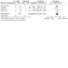
Four RCTs reported short‐term death or stroke (Chimowitz 2011; Gao 2022; Miao 2012; Zaidat 2015). ET plus CMT compared with CMT alone probably increases the risk of short‐term death or stroke (RR 2.93, 95% CI 1.81 to 4.75; P < 0.001, I2 = 0%; 989 participants; moderate‐certainty evidence; Analysis 1.1).
1.1. Analysis.

Comparison 1: Primary outcome: short‐term death or stroke, Outcome 1: Short‐term death or stroke
Subgroup analysis: types of endovascular therapy
We included three RCTs with 919 participants in the subgroup analysis of types of ET: Chimowitz 2011 and Gao 2022 treated participants in the ET group (total n = 400) with a self‐expandable stent (Wingspan), and Zaidat 2015 treated participants in the ET group (n = 58) with a balloon‐mounted stent (PHAROS Vitesse). We excluded Miao 2012 from this subgroup analysis because it used various types of ET without providing separate data for each. There was no statistically significant subgroup effect (P = 0.27), which suggests type of ET did not modify the effect of ET plus CMT in comparison to CMT alone on short‐term death or stroke. However, there were fewer trials and participants in the balloon‐mounted stent subgroup than in the self‐expandable stent subgroup, and detecting differences within these subgroups may be difficult. ET plus CMT compared to CMT alone was associated with a higher risk of short‐term death or stroke in the self‐expandable stent subgroup (RR 2.51, 95% CI 1.46 to 4.32; P < 0.001; 2 RCTs, 808 participants; Analysis 1.2) and in the balloon‐mounted stent subgroup (RR 5.18, 95% CI 1.61 to 16.68; P = 0.006; 1 RCT, 111 participants; Analysis 1.2).
1.2. Analysis.

Comparison 1: Primary outcome: short‐term death or stroke, Outcome 2: Subgroup analysis: types of endovascular therapy
Subgroup analysis: ethnicity
Miao 2012 and Gao 2022 enroled only Chinese people. Neither study found a significant difference in short‐term death or stroke between the ET plus CMT group and the CMT alone group. We did not perform the subgroup analysis of short‐term death or stroke in different ethnicities owing to a lack of data from other ethnic groups. Chimowitz 2011 and Zaidat 2015 enroled people from multiple ethnicities including white, Asian, Black, and Hispanic; however, they did not report outcomes by ethnicity in both treatment arms. We requested individual participant data from the study authors, but these data were unavailable.
Subgroup analysis: time intervals from qualifying event to intervention
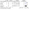
Data from all four RCTs were available. Chimowitz 2011 and Zaidat 2015 performed ET within three weeks of the qualifying event (562 participants); the mean time interval was seven days in Chimowitz 2011 and nine days in Zaidat 2015. Gao 2022 and Miao 2012 performed ET more than three weeks after symptom onset (427 participants); the mean time interval was 34.5 days in Gao 2022 and 261.95 days in Miao 2012. There was no significant subgroup difference (P = 0.67), suggesting that the time interval between the qualifying event and the intervention did not alter the effect of ET plus CMT relative to CMT alone on short‐term death or stroke. However, fewer participants contributed to the subgroup with more than a three‐week delay from symptom onset to treatment, meaning that more research is necessary to detect this phenomenon. The pooled effect estimate for the subgroup with more than three weeks between symptom onset and treatment indicated no clear difference between ET plus CMT and CMT alone (RR 2.37, 95% CI 0.80 to 7.05; P = 0.91; 2 RCTs, 427 participants; Analysis 1.3). In contrast, the pooled effect estimate for the subgroup with a delay of less than three weeks favoured CMT (RR 3.08, 95% CI 1.80 to 5.29; P = 0.001; 2 RCTs, 562 participants; Analysis 1.3).
1.3. Analysis.

Comparison 1: Primary outcome: short‐term death or stroke, Outcome 3: Subgroup analysis: time interval from qualifying event to intervention
Subgroup analysis: location of lesion
There were insufficient data for subgroup analysis by lesion location. Chimowitz 2011, Gao 2022, and Zaidat 2015 enroled people with lesions in several territories, including internal carotid artery, middle cerebral artery, basilar artery, and vertebral artery (intracranial segment). None of these three studies reported outcomes by lesion location. Miao 2012 enroled people with middle cerebral artery stenosis, and found no clear difference between ET plus CMT and CMT alone (RR 2.84, 95% CI 0.12 to 67.36; P = 0.52; 1 RCT, 70 participants).
Secondary outcomes
Short‐term outcomes
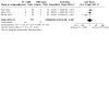
We obtained sufficient data for analysing five short‐term outcomes: ipsilateral stroke, TIA, ischaemic stroke, haemorrhagic stroke, and death. Chimowitz 2011 did not report TIA, but all four studies reported the remaining four outcomes. ET plus CMT compared with CMT alone probably increases the short‐term risk of ipsilateral stroke (RR 3.26, 95% CI 1.94 to 5.48; P < 0.001; 4 RCTs, 989 participants; moderate‐certainty evidence; Analysis 2.1) and ischaemic stroke (RR 2.24, 95% CI 1.30 to 3.87; P = 0.004; 4 RCTs, 989 participants; moderate‐certainty evidence; Analysis 2.2). ET plus CMT compared with CMT alone may increase the short‐term risk of haemorrhagic stroke (RR 13.49, 95% CI 2.59 to 70.15; P = 0.002; 4 RCTs, 989 participants; low‐certainty evidence; Analysis 2.3) and death (RR 5.43, 95% CI 1.21 to 24.40; P = 0.03; 4 RCTs, 989 participants; low‐certainty evidence; Analysis 2.5). It is unclear if ET plus CMT compared with CMT alone affects short‐term risk of TIA (RR 0.79, 95% CI 0.30 to 2.07; P = 0.63; 3 RCTs, 344 participants; moderate‐certainty evidence; Analysis 2.4). There was no heterogeneity among studies in these 30‐day outcomes.
2.1. Analysis.

Comparison 2: Other outcomes, Outcome 1: Short‐term ipsilateral stroke
2.2. Analysis.
Comparison 2: Other outcomes, Outcome 2: Short‐term ischaemic stroke
2.3. Analysis.
Comparison 2: Other outcomes, Outcome 3: Short‐term haemorrhagic stroke
2.5. Analysis.

Comparison 2: Other outcomes, Outcome 5: Short‐term death
2.4. Analysis.

Comparison 2: Other outcomes, Outcome 4: Short‐term TIA
Long‐term outcomes
We obtained sufficient data for analysing seven one‐year outcomes: death or stroke, ipsilateral stroke, TIA, ischaemic stroke, haemorrhage stroke, death, and dependency. Chimowitz 2011 did not report TIA, but all four studies reported the remaining six outcomes. We defined dependency as a modified Rankin Scale of 4 or more points.
ET plus CMT compared with CMT alone probably increases the long‐term risk of death or stroke (RR 1.49, 95% CI 1.12 to 1.99; P <0.001; 4 RCTs, 970 participants; moderate‐certainty evidence; Analysis 2.6). ET plus CMT compared with CMT alone may increase the long‐term risk of haemorrhagic stroke (RR 7.81, 95% CI 1.43 to 42.59; P = 0.02; 3 RCTs, 879 participants; low‐certainty evidence; Analysis 2.9). It is unclear if ET plus CMT compared with CMT alone affects long‐term risk of ipsilateral stroke (RR 1.78, 95% CI 1.00 to 3.17; P = 0.05; 4 RCTs, 970 participants; moderate‐certainty evidence; Analysis 2.7); ischaemic stroke (RR 1.56, 95% CI 0.77 to 3.16; P = 0.21; 4 RCTs, 970 participants; moderate‐certainty evidence; Analysis 2.8); TIA (RR 1.05, 95% CI 0.50 to 2.19; P = 0.89; 3 RCTs, 335 participants; moderate‐certainty evidence; Analysis 2.10); death (RR 1.61, 95% CI 0.77 to 3.38; P = 0.20; 4 RCTs, 951 participants; moderate‐certainty evidence; Analysis 2.11); or dependency (RR 1.51, 95% CI 0.93 to 2.45; P = 0.09; 4 RCTs, 947 participants; moderate‐certainty evidence; Analysis 2.12). For the above analysis, most I2 values were 0%; only the analysis of one‐year ischaemic stroke showed moderate heterogeneity (I2 = 65%). Through sensitivity analysis, we found that the heterogeneity was due to Gao 2022: excluding this trial reduced the I2 value to 12%, but also pushed the effect measure above the line of no effect to favour CMT (RR 2.07, 95% CI 1.37 to 3.13; P < 0.001; 3 RCTs, 632 participants).
2.6. Analysis.

Comparison 2: Other outcomes, Outcome 6: Long‐term death or stroke
2.9. Analysis.

Comparison 2: Other outcomes, Outcome 9: Long‐term haemorrhagic stroke
2.7. Analysis.

Comparison 2: Other outcomes, Outcome 7: Long‐term ipsilateral stroke
2.8. Analysis.

Comparison 2: Other outcomes, Outcome 8: Long‐term ischaemic stroke
2.10. Analysis.
Comparison 2: Other outcomes, Outcome 10: Long‐term TIA
2.11. Analysis.

Comparison 2: Other outcomes, Outcome 11: Long‐term death
2.12. Analysis.

Comparison 2: Other outcomes, Outcome 12: Long‐term dependency
We did not perform a meta‐analysis for restenosis of the involved vessel owing to lack of relevant data in the CMT group. Only Zaidat 2015 reported restenosis in people treated with a stent, finding that 26.5% had least 50% restenosis and 2.9% had at least 70% restenosis at one year of follow‐up. We did not assess health‐related quality of life because the studies provided no relevant data.
Discussion
Summary of main results
This review update included four RCTs with 989 participants. These studies provided moderate‐certainty evidence that CMT was superior to ET in participants with symptomatic ICAS of 70% or greater in terms of short‐ and long‐term death or stroke, short‐term ipsilateral stroke, and short‐term ischaemic stroke. There was low‐certainty evidence favouring CMT over ET for reducing short‐ and long‐term haemorrhagic stroke and short‐term death. However, there were no clear differences between the interventions for long‐term ipsilateral stroke, long‐term ischaemic stroke, short‐ and long‐term TIA, long‐term death, and long‐term dependency. The main reasons for downgrading the certainty of the evidence were early termination of the trials and small sample sizes. One Cochrane Review on angioplasty for ICAS calculated that 950 participants per group would be necessary under alpha of 0.05, power of 80%, expected relative risk reduction of 25%, and less than 7% perioperative risk of death or stroke (Cruz‐Flores 2006). In view of this estimation, the sample size of our review may have been insufficient.
Overall completeness and applicability of evidence
The certainty of evidence for major outcomes ranged from low to moderate, which partially limits the application of our findings to clinical decision‐making. Nonetheless, this review provides the highest level of current evidence. Furthermore, investigators applied different participant selection criteria for ET in the post‐SAMMPRIS trials. Trials with modified inclusion criteria reported a promising perioperative risk of 2% and 4.3%, respectively (Gao 2016; Miao 2015). The ongoing RCTs we identified have adopted the revised inclusion criteria (Cui 2016; Yang 2018). Future studies should clarify the safety and efficacy of ET for selected participants. One major limitation of this review is the small number of trials contributing evidence to the meta‐analysis. Consequently, the tests of heterogeneity have a very low power and do not provide reliable results. Substantial heterogeneity may exist although the tests are negative.
The time interval from the qualifying event to intervention may be a factor affecting the outcomes of ET. According to the subgroup analysis of this review, it may be safer to perform ET more than three weeks after the qualifying event versus less than three weeks after. The rates of short‐term death or stroke were low in studies that performed ET more than three weeks after the qualifying event (Gao 2016; Miao 2015). The cause of the increased risk of death or stroke in people who undergo ET in the early period may relate to instability of plaque and intracranial microcirculation leading to the 'snowploughing' effect (displacement of plaque to perforating arteries) and reperfusion haemorrhage (Luo 2018). Future studies are needed to confirm this speculation.
Quality of the evidence
The certainty of evidence in the included RCTs was low or moderate. The included trials had a high risk of performance bias because they could not blind participants or operators to the intervention. Moreover, three trials were terminated early after interim analysis (Chimowitz 2011; Miao 2012; Zaidat 2015). Miao 2012 had a high risk of attrition bias because the rate of loss to one‐year follow‐up was 30%, and 19.4% of participants transferred from ET to CMT.
Potential biases in the review process
We tried to minimise bias at every stage of the review's development; however, we could not avoid all bias during our review. Because we searched only English and Chinese databases, we may have missed articles in other languages without English abstracts. In addition, we only analysed data from published RCTs, and the lack of data from unpublished articles may have contributed to bias. Moreover, the number of studies related to the topic and the overall sample size were small, which may result in biased analysis results due to low statistical power.
Agreements and disagreements with other studies or reviews
The 2020 version of this review did not favour ET plus CMT over CMT alone (Wang 2020). We included updated data from randomised trials, and reached the same conclusion. Our results are similar to those of Zhang 2017, which meta‐analysed data from two randomised trials and two retrospective cohort studies, finding that ET plus CMT and CMT alone had comparable risks of event‐free survival and recurrent TIA, but that ET was associated with a higher risk of short‐term stroke.
However, two studies published in the late 2010s suggested that ET would benefit selected people with symptomatic intracranial atherosclerosis and cerebral hypoperfusion and people with at least two failures of best medical management (Abualhasan 2019; Padalia 2018). The WEAVE (Wingspan Stent System Post Market Surveillance) trial was an FDA‐authorised post‐market surveillance trial that aimed to reassess the perioperative safety of the Wingspan stent in on‐label patients who had symptomatic intracranial atherosclerosis with 70% to 90% stenosis, a baseline modified Rankin score of less than 3, and at least two failures of best medical management. The result was favourable for ET (in all cases performed more than eight days after the latest event), with a 2.6% rate of perioperative stroke and death and an 8.5% rate of one‐year stroke and death (Alexander 2019; Alexander 2021).
Authors' conclusions
Implications for practice.
This systematic review provides moderate‐certainty evidence that endovascular therapy plus conventional medical treatment compared to conventional medical treatment alone carries a higher risk of short‐term and long‐term stroke and death, short‐term ipsilateral stroke, and short‐term ischaemic stroke in people with recently symptomatic severe intracranial artery stenosis. Further information on treatment decisions is limited by the relatively small sample size.
Implications for research.
There are ongoing debates about whether specific subgroups, for example those treated after a certain delay, may benefit from ET more than those treated acutely, but more high‐certainty evidence is needed to unpick this.
What's new
| Date | Event | Description |
|---|---|---|
| 30 January 2023 | New search has been performed | New search for studies and content updated. We included one new study since the previous version, bringing the total number of includes studies to four, involving 989 participants |
| 30 January 2023 | New citation required but conclusions have not changed | New search for studies and content updated (no change to conclusions) |
History
Protocol first published: Issue 2, 2019 Review first published: Issue 8, 2020
Acknowledgements
We thank the following people, who conducted the editorial process for this article:
Sign‐off Editor (final editorial decision): Peter Langhorne, University of Glasgow
Managing Editor (selected peer reviewers, collated peer‐reviewer comments, provided editorial guidance to authors, edited the article, conducted editorial policy checks and supported editorial team): Hazel Fraser, Cochrane Stroke
Copy Editor (copy editing and production): Julia Turner
Peer‐reviewers (provided comments and recommended an editorial decision): Peter Langhorne, University of Glasgow (clinical/content review and methods review), Dr Amanda Barugh, Associate Editor of Cochrane Stroke (clinical/content review), Aryelly Rodriguez, Edinburgh Clinical Trials Unit (ECTU) at the University of Edinburgh (statistical review).
One peer reviewer provided search peer review but chose not to be publicly acknowledged.
If any readers are aware of additional trials that we have omitted, please write to Prof Liqun Jiao.
Appendices
Appendix 1. CENTRAL search strategy
1. MeSH descriptor: [Vascular Surgical Procedures] this term only 2. MeSH descriptor: [Endovascular Procedures] this term only 3. MeSH descriptor: [Angioplasty] this term only 4. MeSH descriptor: [Angioplasty, Balloon] this term only 5. MeSH descriptor: [Angioplasty, Balloon, Laser‐Assisted] this term only 6. MeSH descriptor: [Cerebral Revascularization] this term only 7. MeSH descriptor: [Blood Vessel Prosthesis] this term only 8. MeSH descriptor: [Blood Vessel Prosthesis Implantation] this term only 9. MeSH descriptor: [Stents] this term only 10. MeSH descriptor: [Dilatation] this term only 11. MeSH descriptor: [Catheterization] this term only 12. (angioplasty or stent* or pta or revasculari?ation or recanali?ation or catheter* or dilatation):ti,ab,kw 13. #1 or #2 or #3 or #4 or #5 or #6 or #7 or #8 or #9 or #10 or #11 or #12 14. MeSH descriptor: [Intracranial Arterial Diseases] explode all trees 15. MeSH descriptor: [Intracranial Arteriosclerosis] this term only 16. MeSH descriptor: [Vertebrobasilar Insufficiency] this term only 17. MeSH descriptor: [Cerebral Arteries] explode all trees 18. MeSH descriptor: [Basilar Artery] this term only 19. MeSH descriptor: [Vertebral Artery Dissection] this term only 20. #17 or #18 or #19 21. MeSH descriptor: [Arterial Occlusive Diseases] explode all trees 22. MeSH descriptor: [Arteriosclerosis] this term only 23. MeSH descriptor: [Constriction, Pathologic] this term only 24. #21 or #22 or #23 25. #20 and #24 26. ((intracranial or intra‐cranial or cerebral arter* or basilar arter* or vertebral arter* or vertebrobasilar or vertebro basilar) near/5 (stenos?s or isch?emia or insufficien* or arteriosclerosis or atherosclerosis or occlus* or obstruct* or plaque*)):ti,ab,kw 27. #14 or #15 or #16 or #25 or #26 28. #13 AND #27 29. MeSH descriptor: [Carotid Artery Diseases] this term only and with qualifier(s): [surgery ‐ SU, therapy ‐ TH] 30. MeSH descriptor: [Carotid Stenosis] this term only and with qualifier(s): [surgery ‐ SU, therapy ‐ TH] 31. MeSH descriptor: [Vertebrobasilar Insufficiency] this term only and with qualifier(s): [surgery ‐ SU, therapy ‐ TH] 32. MeSH descriptor: [Brain Ischemia] this term only and with qualifier(s): [surgery ‐ SU, therapy ‐ TH] 33. MeSH descriptor: [Cerebral Arteries] this term only and with qualifier(s): [surgery ‐ SU] 34. MeSH descriptor: [Basilar Artery] this term only and with qualifier(s): [surgery ‐ SU] 35. MeSH descriptor: [Vertebral Artery] this term only and with qualifier(s): [surgery ‐ SU] 36. #29 or #30 or #31 or #32 or #33 or #34 or #35 37. #28 or #36 in Trials
Appendix 2. MEDLINE Ovid search strategy
1. vascular surgical procedures/ 2. endovascular procedures/ or angioplasty/ or angioplasty, balloon/ or angioplasty, balloon, laser‐assisted/ 3. cerebral revascularization/ or Blood Vessel Prosthesis/ or Blood Vessel Prosthesis Implantation/ 4. stents/ or dilatation/ or catheterization/ 5. (angioplasty or stent$ or pta or revasculari?ation or recanali?ation or catheter$ or dilatation).tw. 6. or/1‐5 7. exp intracranial arterial diseases/ or intracranial arteriosclerosis/ or vertebrobasilar insufficiency/ 8. exp cerebral arteries/ or basilar artery/ or vertebral artery/ 9. exp arterial occlusive diseases/ or arteriosclerosis/ or constriction, pathologic/ 10. 8 and 9 11. ((intracranial or intra‐cranial or cerebral arter$ or basilar arter$ or vertebral arter$ or vertebrobasilar or vertebro basilar) adj5 (stenos?s or isch?emia or insufficien$ or arteriosclerosis or atherosclerosis or occlus$ or obstruct$ or plaque$)).tw. 12. 7 or 10 or 11 13. 6 and 12 14. *Carotid Artery Diseases/su, th [Surgery, Therapy] 15. *Carotid Stenosis/su, th [Surgery, Therapy] 16. *Vertebrobasilar Insufficiency/su, th [Surgery, Therapy] 17. *Brain Ischemia/su, th [Surgery, Therapy] 18. *Cerebral Arteries/su [Surgery] 19. *Basilar Artery/su [Surgery] 20. *Vertebral Artery/su [Surgery] 21. or/14‐20 22. 13 or 21 23. randomized controlled trial.pt. 24. controlled clinical trial.pt. 25. randomized.ab. 26. placebo.ab. 27. randomly.ab. 28. trial.ab. 29. groups.ab. 30. 23 or 24 or 25 or 26 or 27 or 28 or 29 31. 22 and 30
Appendix 3. Embase Ovid search strategy
1. endovascular surgery/ or vascular surgery/ 2. angioplasty/ or bare metal stenting/ or carotid angioplasty/ or carotid artery stenting/ or patch angioplasty/ 3. cerebral revascularization/ 4. blood vessel prosthesis/ or artery prosthesis/ 5. balloon dilatation/ or stent/ or exp metal stent/ or exp self expanding stent/ 6. (angioplasty or stent$ or pta or revasculari?ation or recanali?ation or catheter$ or dilatation).tw. 7. or/1‐6 8. cerebral artery disease/ or brain atherosclerosis/ or vertebrobasilar insufficiency/ 9. exp brain artery/ or vertebral artery/ 10. peripheral occlusive artery disease/ or atherosclerosis/ or arteriosclerosis/ or atherosclerotic plaque/ or brain atherosclerosis/ or carotid atherosclerosis/ 11. 9 and 10 12. ((intracranial or intra‐cranial or cerebral arter$ or basilar arter$ or vertebral arter$ or vertebrobasilar or vertebro basilar) adj5 (stenos? s or isch?emia or insufficien$ or arteriosclerosis or atherosclerosis or occlus$ or obstruct$ or plaque$)).tw. 13. 8 or 11 or 12 14. 7 and 13 15. carotid artery disease/su, th [Surgery, Therapy] 16. carotid artery obstruction/su, th [Surgery, Therapy] 17. vertebrobasilar insufficiency/su, th [Surgery, Therapy] 18. brain ischemia/su, th [Surgery, Therapy] 19. brain artery/su [Surgery] 20. basilar artery/su [Surgery] 21. vertebral artery/su [Surgery] 22. or/15‐21 23. 14 or 22 24. Randomized Controlled Trial/ or "randomized controlled trial (topic)"/ 25. randomisation/ 26. Controlled clinical trial/ or "controlled clinical trial (topic)"/ 27. control group/ or controlled study/ 28. clinical trial/ or "clinical trial (topic)"/ or phase 1 clinical trial/ or phase 2 clinical trial/ or phase 3 clinical trial/ or phase 4 clinical trial/ 29. Crossover Procedure/ 30. Double Blind Procedure/ 31. Single Blind Procedure/ or triple blind procedure/ 32. placebo/ or placebo effect/ 33. (random$ or RCT or RCTs).tw. 34. controlled adj5 (trial$ or stud$)).tw. 35. (clinical$ adj5 trial$).tw. 36. ((control or treatment or experiment$ or intervention) adj5 (group$ or subject$ or patient$)).tw. 37. (quasi‐random$ or quasi random$ or pseudo‐random$ or pseudo random$).tw. 38. ((control or experiment$ or conservative) adj5 (treatment or therapy or procedure or manage$)).tw. 39. ((singl$ or doubl$ or tripl$ or trebl$) adj5 (blind$ or mask$)).tw. 40. (cross‐over or cross over or crossover).tw. 41. (placebo$ or sham).tw. 42. trial.ti. 43. (assign$ or allocat$).tw. 44. controls.tw. 45. or/24‐44 46. 23 and 45
Appendix 4. Science Citation Index Web of Science search strategy
1. TS=(angioplasty OR stent* OR pta OR revascularization OR revascularisation OR recanalisation OR recanalization OR recanaliation OR catheter* OR dilatation) 2. TS=(intracranial OR intra‐cranial OR cerebral arter* OR basilar arter* OR vertebral arter* OR vertebrobasilar OR vertebro basilar) AND TS=(stenosis OR stenosis OR ischemia OR ischaemia OR insufficien* OR arteriosclerosis OR atherosclerosis OR occlus* OR obstruct* OR plaque*) 3. #1 AND #2 4. TS=(randomized OR placebo OR randomly OR trial OR groups) 5. #3 AND #4
Appendix 5. Scopus search strategy
1. INDEXTERMS("endovascular procedures" OR angioplasty OR "vascular surgical procedures" OR "cerebral revascularization" OR "Blood Vessel Prosthesis" OR "Blood Vessel Prosthesis Implantation" OR "balloon dilatation" OR "stents" OR "dilatation" OR "catheterization" ) 2. TITLE‐ABS ( angioplasty OR stent* OR pta OR revasculari?ation OR recanali?ation OR catheter* OR dilatation ) 3. #1 OR #2 4. INDEXTERMS("intracranial arterial diseases" OR "intracranial arteriosclerosis" OR "vertebrobasilar insufficiency") 5. INDEXTERMS("cerebral arteries" OR "basilar artery" OR "vertebral artery" ) 6. INDEXTERMS("arterial occlusive diseases" OR arteriosclerosis OR "constriction, pathologic") 7. #5 AND #6 8. TITLE‐ABS ( intracranial OR intra‐cranial OR "cerebral arter*" OR "basilar arter*" OR "vertebral arter*" OR vertebrobasilar OR "vertebro basilar" ) W/5 ( stenos?s OR isch?emia OR insufficien* OR arteriosclerosis OR atherosclerosis OR occlus* OR obstruct* OR plaque* ) 9. #4 OR #7 OR #8 10. #3 AND #9 11. TITLE‐ABS ( randomized OR placebo OR randomly OR trial OR groups ) 12. INDEXTERMS ( "randomized controlled trial" OR "controlled clinical trial" ) 13. #11 OR #12 14. #10 AND #13
Appendix 6. Academic Source Complete EBSCO search strategy
1. SU endovascular procedures OR SU angioplasty OR SU vascular surgical procedures OR SU cerebral revascularization OR SU Blood Vessel Prosthesis OR SU Blood Vessel Prosthesis Implantation OR SU balloon dilatation OR SU stents OR SU dilatation OR SU catheterization 2. angioplasty OR stent* OR pta OR revascularization OR revascularisation OR recanalisation OR recanalization OR recanaliation OR catheter* OR dilatation 3. #1 OR #2 4. SU intracranial arterial diseases OR SU intracranial arteriosclerosis OR SU vertebrobasilar insufficiency 5. SU cerebral arteries OR SU basilar artery OR SU vertebral artery 6. SU arterial occlusive diseases OR SU arteriosclerosis OR SU constriction, pathologic 7. #5 AND #6 8. (intracranial OR intra‐cranial OR cerebral arter* OR basilar arter* OR vertebral arter* OR vertebrobasilar OR vertebro basilar) AND (stenosis OR stenosis OR ischemia OR ischaemia OR insufficien* OR arteriosclerosis OR atherosclerosis OR occlus* OR obstruct* OR plaque*) 9. #4 OR #7 OR #8 10. #3 AND #9 11. SU randomized controlled trial OR SU controlled clinical trial OR AB randomized OR AB placebo OR AB randomly OR AB trial OR AB groups 12. #10 AND #11
Appendix 7. China Biological Medicine Database search strategy
1. ((((((((("血管内操作"[不加权:不扩展]) OR "血管成形术"[不加权:扩展]) OR "血管外科手术"[不加权:不扩展]) OR "脑血管重建术"[不加权:不扩展]) OR "人工血管"[不加权:不扩展]) OR "血管假体植入"[不加权:不扩展]) OR "血管成形术, 气囊"[不加权:不扩展]) OR "支架"[不加权:不扩展]) OR "扩张术"[不加权:不扩展]) OR "导管插入术"[不加权:不扩展] 2. 血管 OR 血运 3. 重建 OR 再建 OR 再生 OR 再造 OR 重造 OR 重生 OR 成形 4. (#3) AND (#2) 5. 导管 OR 扩张 OR 膨胀 OR 支架 OR PTA 6. (#5) OR (#4) OR (#1) 7. (("颅内动脉疾病"[不加权:扩展]) OR "颅内动脉硬化"[不加权:不扩展]) OR "椎底动脉供血不足"[不加权:不扩展] 8. (("脑动脉"[不加权:扩展]) OR "基底动脉"[不加权:不扩展]) OR "椎动脉"[不加权:不扩展] 9. (("动脉闭塞性疾病"[不加权:扩展]) OR "动脉硬化"[不加权:不扩展]) OR "缩窄, 病理性"[不加权:不扩展] 10. (#9) AND (#8) 11. 颅内动脉 OR 脑动脉 OR 基底动脉 OR 椎动脉 OR 椎基底动脉 12. 狭窄 OR 缺血 OR 供血不足 OR 动脉粥样硬化 OR 动脉硬化 OR 粥样硬化 OR 阻塞 OR 闭塞 OR 梗阻 OR 斑块 13. (#12) AND (#11) 14. (#13) OR (#10) OR (#7) 15. (#14) AND (#6) 16. (((((("颈动脉疾病/外科学"[加权:不扩展]) OR "颈动脉狭窄/外科学"[加权:不扩展]) OR "椎底动脉供血不足/外科学"[加权:不扩展]) OR "脑缺血/外科学"[加权:不扩展]) OR "脑动脉/外科学"[加权:不扩展]) OR "基底动脉/外科学"[加权:不扩展]) OR "椎动脉/外科学"[加权:不扩展] 17. ("随机对照试验"[不加权:扩展]) OR "临床对照试验"[不加权:扩展] 18. (((("随机"[摘要:智能]) OR "安慰剂"[摘要:智能]) OR "试验"[摘要:智能]) OR "分组"[摘要:智能]) OR "对照"[摘要:智能] 19. (#18) OR (#17) 20. (#16) OR (#15) 21. (#20) AND (#19) 22. ((#30) AND (#19)) AND ( 随机对照试验[文献类型]) 23. 动物[特征词] NOT 人类[特征词] 24. #22 NOT #23
Appendix 8. ClinicalTrials.gov search strategy
1. (angioplasty OR stent OR surgery OR revascular* OR recanal* OR catheter* OR dilatation ) | Interventional Studies | intracranial artery OR cerebral artery OR vertebral OR basilar OR vertebrobasilar
Appendix 9. WHO ICTRP search strategy
1. intracranial AND angioplasty OR intracranial AND stent OR intracranial AND PTA OR intracranial AND revascularization OR intracranial AND revascularisation OR intracranial AND recanalisation OR intracranial AND recanalization OR intracranial AND recanaliation OR intracranial AND catheterisation OR intracranial AND dilatation OR intracranial AND endovascular procedures OR intracranial AND surgical OR intracranial AND blood vessel prosthesis 2. cerebral arteries AND angioplasty OR cerebral arteries AND stent OR cerebral arteries AND PTA OR cerebral arteries AND revascularization OR cerebral arteries AND revascularisation OR cerebral arteries AND recanalisation OR cerebral arteries AND recanalization OR cerebral arteries AND recanaliation OR cerebral arteries AND catheterisation OR cerebral arteries AND dilatation OR cerebral arteries AND endovascular procedures OR cerebral arteries AND surgical OR cerebral arteries AND blood vessel prosthesis 3. vertebral AND angioplasty OR vertebral AND stent OR vertebral AND PTA OR vertebral AND revascularization OR vertebral AND revascularisation OR vertebral AND recanalisation OR vertebral AND recanalization OR vertebral AND recanaliation OR vertebral AND catheterisation OR vertebral AND dilatation OR vertebral AND endovascular procedures OR vertebral AND surgical OR vertebral AND blood vessel prosthesis 4. basilar AND angioplasty OR basilar AND stent OR basilar AND PTA OR basilar AND revascularization OR basilar AND revascularisation OR basilar AND recanalisation OR basilar AND recanalization OR basilar AND recanaliation OR basilar AND catheterisation OR basilar AND dilatation OR basilar AND endovascular procedures OR basilar AND vascular surgical procedures OR basilar AND blood vessel prosthesis
Data and analyses
Comparison 1. Primary outcome: short‐term death or stroke.
| Outcome or subgroup title | No. of studies | No. of participants | Statistical method | Effect size |
|---|---|---|---|---|
| 1.1 Short‐term death or stroke | 4 | 989 | Risk Ratio (M‐H, Fixed, 95% CI) | 2.93 [1.81, 4.75] |
| 1.2 Subgroup analysis: types of endovascular therapy | 3 | 919 | Risk Ratio (M‐H, Fixed, 95% CI) | 2.93 [1.80, 4.78] |
| 1.2.1 Self‐expandable stent plus CMT vs CMT alone | 2 | 808 | Risk Ratio (M‐H, Fixed, 95% CI) | 2.51 [1.46, 4.32] |
| 1.2.2 Balloon‐mounted stent plus CMT vs CMT alone | 1 | 111 | Risk Ratio (M‐H, Fixed, 95% CI) | 5.18 [1.61, 16.68] |
| 1.3 Subgroup analysis: time interval from qualifying event to intervention | 4 | 989 | Risk Ratio (M‐H, Fixed, 95% CI) | 2.93 [1.81, 4.75] |
| 1.3.1 3 weeks or less | 2 | 562 | Risk Ratio (M‐H, Fixed, 95% CI) | 3.08 [1.80, 5.29] |
| 1.3.2 More than 3 weeks | 2 | 427 | Risk Ratio (M‐H, Fixed, 95% CI) | 2.37 [0.80, 7.05] |
Comparison 2. Other outcomes.
| Outcome or subgroup title | No. of studies | No. of participants | Statistical method | Effect size |
|---|---|---|---|---|
| 2.1 Short‐term ipsilateral stroke | 4 | 989 | Risk Ratio (M‐H, Fixed, 95% CI) | 3.26 [1.94, 5.48] |
| 2.2 Short‐term ischaemic stroke | 4 | 989 | Risk Ratio (M‐H, Fixed, 95% CI) | 2.24 [1.30, 3.87] |
| 2.3 Short‐term haemorrhagic stroke | 4 | 989 | Risk Ratio (M‐H, Fixed, 95% CI) | 13.49 [2.59, 70.15] |
| 2.4 Short‐term TIA | 3 | 344 | Risk Ratio (M‐H, Fixed, 95% CI) | 0.79 [0.30, 2.07] |
| 2.5 Short‐term death | 4 | 989 | Risk Ratio (M‐H, Fixed, 95% CI) | 5.43 [1.21, 24.40] |
| 2.6 Long‐term death or stroke | 4 | 970 | Risk Ratio (M‐H, Fixed, 95% CI) | 1.49 [1.12, 1.99] |
| 2.7 Long‐term ipsilateral stroke | 4 | 970 | Risk Ratio (M‐H, Random, 95% CI) | 1.78 [1.00, 3.17] |
| 2.8 Long‐term ischaemic stroke | 4 | 978 | Risk Ratio (M‐H, Random, 95% CI) | 1.56 [0.77, 3.16] |
| 2.9 Long‐term haemorrhagic stroke | 3 | 879 | Risk Ratio (M‐H, Fixed, 95% CI) | 7.81 [1.43, 42.59] |
| 2.10 Long‐term TIA | 3 | 335 | Risk Ratio (M‐H, Fixed, 95% CI) | 1.05 [0.50, 2.19] |
| 2.11 Long‐term death | 4 | 951 | Risk Ratio (M‐H, Fixed, 95% CI) | 1.61 [0.77, 3.38] |
| 2.12 Long‐term dependency | 4 | 947 | Risk Ratio (M‐H, Fixed, 95% CI) | 1.51 [0.93, 2.45] |
Characteristics of studies
Characteristics of included studies [ordered by study ID]
Chimowitz 2011.
| Study characteristics | ||
| Methods | Multicentre open‐label RCT with 3‐year follow‐up | |
| Participants | Estimated sample size: 764 Number randomised: 451 Inclusion criteria
Exclusion criteria
|
|
| Interventions | ET plus CMT versus CMT alone | |
| Outcomes | Primary outcomes
Secondary outcomes
|
|
| Funding source | US National Institute of Neurological Disorders and Stroke | |
| Notes | ClinicalTrials.gov identifier: NCT00576693 | |
| Risk of bias | ||
| Bias | Authors' judgement | Support for judgement |
| Random sequence generation (selection bias) | Low risk | Quote: "The sequence of treatment assignments are stratified by centre and produced at the Statistical Coordinating centre using a pseudo‐random number generator with randomly permuted block sizes." |
| Allocation concealment (selection bias) | Low risk | Quote: "Patients are randomized (1:1) to aggressive medical management alone or to PTAS plus aggressive medical management." Comment: the study used software that carried out allocations in a closed environment, and nobody knew the allocation until after enrolment had been confirmed. |
| Blinding of participants and personnel (performance bias) All outcomes | High risk | As the 2 treatments were very different, blinding of participants and personnel was not possible. |
| Blinding of outcome assessment (detection bias) All outcomes | Low risk | Quote: "All potential endpoints are adjudicated by independent panels of neurologists and cardiologists who are unaware of treatment assignments." |
| Incomplete outcome data (attrition bias) All outcomes | Low risk | Incomplete outcome data adequately resolved and unlikely to seriously alter the results with ITT analysis. |
| Selective reporting (reporting bias) | Low risk | All study outcomes were prespecified in the published protocol. |
| Other bias | Low risk | Although the study was stopped early, the interim analysis already showed superiority of CMT over ET, and this result was unlikely to change. |
Gao 2022.
| Study characteristics | ||
| Methods | Multicentre, randomised, open‐label, outcome assessor‐blinded trial with 3‐year follow‐up | |
| Participants | Estimated sample size: 380 Number randomised: 380 Inclusion criteria:
Exclusion criteria
|
|
| Interventions | ET plus CMT versus CMT alone | |
| Outcomes | Primary endpoints
Secondary endpoints
|
|
| Funding source | Funded by a research grant (2011BAI08B04) from the National Health Commission of the People's Republic of China. Stryker Neurovascular provided supplemental funding for third‐party site monitoring and auditing. | |
| Notes | ClinicalTrials.gov Identifier: NCT01763320 | |
| Risk of bias | ||
| Bias | Authors' judgement | Support for judgement |
| Random sequence generation (selection bias) | Low risk | Quote: "IVRS (Interactive Voice Response System, Clinicalsoft Company Limited) will be used for patient randomization." |
| Allocation concealment (selection bias) | Low risk | Quote: "Patients meeting the inclusion criteria will be randomized (1:1) to medical therapy alone or medical therapy plus stenting using Wingspan." Comment: the study used software that carried out allocations in a closed environment, and nobody knew the allocation until after enrolment had been confirmed. |
| Blinding of participants and personnel (performance bias) All outcomes | High risk | As the 2 treatments were very different, blinding of participants and personnel was not possible. |
| Blinding of outcome assessment (detection bias) All outcomes | Low risk | Quote: "An imaging database was established to facilitate central reading by an independent imaging core lab (IsCore Image Corelab)." |
| Incomplete outcome data (attrition bias) All outcomes | Low risk | Incomplete outcome data adequately resolved and unlikely to seriously alter the results with ITT analysis. |
| Selective reporting (reporting bias) | Low risk | All study outcomes were prespecified in the published protocol. |
| Other bias | High risk | The industry funding source may be a source of bias |
Miao 2012.
| Study characteristics | ||
| Methods | Single‐centre RCT with 1‐year follow‐up | |
| Participants | Estimated sample size: 184 Number randomised: 70 Inclusion criteria
Exclusion criteria
|
|
| Interventions | ET plus CMT versus CMT alone | |
| Outcomes | Primary outcomes
Secondary outcomes
|
|
| Funding source | Funded by the Research Fund of Capital Medical Development (2007–2069). | |
| Notes | Clinical Trial Registration URL: www.chictr.org/en/ Unique identifier: ChiCTR‐TRC‐12001989 | |
| Risk of bias | ||
| Bias | Authors' judgement | Support for judgement |
| Random sequence generation (selection bias) | Low risk | Quote: "The randomisation numbers were produced by Randlist software (Version 1.2 \Seed: [1497369600]; http://www.randomisation.net), 4 numbers as a block." |
| Allocation concealment (selection bias) | Low risk | Quote: "Patients were randomised to either the PTAS or medical group." Comment: the study used software that carried out allocations in a closed environment, and nobody knew the allocation until after enrolment had been confirmed. |
| Blinding of participants and personnel (performance bias) All outcomes | High risk | As the 2 treatments were very different, blinding of participants and personnel was not possible. |
| Blinding of outcome assessment (detection bias) All outcomes | Low risk | Quote: "Investigators who did not participate in the enrolment of the patients and were blinded to the treatment assignment..." |
| Incomplete outcome data (attrition bias) All outcomes | High risk | There was a high rate of loss to 1‐year follow‐up (49/70, 30%), and 19.4% of participants randomised to the ET group refused ET. |
| Selective reporting (reporting bias) | Low risk | All study outcomes were prespecified in the registered information. |
| Other bias | High risk | The study was stopped early; results of an interim analysis showed similar safety and efficacy for CMT and ET. However, the final results could have changed if recruitment had continued. |
Zaidat 2015.
| Study characteristics | ||
| Methods | Multicentre RCT with 1‐year follow‐up | |
| Participants | Estimated sample size: 250 Number randomised: 112 Inclusion criteria
Exclusion criteria
|
|
| Interventions | ET plus CMT versus CMT alone | |
| Outcomes | Primary endpoints
Secondary endpoints
|
|
| Funding source | Initiated and funded by Micrus Endovascular | |
| Notes | ClinicalTrials.gov identifier: NCT00816166 | |
| Risk of bias | ||
| Bias | Authors' judgement | Support for judgement |
| Random sequence generation (selection bias) | Low risk | Quote: "Patients meeting the final enrolment criteria after angiogram were randomly assigned 1:1 to either treatment with medical therapy alone or to medical therapy and PHAROS Vitesse neurovascular stent by a telephonic interactive voice response system (BioClinica Inc, Newtown, PA, USA). Randomisation was stratified by 2 factors: enrolment site and age (18–55 versus 56–85 years)..." |
| Allocation concealment (selection bias) | Low risk | Quote: "Patients meeting the final enrolment criteria after angiogram were randomly assigned 1:1 to either treatment with medical therapy alone or to medical therapy and PHAROS Vitesse neurovascular stent by a telephonic interactive voice response system (BioClinica Inc, Newtown, PA, USA). Randomisation was stratified by 2 factors: enrolment site and age (18–55 versus 56–85 years)..." Comment: the study used software that carried out allocations in a closed environment, and nobody knew the allocation until after enrolment had been confirmed. |
| Blinding of participants and personnel (performance bias) All outcomes | High risk | Quote: "The current trial was not double‐blinded due to the lack of feasibility of masking the stent group..." |
| Blinding of outcome assessment (detection bias) All outcomes | Low risk | Quote: "... end point assessment by an independent neurologist who was not involved in the procedure reduced potential bias..." |
| Incomplete outcome data (attrition bias) All outcomes | Low risk | Incomplete outcome data adequately resolved and unlikely to seriously alter the results with ITT analysis. |
| Selective reporting (reporting bias) | Low risk | All study outcomes were prespecified in the published protocol. |
| Other bias | High risk | Although the study was stopped early, the interim analysis showed superiority of CMT over ET and this result was unlikely to change. The industry funding source may be a source of bias. |
BI: Barthel Index; CMT: conventional medical treatment; CT: computed tomography; DWI: diffusion‐weighted imaging; ET: endovascular therapy; ITT: intention‐to‐treat; MCA: middle cerebral artery; MoCA: Montreal Cognitive Assessment; mRS: modified Rankin Scale; NIHSS: National Institutes of Health Stroke Scale; PTAS: percutaneous transluminal angioplasty and stenting; RCT: randomised controlled trial; TIA: transient ischaemic attack.
Characteristics of excluded studies [ordered by study ID]
| Study | Reason for exclusion |
|---|---|
| Compter 2015 | The trial recruited 115 people with vertebral atherosclerotic stenosis, but only 16.5% of participants had intracranial vertebral stenosis, and we were unable to obtain detailed data for these participants. |
| Coward 2007 | The trial enroled only 16 people with vertebral atherosclerosis, including the extracranial and intracranial artery, to compare ET versus best medical treatment. We were unable to obtain detailed data for the participants with ICAS. |
| Markus 2017 | The trial recruited 179 people with vertebral atherosclerotic stenosis, but only 17.3% of participants had intracranial vertebral stenosis, and we were unable to obtain detailed data for these participants. |
ET: endovascular therapy; ICAS: intracranial artery stenosis.
Characteristics of ongoing studies [ordered by study ID]
Cui 2016.
| Study name | Angioplasty and stenting for patients with symptomatic intracranial atherosclerosis |
| Methods | Multicentre randomised trial in 3 high‐volume centres |
| Participants | Estimated sample size: 539 Inclusion criteria
Exclusion criteria
|
| Interventions | PTAS plus aggressive medical management after ≥ 3 weeks versus aggressive medical management alone |
| Outcomes | Primary outcome
Secondary outcomes
|
| Starting date | April 2016 |
| Contact information | ClinicalTrials.gov identifier: NCT02689037. More details in the published protocol: DOI: 10.1136/bmjopen‐2016‐012175 |
| Notes | This study is financially supported by a Health Research Fund from People's Liberation Army (No. 12MA100). The study sponsor and funders play no role in study design, collection, management, analysis, and interpretation of data, writing of the report, or the decision to submit the report for publication. |
Yang 2018.
| Study name | A randomised trial of Stenting for Symptomatic Intracranial Stenosis |
| Methods | Multicentre randomised trial in 22 research settings |
| Participants | Estimated sample size: 704 Inclusion criteria Age 18–80 years
Exclusion criteria
|
| Interventions | PTAS plus aggressive medical management after ≥ 3 weeks versus aggressive medical management alone |
| Outcomes | Primary outcomes
Secondary outcomes:
|
| Starting date | May 2018 |
| Contact information | ChiCTR1800015766 with URL www.chictr.org.cn/showproj.aspx?proj=26742 |
| Notes | This study is self‐financed. |
BI: Barthel Index; C‐NIHSS: Chinese version of National Institutes of Health Stroke Scale; EQ‐5D: EuroQol five‐dimension questionnaire; ICAS: intracranial artery stenosis; MCA: middle cerebral artery; mRS: modified Rankin Scale; NIHSS: National Institutes of Health Stroke Scale; PTAS: percutaneous transluminal angioplasty and stenting; TIA: transient ischaemic attack.
Differences between protocol and review
None (Wang 2019).
Contributions of authors
JL, TW, and KY contributed equally to this review. JL, TW, XW, and LJ drafted the protocol. JL, TW, and XW developed the search strategy. JL, XW, RX, and HG searched for trials. XZ and JW obtained copies of relevant references. JL, TW, RY, and YM selected trials. JL, KY, and PG extracted and entered data. JL, TW, KY, PG, YM, and LJ analysed the data, interpreted the results, and wrote the final review.
Sources of support
Internal sources
No sources of support provided
External sources
No sources of support provided
Declarations of interest
JL: none TW: none KY: none XW: none RX: none HG: none XZ: none JW: none RY: none PG: none YM: none LJ: none
Joint first author
New search for studies and content updated (no change to conclusions)
References
References to studies included in this review
Chimowitz 2011 {published data only}
- Chimowitz MI, Lynn MJ, Derdeyn CP, Turan TN, Fiorella D, Lane BF, et al. Stenting versus aggressive medical therapy for intracranial arterial stenosis. New England Journal of Medicine 2011;365(11):993-1003. [DOI] [PMC free article] [PubMed] [Google Scholar]
Gao 2022 {published data only}
- Gao P, Wang T, Wang D, Liebeskind DS, Shi H, Li T, et al. Effect of stenting plus medical therapy vs medical therapy alone on risk of stroke and death in patients with symptomatic intracranial stenosis: the CASSISS randomized clinical trial. JAMA 2022;328(6):534-42. [DOI] [PMC free article] [PubMed] [Google Scholar]
Miao 2012 {published data only}
- Miao Z, Jiang L, Wu H, Bao Y, Jiao L, Li S, et al. Randomized controlled trial of symptomatic middle cerebral artery stenosis: endovascular versus medical therapy in a Chinese population. Stroke 2012;43(12):3284-90. [DOI] [PubMed] [Google Scholar]
Zaidat 2015 {published data only}
- Zaidat OO, Fitzsimmons BF, Woodward BK, Wang Z, Killer-Oberpfalzer M, Wakhloo A, et al. Effect of a balloon-expandable intracranial stent vs medical therapy on risk of stroke in patients with symptomatic intracranial stenosis: the VISSIT randomized clinical trial. JAMA 2015;313(12):1240-8. [DOI] [PubMed] [Google Scholar]
References to studies excluded from this review
Compter 2015 {published data only}
- Compter A, Worp HB, Schonewille WJ, Vos JA, Boiten J, Nederkoorn PJ, et al. Stenting versus medical treatment in patients with symptomatic vertebral artery stenosis: a randomised open-label phase 2 trial. Lancet Neurology 2015;14(6):606-14. [DOI] [PubMed] [Google Scholar]
Coward 2007 {published data only}
- Coward LJ, McCabe DJ, Ederle J, Featherstone RL, Clifton A, Brown MM. Long-term outcome after angioplasty and stenting for symptomatic vertebral artery stenosis compared with medical treatment in the Carotid And Vertebral Artery Transluminal Angioplasty Study (CAVATAS): a randomized trial. Stroke 2007;38(5):1526-30. [DOI] [PubMed] [Google Scholar]
Markus 2017 {published data only}
- Markus HS, Larsson SC, Kuker W, Schulz UG, Ford I, Rothwell PM, et al. Stenting for symptomatic vertebral artery stenosis: the Vertebral Artery Ischaemia Stenting Trial. Neurology 2017;89(12):1229-36. [DOI] [PMC free article] [PubMed] [Google Scholar]
References to ongoing studies
Cui 2016 {published data only}
- Cui XP, Lin M, Mu JS, Ye JX, He WQ, Fu ML, et al. Angioplasty and stenting for patients with symptomatic intracranial atherosclerosis: study protocol of a randomised controlled trial. BMJ Open 2016;6(11). [DOI] [PMC free article] [PubMed]
Yang 2018 {published data only}
- Yang QW, Shuai J. A multicenter randomized trial of stenting plus standard medical treatment versus standard medical treatment alone for symptomatic intracranial atherosclerotic stenosis [A randomized trial of Stenting for Symptomatic Intracranial STenosis (ASSIST)]. Chinese Clinical Trial Registry 2018. [ChiCTR1800015766]
Additional references
Abou‐Chebl 2012
- Abou-Chebl A, Steinmetz H. Critique of "Stenting versus aggressive medical therapy for intracranial arterial stenosis" by Chimowitz et al in the New England Journal of Medicine. Stroke 2012;43(2):616-20. [DOI] [PubMed] [Google Scholar]
Abualhasan 2019
- Abualhasan A, Abd-Allah F, Pero G, Sobh K, Mansour O, El-Serafy O, et al. Intracranial stenting: is it still an option for treatment of patients with intracranial atherosclerosis? Frontiers in Neurology 2019;10:1248. [DOI] [PMC free article] [PubMed] [Google Scholar]
Alexander 2019
- Alexander MJ, Zauner A, Chaloupka JC, Baxter B, Callison RC, Gupta R, et al. WEAVE Trial: final results in 152 on-label patients. Stroke 2019;50(4):889-94. [DOI] [PubMed] [Google Scholar]
Alexander 2021
- Alexander MJ, Zauner A, Gupta R, Alshekhlee A, Fraser JF, Toth G, et al. The WOVEN trial: Wingspan One-year Vascular Events and Neurologic outcomes. Journal of Neurointerventional Surgery 2021;13(4):307-10. [DOI] [PubMed] [Google Scholar]
An 2002
- An H, Lin W. Cerebral oxygen extraction fraction and cerebral venous blood volume measurements using MRI: effects of magnetic field variation. Magnetic Resonance in Medicine 2002;47(5):958-66. [PMID: ] [DOI] [PubMed] [Google Scholar]
Aoki 1995
- Aoki S, Shirouzu I, Sasaki Y, Okubo T, Hayashi N, Machida T, et al. Enhancement of the intracranial arterial wall at MR imaging: relationship to cerebral atherosclerosis. Radiology 1995;194(2):477-81. [PMID: ] [DOI] [PubMed] [Google Scholar]
Arenillas 2011
- Arenillas JF. Intracranial atherosclerosis: current concepts. Stroke 2011;42(1 Suppl):S20-3. [DOI] [PubMed] [Google Scholar]
Atkins 2004
- Atkins D, Best D, Briss PA, Eccles M, Falck-Ytter Y, Flottorp S, et al. GRADE Working Group. Grading quality of evidence and strength of recommendations. BMJ 2004;328(7454):1490. [DOI] [PMC free article] [PubMed] [Google Scholar]
Banerjee 2017
- Banerjee C, Chimowitz MI. Stroke caused by atherosclerosis of the major intracranial arteries. Circulation Research 2017;120(3):502-13. [DOI] [PMC free article] [PubMed] [Google Scholar]
Bang 2014
- Bang OY. Intracranial atherosclerosis: current understanding and perspectives. Journal of Stroke 2014;16(1):27-35. [DOI] [PMC free article] [PubMed] [Google Scholar]
Benjamin 2017
- Benjamin EJ, Blaha MJ, Chiuve SE, Cushman M, Das SR, Deo R, American Heart Association Statistics Committee and Stroke Statistics Subcommittee. Heart disease and stroke statistics – 2017 update: a report from the American Heart Association. Circulation 2017;135(10):e146-603. [DOI] [PMC free article] [PubMed] [Google Scholar]
Berkefeld 2009
- Berkefeld J, Zanella FE. Intracranial stenting of atherosclerotic stenoses: current status and perspectives. Klinische Neuroradiologie 2009;19(1):38-44. [DOI] [PubMed] [Google Scholar]
Boissel 1999
- Boissel JP, Cucherat M, Li W, Chatellier G, Gueyffier F, Buyse M, et al. The problem of therapeutic efficacy indices. 3. Comparison of the indices and their use [Apercu sur la problematique des indices d'efficacite therapeutique, 3: comparaison des indices et utilisation. Groupe d'Etude des Indices D'efficacite]. Therapie 1999;54(4):405-11. [PMID: ] [PubMed] [Google Scholar]
Carson 2011
- Carson AP, Howard G, Burke GL, Shea S, Levitan EB, Muntner P. Ethnic differences in hypertension incidence among middle-aged and older adults: the multi-ethnic study of atherosclerosis. Hypertension 2011;57(6):1101-7. [PMID: ] [DOI] [PMC free article] [PubMed] [Google Scholar]
Chatterjee 2015
- Chatterjee AR, Derdeyn CP. Stenting in intracranial stenosis: current controversies and future directions. Current Atherosclerosis Reports 2015;17(8):48. [DOI] [PubMed] [Google Scholar]
Chimowitz 2005
- Chimowitz MI, Lynn MJ, Howlett-Smith H, Stern BJ, Hertzberg VS, Frankel MR, et al. Comparison of warfarin and aspirin for symptomatic intracranial arterial stenosis. New England Journal of Medicine 2005;352(13):1305-16. [PMID: ] [DOI] [PubMed] [Google Scholar]
Chimowitz 2015
- Chimowitz MI, Derdeyn CP. Endovascular therapy for atherosclerotic intracranial arterial stenosis: back to the drawing board. JAMA 2015;313(12):1219-20. [PMID: ] [DOI] [PubMed] [Google Scholar]
Chu 2004
- Chu B, Kampschulte A, Ferguson MS, Kerwin WS, Yarnykh VL, O'Brien KD, et al. Hemorrhage in the atherosclerotic carotid plaque: a high-resolution MRI study. Stroke 2004;35(5):1079-84. [PMID: ] [DOI] [PubMed] [Google Scholar]
Connors 2014
- Connors JJ 3rd, Wojak JC, Hoppe BH. The technique of endovascular intracranial revascularization. Frontiers in Neurology 2014;5:246. [PMID: ] [DOI] [PMC free article] [PubMed] [Google Scholar]
Cruz‐Flores 2006
- Cruz‐Flores S, Diamond AL. Angioplasty for intracranial artery stenosis. Cochrane Database of Systematic Reviews 2006, Issue 3. Art. No: CD004133. [DOI: 10.1002/14651858.CD004133.pub2] [PMID: ] [DOI] [PMC free article] [PubMed] [Google Scholar]
Derdeyn 1998
- Derdeyn CP, Powers WJ, Grubb RL Jr. Hemodynamic effects of middle cerebral artery stenosis and occlusion. American Journal of Neuroradiology 1998;19(8):1463-9. [PMID: ] [PMC free article] [PubMed] [Google Scholar]
Derdeyn 1999
- Derdeyn CP, Grubb RL Jr, Powers WJ. Cerebral hemodynamic impairment: methods of measurement and association with stroke risk. Neurology 1999;53(2):251-9. [PMID: ] [DOI] [PubMed] [Google Scholar]
Derdeyn 2007
- Derdeyn CP. Mechanisms of ischemic stroke secondary to large artery atherosclerotic disease. Neuroimaging Clinics of North America 2007;17(3):303-11, vii-viii. [PMID: ] [DOI] [PubMed] [Google Scholar]
Derdeyn 2013
- Derdeyn C, Fiorella D, Lynn M, Rumboldt Z, Cloft H, Gibson D, et al. Mechanisms of stroke after intracranial angioplasty and stenting in the SAMMPRIS trial. Neurosurgery 2013;72(5):777-95. [DOI] [PMC free article] [PubMed] [Google Scholar]
Derdeyn 2014
- Derdeyn CP, Chimowitz MI, Lynn MJ, Fiorella D, Turan TN, Janis LS, et al. Aggressive medical treatment with or without stenting in high-risk patients with intracranial artery stenosis (SAMMPRIS): the final results of a randomised trial. Lancet 2014;383(9914):333-41. [DOI] [PMC free article] [PubMed] [Google Scholar]
Easton 2009
- Easton JD, Saver JL, Albers GW, Alberts MJ, Chaturvedi S, Feldmann E, et al. Definition and evaluation of transient ischemic attack: a scientific statement for healthcare professionals from the American Heart Association/American Stroke Association Stroke Council; Council on Cardiovascular Surgery and Anesthesia; Council on Cardiovascular Radiology and Intervention; Council on Cardiovascular Nursing; and the Interdisciplinary Council on Peripheral Vascular Disease. The American Academy of Neurology affirms the value of this statement as an educational tool for neurologists. Stroke 2009;40(6):2276-93. [DOI] [PubMed]
EC/IC Bypass Study Group 1985
- EC/IC Bypass Study Group. Failure of extracranial-intracranial arterial bypass to reduce the risk of ischemic stroke. Results of an international randomized trial. New England Journal of Medicine 1985;313(19):1191-200. [DOI] [PubMed] [Google Scholar]
Feldmann 1990
- Feldmann E, Daneault N, Kwan E, Ho KJ, Pessin MS, Langenberg P, et al. Chinese-white differences in the distribution of occlusive cerebrovascular disease. Neurology 1990;40(10):1541-5. [PMID: ] [DOI] [PubMed] [Google Scholar]
Feldmann 2007
- Feldmann E, Wilterdink JL, Kosinski A, Lynn M, Chimowitz MI, Sarafin J, et al. The Stroke Outcomes and Neuroimaging of Intracranial Atherosclerosis (SONIA) trial. Neurology 2007;68(24):2099-106. [PMID: ] [DOI] [PubMed] [Google Scholar]
Gao 2016
- Gao P, Wang D, Zhao Z, Cai Y, Li T, Shi H, et al. Multicenter prospective trial of stent placement in patients with symptomatic high-grade intracranial stenosis. American Journal of Neuroradiology 2016;37(7):1275-80. [DOI] [PMC free article] [PubMed] [Google Scholar]
Gorelick 2008
- Gorelick PB, Wong KS, Bae HJ, Pandey DK. Large artery intracranial occlusive disease: a large worldwide burden but a relatively neglected frontier. Stroke 2008;39(8):2396-9. [PMID: ] [DOI] [PubMed] [Google Scholar]
GRADEpro GDT [Computer program]
- GRADEpro GDT. Hamilton (ON): McMaster University (developed by Evidence Prime), (accessed 14 August 2022). Available at gradepro.org.
Gupta 2006
- Gupta R, Al-Ali F, Thomas AJ, Horowitz MB, Barrow T, Vora NA, et al. Safety, feasibility, and short-term follow-up of drug-eluting stent placement in the intracranial and extracranial circulation. Stroke 2006;37(10):2562-6. [PMID: ] [DOI] [PubMed] [Google Scholar]
Hankey 2002
- Hankey GJ. Warfarin-Aspirin Recurrent Stroke Study (WARSS) trial: is warfarin really a reasonable therapeutic alternative to aspirin for preventing recurrent noncardioembolic ischemic stroke? Stroke 2002;33(6):1723-6. [PMID: ] [DOI] [PubMed] [Google Scholar]
Hatsukami 2000
- Hatsukami TS, Ross R, Polissar NL, Yuan C. Visualization of fibrous cap thickness and rupture in human atherosclerotic carotid plaque in vivo with high-resolution magnetic resonance imaging. Circulation 2000;102(9):959-64. [PMID: ] [DOI] [PubMed] [Google Scholar]
Higgins 2011
- Higgins JP, Green S, editor(s). Cochrane Handbook for Systematic Reviews of Interventions Version 5.1.0 [updated March 2011]. The Cochrane Collaboration, 2011. Available from training.cochrane.org/handbook/archive/v5.1/.
Higgins 2022
- Higgins JP, Thomas J, Chandler J, Cumpston MS, Li T, Page MJ, Welch VA, editor(s). Cochrane Handbook for Systematic Reviews of Interventions Version 6.3 (updated February 2022). Cochrane, 2022. Available from: training.cochrane.org/handbook.
Hirai 2002
- Hirai T, Korogi Y, Ono K, Nagano M, Maruoka K, Uemura S, et al. Prospective evaluation of suspected stenoocclusive disease of the intracranial artery: combined MR angiography and CT angiography compared with digital subtraction angiography. American Journal of Neuroradiology 2002;23(1):93-101. [PMID: ] [PMC free article] [PubMed] [Google Scholar]
Holmstedt 2013
- Holmstedt CA, Turan TN, Chimowitz MI. Atherosclerotic intracranial arterial stenosis: risk factors, diagnosis, and treatment. Lancet Neurology 2013;12(11):1106-14. [DOI] [PMC free article] [PubMed] [Google Scholar]
Hou 2009
- Hou WH, Liu X, Duan YY, Wang J, Sun SG, Deng JP, et al. Evaluation of transcranial color-coded duplex sonography for cerebral artery stenosis or occlusion. Cerebrovascular Diseases 2009;27(5):479-84. [PMID: ] [DOI] [PubMed] [Google Scholar]
Huang 2014
- Huang Y, Cai X, Li Y, Su L, Mai W, Wang S, et al. Prehypertension and the risk of stroke: a meta-analysis. Neurology 2014;82(13):1153-61. [PMID: ] [DOI] [PubMed] [Google Scholar]
Jiang 2007
- Jiang WJ, Xu XT, Du B, Dong KH, Jin M, Wang QH, et al. Comparison of elective stenting of severe vs moderate intracranial atherosclerotic stenosis. Neurology 2007;68(6):420-6. [PMID: ] [DOI] [PubMed] [Google Scholar]
Kasner 2006
- Kasner SE, Chimowitz MI, Lynn MJ, Howlett-Smith H, Stern BJ, Hertzberg VS, et al. Predictors of ischemic stroke in the territory of a symptomatic intracranial arterial stenosis. Circulation 2006;113(4):555-63. [PMID: ] [DOI] [PubMed] [Google Scholar]
Kernan 2014
- Kernan WN, Ovbiagele B, Black HR, Bravata DM, Chimowitz MI, Ezekowitz MD, et al. Guidelines for the prevention of stroke in patients with stroke and transient ischemic attack: a guideline for healthcare professionals from the American Heart Association/American Stroke Association. Stroke 2014;45(7):2160-236. [DOI] [PubMed] [Google Scholar]
Kleindorfer 2021
- Kleindorfer DO, Towfighi A, Chaturvedi S, Cockroft KM, Gutierrez J, Lombardi-Hill D, et al. 2021 Guideline for the Prevention of Stroke in Patients With Stroke and Transient Ischemic Attack: a guideline From the American Heart Association/American Stroke Association. Stroke 2021;52:e364-e467. [PMID: ] [DOI] [PubMed] [Google Scholar]
Kurre 2012
- Kurre W, Brassel F, Bruning R, Buhk J, Eckert B, Horner S, et al. Complication rates using balloon-expandable and self-expanding stents for the treatment of intracranial atherosclerotic stenoses: analysis of the INTRASTENT multicentric registry. Neuroradiology 2012;54(1):43-50. [PMID: ] [DOI] [PubMed] [Google Scholar]
Leung 1993
- Leung SY, Ng TH, Yuen ST, Lauder IJ, Ho FC. Pattern of cerebral atherosclerosis in Hong Kong Chinese. Severity in intracranial and extracranial vessels. Stroke 1993;24(6):779-86. [PMID: ] [DOI] [PubMed] [Google Scholar]
Liu 1996
- Liu HM, Tu YK, Yip PK, Su CT. Evaluation of intracranial and extracranial carotid steno-occlusive diseases in Taiwan Chinese patients with MR angiography: preliminary experience. Stroke 1996;27(4):650-3. [DOI] [PubMed] [Google Scholar]
Luo 2018
- Luo J, Wang T, Gao P, Krings T, Jiao L. Endovascular treatment of intracranial atherosclerotic stenosis: current debates and future prospects. Frontiers in Neurology 2018;9:666. [PMID: ] [DOI] [PMC free article] [PubMed] [Google Scholar]
Ma 2013
- Ma N, Mo DP, Gao F, Miao ZR. Endovascular recanalization for chronic symptomatic middle cerebral artery total occlusion. Journal of Neurointerventional Surgery 2013;5(3):e15. [PMID: ] [DOI] [PMC free article] [PubMed] [Google Scholar]
Manzano 2012
- Manzano JJ, De Silva DA, Pascual JL, Chang HM, Wong MC, Chen CP. Associations of ankle-brachial index (ABI) with cerebral arterial disease and vascular events following ischemic stroke. Atherosclerosis 2012;223(1):219-22. [PMID: ] [DOI] [PubMed] [Google Scholar]
Marks 2012
- Marks MP. Is there a future for endovascular treatment of intracranial atherosclerotic disease after Stenting and Aggressive Medical Management for Preventing Recurrent Stroke and Intracranial Stenosis (SAMMPRIS)? Stroke 2012;43(2):580-4. [PMID: ] [DOI] [PubMed] [Google Scholar]
Miao 2015
- Miao Z, Zhang Y, Shuai J, Jiang C, Zhu Q, Chen K, et al. Thirty-day outcome of a multicenter registry study of stenting for symptomatic intracranial artery stenosis in China. Stroke 2015;46(10):2822-9. [DOI] [PubMed] [Google Scholar]
Natarajan 2010
- Natarajan SK, Ogilvy CS, Hopkins LN, Siddiqui AH, Levy EI. Initial experience with an everolimus-eluting, second-generation drug-eluting stent for treatment of intracranial atherosclerosis. Journal of Neurointerventional Surgery 2010;2(2):104-9. [PMID: ] [DOI] [PubMed] [Google Scholar]
Padalia 2018
- Padalia A, Sambursky JA, Skinner C, Moureiden M. Percutaneous transluminal angioplasty with stent placement versus best medical therapy alone in symptomatic intracranial arterial stenosis: a best evidence review. Cureus 2018;10(7):e2988. [DOI] [PMC free article] [PubMed] [Google Scholar]
Page 2021
- Page MJ, McKenzie JE, Bossuyt PM, Boutron I, Hoffmann TC, Mulrow CD, et al. The PRISMA 2020 statement: an updated guideline for reporting systematic reviews. Systematic Reviews 2021;10(1):1-11. [PMID: ] [DOI] [PMC free article] [PubMed] [Google Scholar]
Patel 2013
- Patel IJ, Bezerra HG, Costa MA, Fujino Y, Hsu DP. Novel use of optical coherence tomography. Journal of Neurointerventional Surgery 2013;5(6):e41. [PMID: ] [DOI] [PubMed] [Google Scholar]
Qureshi 2009
- Qureshi AI, Feldmann E, Gomez CR, Johnston SC, Kasner SE, Quick DC, et al. Intracranial atherosclerotic disease: an update. Annals of Neurology 2009;66(6):730-8. [DOI] [PubMed] [Google Scholar]
Qureshi 2014
- Qureshi AI, Caplan LR. Intracranial atherosclerosis. Lancet 2014;383(9921):984-98. [DOI] [PubMed] [Google Scholar]
Resch 1970
- Resch JA, Loewenson RB, Baker AB. Physical factors in the pathogenesis of cerebral atherosclerosis. Stroke 1970;1(2):77-85. [PMID: ] [DOI] [PubMed] [Google Scholar]
Review Manager 2020 [Computer program]
- Review Manager 5 (RevMan 5). Version 5.4. Copenhagen: Nordic Cochrane Centre, The Cochrane Collaboration, 2020.
Ritz 2014
- Ritz K, Denswil NP, Stam OC, Lieshout JJ, Daemen MJ. Cause and mechanisms of intracranial atherosclerosis. Circulation 2014;130(16):1407-14. [PMID: ] [DOI] [PubMed] [Google Scholar]
Saam 2005
- Saam T, Ferguson MS, Yarnykh VL, Takaya N, Xu D, Polissar NL, et al. Quantitative evaluation of carotid plaque composition by in vivo MRI. Arteriosclerosis, Thrombosis, and Vascular Biology 2005;25(1):234-9. [PMID: ] [DOI] [PubMed] [Google Scholar]
Sacco 1995
- Sacco RL, Kargman DE, Gu Q, Zamanillo MC. Race-ethnicity and determinants of intracranial atherosclerotic cerebral infarction. The Northern Manhattan Stroke Study. Stroke 1995;26(1):14-20. [DOI] [PubMed] [Google Scholar]
Sacco 2013
- Sacco R, Kasner S, Broderick J, Caplan L, Connors J, Culebras A, et al. An updated definition of stroke for the 21st century: a statement for healthcare professionals from the American Heart Association/American Stroke Association. Stroke 2013;44(7):2064-89. [PMID: ] [DOI] [PMC free article] [PubMed] [Google Scholar]
Saini 2021
- Saini V, Guada L, Yavagal DR. Global epidemiology of stroke and access to acute ischemic stroke interventions. Neurology 2021;97(20 Suppl 2):S6-S16. [DOI] [PubMed] [Google Scholar]
Sloan 2004
- Sloan MA, Alexandrov AV, Tegeler CH, Spencer MP, Caplan LR, Feldmann E, et al. Assessment: transcranial Doppler ultrasonography: report of the Therapeutics and Technology Assessment Subcommittee of the American Academy of Neurology. Neurology 2004;62(9):1468-81. [PMID: ] [DOI] [PubMed] [Google Scholar]
Solberg 1972
- Solberg LA, McGarry PA. Cerebral atherosclerosis in Negroes and Caucasians. Atherosclerosis 1972;16(2):141-54. [PMID: ] [DOI] [PubMed] [Google Scholar]
Stevens 2008
- Stevens J, Truesdale KP, Katz EG, Cai J. Impact of body mass index on incident hypertension and diabetes in Chinese Asians, American Whites, and American Blacks: the People's Republic of China Study and the Atherosclerosis Risk in Communities Study. American Journal of Epidemiology 2008;167(11):1365-74. [PMID: ] [DOI] [PMC free article] [PubMed] [Google Scholar]
Tsivgoulis 2016
- Tsivgoulis G, Katsanos AH, Magoufis G, Kargiotis O, Papadimitropoulos G, Vadikolias K, et al. Percutaneous transluminal angioplasty and stenting for symptomatic intracranial arterial stenosis: a systematic review and meta-analysis. Therapeutic Advances in Neurological Disorders 2016;9(5):351-8. [DOI] [PMC free article] [PubMed] [Google Scholar]
Turan 2010
- Turan TN, Makki AA, Tsappidi S, Cotsonis G, Lynn MJ, Cloft HJ, et al. Risk factors associated with severity and location of intracranial arterial stenosis. Stroke 2010;41(8):1636-40. [PMID: ] [DOI] [PMC free article] [PubMed] [Google Scholar]
Turan 2015
- Turan TN, LeMatty T, Martin R, Chimowitz MI, Rumboldt Z, Spampinato MV, et al. Characterization of intracranial atherosclerotic stenosis using high-resolution MRI study – rationale and design. Brain and Behavior 2015;5(12):e00397. [PMID: ] [DOI] [PMC free article] [PubMed] [Google Scholar]
Van der Kolk 2015
- Van der Kolk AG, Zwanenburg JJ, Denswil NP, Vink A, Spliet WG, Daemen MJ, et al. Imaging the intracranial atherosclerotic vessel wall using 7T MRI: initial comparison with histopathology. American Journal of Neuroradiology 2015;36(4):694-701. [PMID: ] [DOI] [PMC free article] [PubMed] [Google Scholar]
Waddy 2009
- Waddy SP, Cotsonis G, Lynn MJ, Frankel MR, Chaturvedi S, Williams JE, et al. Racial differences in vascular risk factors and outcomes of patients with intracranial atherosclerotic arterial stenosis. Stroke 2009;40(3):719-25. [PMID: ] [DOI] [PMC free article] [PubMed] [Google Scholar]
Wang 2014
- Wang Y, Zhao X, Liu L, Soo YO, Pu Y, Pan Y, et al. Prevalence and outcomes of symptomatic intracranial large artery stenoses and occlusions in China: the Chinese Intracranial Atherosclerosis (CICAS) Study. Stroke 2014;45(3):663-9. [DOI] [PubMed] [Google Scholar]
White 2005
- White H, Boden-Albala B, Wang C, Elkind MS, Rundek T, Wright CB, et al. Ischemic stroke subtype incidence among whites, blacks, and Hispanics: the Northern Manhattan Study. Circulation 2005;111(10):1327-31. [DOI] [PubMed] [Google Scholar]
WHO 2017
- World Health Organization. Global Health Observatory (GHO) data. www.who.int/gho/en (accessed prior to 7 February 2019).
Wityk 1996
- Wityk RJ, Lehman D, Klag M, Coresh J, Ahn H, Litt B. Race and sex differences in the distribution of cerebral atherosclerosis. Stroke 1996;27(11):1974-80. [PMID: ] [DOI] [PubMed] [Google Scholar]
Wong 2006
- Wong LK. Global burden of intracranial atherosclerosis. International Journal of Stroke 2006;1(3):158-9. [PMID: ] [DOI] [PubMed] [Google Scholar]
Wong 2007
- Wong KS, Ng PW, Tang A, Liu R, Yeung V, Tomlinson B. Prevalence of asymptomatic intracranial atherosclerosis in high-risk patients. Neurology 2007;68(23):2035-8. [PMID: ] [DOI] [PubMed] [Google Scholar]
Yu 2014
- Yu SC, Leung TW, Lee KT, Wong LK. Angioplasty and stenting of intracranial atherosclerosis with the Wingspan system: 1-year clinical and radiological outcome in a single Asian center. Journal of Neurointerventional Surgery 2014;6(2):96-102. [DOI] [PMC free article] [PubMed] [Google Scholar]
Zacharatos 2010
- Zacharatos H, Hassan AE, Qureshi AI. Intravascular ultrasound: principles and cerebrovascular applications. American Journal of Neuroradiology 2010;31(4):586-97. [PMID: ] [DOI] [PMC free article] [PubMed] [Google Scholar]
Zhang 2017
- Zhang Q, Dong K, Song H. Comparison of stent versus medical therapy for symptomatic patients with intracranial atherosclerotic stenosis: a meta-analysis. Journal of the Neurological Sciences 2017;372:272-8. [DOI] [PubMed] [Google Scholar]
References to other published versions of this review
Wang 2019
- Wang T, Yang K, Wang X, Luo J, Gao P, Ma Y, et al. Endovascular therapy versus medical treatment for symptomatic intracranial artery stenosis. Cochrane Database of Systematic Reviews 2019, Issue 2. Art. No: CD013267. [DOI: 10.1002/14651858.CD013267] [DOI] [PMC free article] [PubMed] [Google Scholar]
Wang 2020
- Wang T, Luo JC, Wang X, Yang K, Jadhav V, Gao P, et al. Endovascular therapy versus medical treatment for symptomatic intracranial artery stenosis. Cochrane Database of Systematic Reviews 2020, Issue 8. Art. No: CD013267. [DOI: 10.1002/14651858.CD013267.pub2] [DOI] [PMC free article] [PubMed] [Google Scholar]