Abstract
Noninvasive stress testing is routinely indicated and preferable in the diagnosis of coronary artery disease. We present the case of a patient who developed Takotsubo syndrome/cardiomyopathy (TTS) as a result of an exercise stress echocardiography, as well as a literature review of comparable cases. An abnormal stress test necessitated coronary angiography, which revealed nonobstructive coronaries with apical left ventricular ballooning and a decreased ejection fraction (EF), both of which are concerning for TTS. The patient was medically managed with metoprolol and lisinopril, with improvement in the EF on the follow-up echocardiogram.
Keywords: exertional chest pain, myocardial ischemia, trademill exercise test, takotsubo cardiomyopathy, stress ind cardiomyopathy
Introduction
Coronary artery disease (CAD) remains the leading cause of mortality in the United States and across the world [1]. Noninvasive diagnostic testing (either exercise or pharmacological) is preferred and plays an important role in the diagnosis of cardiovascular disorders [2]. In addition to providing predictive value [2], the exercise stress test is inexpensive, widely accessible, and reasonably safe [3]. In this case report, we highlight the case of a patient who experienced Takotsubo syndrome/cardiomyopathy (TTS) during an exercise stress echocardiography.
Case presentation
A 62-year-old Caucasian woman with no significant past medical history presented to the office for evaluation of her chest pain, which had been ongoing for about six months. The chest pain was retrosternal, worse with exertion, and associated with shortness of breath. The patient endorsed some significant stressors in her life for the preceding six months. The physical examination was unremarkable. The jugular venous pressure was normal, and the cardiac sounds were normal with no murmurs.
Investigations
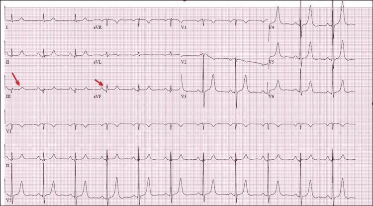
EKG was notable for a normal sinus rhythm with criteria for left ventricular (LV) hypertrophy and a possible inferior infarct (Figure 1).
Figure 1. EKG showing sinus rhythm and possible inferior infarct.
ST-segment elevation in the inferior lead (red arrows).
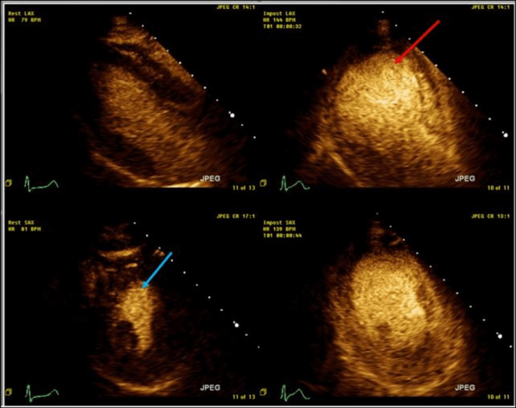
The baseline echocardiogram (echo) showed normal LV systolic function with an ejection fraction (EF) of 60% and concentric LV hypertrophy. She subsequently had an exercise stress echocardiogram. At the peak of exercise, the LV appeared dilated with global hypokinesis and an EF of about 25% (Figure 2).
Figure 2. Echocardiographic images of the left ventricle at rest and during stress.
Echocardiographic images of the left ventricle demonstrate adequate thickening and contraction of the myocardium at rest (blue arrow) as compared to exercise-induced global dilatation and apical ballooning of the left ventricle at stress (red arrow).
Along with this, the patient also had chest pain, which resolved by the end of recovery.
Management
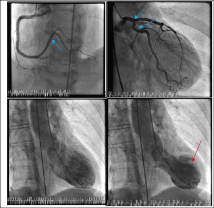
The abnormal stress test prompted a diagnostic angiogram, which revealed nonobstructive coronary artery disease (CAD). The left ventriculogram revealed an EF of about 25% with a Takotsubo-appearing LV with apical ballooning and basilar hyperkinesis (Figure 3).
Figure 3. Coronary angiogram with left ventriculogram demonstrating nonobstructive coronary artery disease (blue arrows), apical ballooning and Takotsubo-appearing left ventricle (red arrow), respectively.
The abnormal tests were thus attributed to the exercise portion of the stress test, as her baseline echocardiogram was normal. There was no clinical suspicion for other inciting factors, such as pheochromocytoma and myopericarditis, and therefore, an exhaustive workup was not pursued. The patient was prescribed metoprolol and lisinopril for stress cardiomyopathy.
Follow-up
The patient had a repeat echo two months following the diagnosis of TTS, which showed complete recovery of LV function with an EF of 60-65% and normal wall motion.
Discussion
First described in 1990 in the Japanese population, Takotsubo cardiomyopathy or stress cardiomyopathy can be caused by various inciting factors. Most common among these are emotional triggers, physical activities, and neurological illness, as well as other medical conditions and procedures. The diagnosis is based on the Mayo Clinic criteria [4].
Although previously thought to be a benign condition, it has now come to light that patient with TTS carries a similar short-term and long-term mortality as those of age- and sex-matched patients with acute coronary syndrome (ACS). The prognosis depends on the inciting stress factors, such that those with physical activities and medical conditions or procedures as the inciting factors have the worst long-term outcomes, while those with emotional triggers have relatively better long-term outcomes [5]. Among the various physical stressors, exercise has been described in the literature as a relatively uncommon etiology of TTS [6]. Here, we review all reported cases and clinical features of exercise-induced TTS available in the literature (Table 1)
Table 1. Review of the reported cases and clinical features of exercise-induced Takotsubo cardiomyopathy available in the literature.
DES: dobutamine stress echocardiogram; EF: ejection fraction; METS: metabolic equivalents; MHR: maximum heart rate; SPECT: single photon emission computerized tomography; WMA: wall motion abnormality.
| Study | Age | Sex | WMA and EF | Symptom | EKG changes | Stress intensity | Troponin | Coronary angiogram results | Follow-up | |
| Magri et al. [7] | 69 | Female | Apical akinesis, anterior hypokinesis, and mild impairment of global systolic function | Chest pain, emotional stressors | ST elevations in I, aVL, V2, V3 and ST depression in III, aVF, V4-V6 | Three METS | 9.7 ng/mL | Normal | Complete resolution of echo findings after one month | |
| Irwin et al. [8] | 81 | Female | Akinesis of midventricular and apical segments. Sparing of the base. EF 38% | Chest pain | ST elevation in the anterolateral leads 15 min post-exercise | 89% of MHR | 0.86 µg/L | Mild diffuse atherosclerosis | Improvement in mid-apical systolic function after seven days; EF 56% | |
| Dhoble et al. [9] (case no. 1) | 50 | Female | Apical akinesis and mid-ventricular hypokinesis; EF 50% | Chest pain | < 1 mm J point elevation in II, III, and aVF | 7.2 METS | 0.22 UI/L | Normal | A dobutamine stress echocardiogram (DES) performed three days later was normal, with EF of 70% | |
| Dhoble et al. [9] (case no. 2) | 75 | Male | Akinetic basal segment with normal apical segments; EF 55% | Pre-operative evaluation for carotid stenosis | ST-elevation | 4.1 METS | 0.78 UI/L | Normal | DES performed the next day was normal, with EF of 70% | |
| Digne et al. [10] | 66 | Female | Akinesis of the septo-apical region | Chest pain and emotional stress | Biphasic T waves from V1-V5 | 85% of MHR | 0.22 UI/L | Normal | Complete resolution after two weeks; EF of 60% | |
| López-Cuenca et al. [11] | 53 | Female | Midventricular, apical WMA; EF 45-50% | Chest pain and dyspnea | T wave depression | 63% of MHR | 0.19 ng/mL | Normal | Complete resolution; EF of 65% | |
| Dorfman et al. [6] | 71 | Female | Apical dyskinesis on SPECT | Chest pain | ST depression in V5 and V6 | Target heart rate achieved | 0.23 UI/L | Normal | Complete resolution on echo one month later | |
| Cantor et al. [12] | 77 | Female | Mid-cavitary hypokinesis with basal and apical hyperkinesis | Palpitations | ST elevations in I, aVL, V5, V6; ST depression in III, aVF, and V1-V3 | 4.5 METS | 11.17 ng/mL | Nonobstructive CAD | Resolution of WMA on echo obtained two weeks later | |
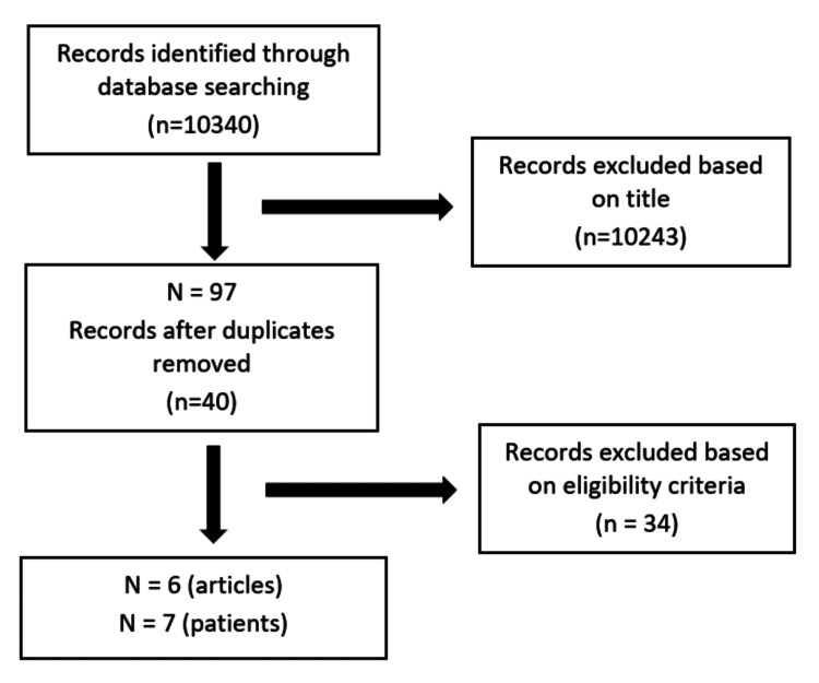
We searched MEDLINE (via PubMed) and Google Scholar up to August 12, 2018, and all the studies imported are shown in Figure 4.
Our review reveals that exercise stress test-induced TTS is indeed a real phenomenon. TTS caused by other physically strenuous activities has also been reported in the literature [13]. As mentioned earlier, since TTS associated with physical activities is associated with similar short-term and long-term outcomes [14] as in age- and sex-matched controls with ACS, the findings of TTS associated with exercise stress tests could potentially carry prognostic information [15]. Conversely, to avoid false positive exercise stress echocardiograms, consideration can be made toward obtaining another form of ischemic evaluation such as a regadenoson myocardial perfusion imaging, especially in specific populations such as the post-menopausal woman with multiple stressors in life who are predisposed to TTS [16].
Figure 4. The inclusion and exclusion process of the studies per the criteria.
Eligibility criteria for study selection include (1) development of TTS after the exercise stress test, (2) absence of other known comorbidities that can cause cardiomyopathy, and (3) articles in the English language.
Conclusions
Takotsubo cardiomyopathy can sometimes be precipitated by exercise stress tests. The presenting symptoms and ECG abnormalities are very similar to those of typical TTS (chest pain and ST-segment elevation). Clinicians must be aware of this risk during the examination. As TTS can be caused by the exercise element of a stress test, individuals who are susceptible to TTS may benefit from an alternative stress modality.
The content published in Cureus is the result of clinical experience and/or research by independent individuals or organizations. Cureus is not responsible for the scientific accuracy or reliability of data or conclusions published herein. All content published within Cureus is intended only for educational, research and reference purposes. Additionally, articles published within Cureus should not be deemed a suitable substitute for the advice of a qualified health care professional. Do not disregard or avoid professional medical advice due to content published within Cureus.
The authors have declared that no competing interests exist.
Human Ethics
Consent was obtained or waived by all participants in this study
References
- 1.Trend analysis of cardiovascular disease mortality, incidence, and mortality-to-incidence ratio: results from global burden of disease study 2017. Amini M, Zayeri F, Salehi M. BMC Public Health. 2021;21:401. doi: 10.1186/s12889-021-10429-0. [DOI] [PMC free article] [PubMed] [Google Scholar]
- 2.Stress testing and noninvasive coronary imaging: what's the best test for my patient? Matta M, Harb SC, Cremer P, Hachamovitch R, Ayoub C. Cleve Clin J Med. 2021;88:502–515. doi: 10.3949/ccjm.88a.20068. [DOI] [PubMed] [Google Scholar]
- 3.Overview of exercise stress testing. Kharabsheh SM, Al-Sugair A, Al-Buraiki J, Al-Farhan J. Ann Saudi Med. 2006;26:1–6. doi: 10.5144/0256-4947.2006.1. [DOI] [PMC free article] [PubMed] [Google Scholar]
- 4.Apical ballooning syndrome (Tako-Tsubo or stress cardiomyopathy): a mimic of acute myocardial infarction. Prasad A, Lerman A, Rihal CS. Am Heart J. 2008;155:408–417. doi: 10.1016/j.ahj.2007.11.008. [DOI] [PubMed] [Google Scholar]
- 5.Long-term prognosis of patients with Takotsubo syndrome. Ghadri JR, Kato K, Cammann VL, et al. https://pubmed.ncbi.nlm.nih.gov/30115226/ J Am Coll Cardiol. 2018;72:874–882. doi: 10.1016/j.jacc.2018.06.016. [DOI] [PubMed] [Google Scholar]
- 6.Takotsubo cardiomyopathy induced by treadmill exercise testing: an insight into the pathophysiology of transient left ventricular apical (or midventricular) ballooning in the absence of obstructive coronary artery disease. Dorfman T, Aqel R, Allred J, Woodham R, Iskandrian AE. J Am Coll Cardiol. 2007;49:1223–1225. doi: 10.1016/j.jacc.2006.12.033. [DOI] [PubMed] [Google Scholar]
- 7.Broken heart during treadmill exercise testing: an unusual cause of ST-segment elevation. Magri CJ, Sammut MA, Fenech A, et al. https://www.hellenicjcardiol.org/archive/full_text/2011/4/2011_4_377.pdf. Hellenic J Cardiol. 2011;52:377–380. [PubMed] [Google Scholar]
- 8.Apical ballooning syndrome following exercise treadmill testing. Irwin R, Mamas M, El-Omar M. https://pubmed.ncbi.nlm.nih.gov/21747667/ Exp Clin Cardiol. 2011;16:57–61. [PMC free article] [PubMed] [Google Scholar]
- 9.Transient left ventricular apical ballooning and exercise induced hypertension during treadmill exercise testing: is there a common hypersympathetic mechanism? Dhoble A, Abdelmoneim SS, Bernier M, Oh JK, Mulvagh SL. Cardiovasc Ultrasound. 2008;6:37. doi: 10.1186/1476-7120-6-37. [DOI] [PMC free article] [PubMed] [Google Scholar]
- 10.Tako-Tsubo syndrome after an exercise echocardiography. Digne F, Paillole C, Pillière R, Elayi SC, Gérardin B, Dib JC, Dahan M. Int J Cardiol. 2008;127:420–422. doi: 10.1016/j.ijcard.2007.04.088. [DOI] [PubMed] [Google Scholar]
- 11.An unusual complication of exercise echocardiography. López-Cuenca A, Gimeno JR, Pinar E, Valdés Chávarri M. Rev Esp Cardiol. 2011;64:82–83. doi: 10.1016/j.recesp.2010.10.004. [DOI] [PubMed] [Google Scholar]
- 12.Mid-left ventricular ballooning variant Takotsubo syndrome induced by treadmill exercise stress testing. Cantor G, Teressa G. Case Rep Cardiol. 2018;2018:5282747. doi: 10.1155/2018/5282747. [DOI] [PMC free article] [PubMed] [Google Scholar]
- 13.Adrift while swimming and Takotsubo syndrome: the vagotonia connection. Madias JE. Int J Cardiol. 2014;177:123. doi: 10.1016/j.ijcard.2014.09.129. [DOI] [PubMed] [Google Scholar]
- 14.Short- and long-term prognosis of patients with Takotsubo syndrome based on different triggers: importance of the physical nature. Uribarri A, Núñez-Gil IJ, Conty DA, et al. J Am Heart Assoc. 2019;8:0. doi: 10.1161/JAHA.119.013701. [DOI] [PMC free article] [PubMed] [Google Scholar]
- 15.Characteristics and outcomes of patients with Takotsubo syndrome: incremental prognostic value of baseline left ventricular systolic function. Alashi A, Isaza N, Faulx J, et al. https://pubmed.ncbi.nlm.nih.gov/32755253/ J Am Heart Assoc. 2020;9:0. doi: 10.1161/JAHA.120.016537. [DOI] [PMC free article] [PubMed] [Google Scholar]
- 16.Are some false-positive stress echocardiograms a forme fruste variety of apical ballooning syndrome? Form AM, Prasad A, Pellikka PA, et al. https://www.ajconline.org/article/S0002-9149(09)00471-8/fulltext. Am J Cardiol. 2009;8:1434–1438. doi: 10.1016/j.amjcard.2009.01.352. [DOI] [PubMed] [Google Scholar]


