

Abstract
Zinc intake has reduced hospitalizations in patients with ulcerative colitis (UC), highlighting the need to maintain blood zinc levels. This prospective study investigated whether the promotion of zinc intake and a Japanese diet (high in n-3 fatty acids) could induce clinical remission in patients with mild active UC. Patients with mild active UC were randomly assigned to either (1) continue an unrestricted diet or (2) receive nutritional guidance promoting zinc intake and a Japanese diet. The primary endpoint was clinical remission at 24 weeks. Secondary endpoints were the Ulcerative Colitis Endoscopic Index of Severity (UCEIS) scores, Clinical Activity Index (CAI), Geboes Histopathology Score (GHS), and biomarkers, including zinc levels, measured at 12 and 24 weeks. Nutritional assessments were performed using the Food Frequency Questionnaire. The CAI, UCEIS, and GHS scores were significantly lower in the intervention group than in the control group, with a significantly higher proportion of patients achieving clinical remission. Furthermore, the intervention group exhibited weight gain and significantly increased blood zinc levels. The combination of promoting dietary zinc intake and a Japanese diet rich in n-3 fatty acids can induce clinical remission in patients with mild active UC.
Keywords: ulcerative colitis, zinc, n-3 fatty acids
Introduction
Both gut bacteria and dietary antigens play important roles in the pathogenesis of inflammatory bowel disease (IBD), and several studies have investigated the involvement of intestinal bacteria and dietary antigens in mucosal immunity.(1–4) Research has demonstrated that changes in metabolites caused by diversity in the intestinal microbiota can affect the digestion and metabolism of food, resulting in significant changes to the intestinal environment. Accordingly, a low-fat diet has been reported to maintain remission and lead to favorable outcomes in patients with Crohn’s disease (CD), while high-fat formulas have been associated with poor therapeutic outcomes.(5) However, in contrast to those with CD, patients with ulcerative colitis (UC) may not experience remission-inducing effects following nutritional therapy,(6) and there is little evidence supporting nutritional therapy for the maintenance of remission in patients with UC. Therefore, unnecessary dietary restrictions should not be imposed in the maintenance phase of treatment for UC.
Patients with IBD tend to exhibit deficiencies of various micronutrients because of intestinal dysbiosis caused by diarrhea and inadequate food intake, which result from the anorexia associated with disease activity.(7) For example, Schneider et al.(8) revealed that the serum concentrations of copper and zinc are insufficient in a substantial proportion of patients with IBD. Other studies have reported that intestinal mucosal permeability increases in patients with IBD,(9) while zinc intake has been shown to strengthen the function of the intestinal barrier via its effects on tight junctions.(10) Furthermore, the administration of zinc in patients with UC increases the blood levels of thymulin, which plays a role in immunity, decreases the activity of natural killer cells, and normalizes intestinal permeability.(11) Finally, a multicenter study of dietary habits in patients with new-onset UC reported that the odds of new-onset UC were significantly lower in populations with high zinc intake before onset than in those with low zinc intake.(12)
Considering that supplementation with zinc preparations often causes side effects such as stomach pain and nausea, which may make continuous zinc supplementation difficult, a zinc-rich diet has been recommended over supplementation.(13)
In the past 50 years, the availability of preserved foods rich in n-6 polyunsaturated fatty acids has led to drastic changes in dietary habits. The rapid increase in the number of patients with IBD is undoubtedly related to these changes in environmental and dietary factors. Certain n-3 fatty acids, including eicosapentaenoic acid (EPA), docosahexaenoic acid (DHA), and alpha-linolenic acid, which are present in seafood and marine products, have been reported to have anti-inflammatory effects. Although there are currently no consistent results suggesting that n-3 fatty acids can suppress intestinal inflammation and induce UC remission,(14) a higher intake of n-3 fatty acids has been shown to result in a lower incidence of UC (hazard ratio, 0.72). Conversely, increased intake of linoleic acid, an n-6 fatty acid, and decreased intake of DHA, an n-3 fatty acid, have been associated with an increased risk of developing UC. Therefore, a decrease in the ratio of n-3 to n-6 fatty acids and increased consumption of red meat have been reported to increase the risk of UC.(15,16)
In this study, we investigated whether the combination of nutritional guidance encouraging zinc intake and a Japanese diet rich in n-3 fatty acids could induce remission in patients with mild active UC who were undergoing standard treatment in our department. In addition, we prospectively examined changes in endoscopic findings, inflammatory blood biomarkers, and clinical symptoms to evaluate the effectiveness of this nutritional intervention in patients with mild active UC.
Materials and Methods
Study design and ethical considerations
This was a prospective, randomized, controlled trial involving 20 patients registered in an institution in Japan (clinical trial registration: UMIN000046664). The study period was from August 2018 to March 2021. The study protocol complied with the tenets of the revised Declaration of Helsinki (1989) and was approved by the institutional review board of our institution. Written informed consent was obtained from all participating patients.
Nutritional guidance
The Nutrition Department of our hospital provided three 30-min nutritional guidance sessions to patients with mild active UC. Apart from dietary intake of zinc (Table 1), patients were encouraged to eat fish rich in n-3 fatty acids (Table 2), flaxseed and egoma oils, and a Japanese diet. Clinical symptoms and endoscopic findings were evaluated using the Clinical Activity Index (CAI), Ulcerative Colitis Endoscopic Index of Severity (UCEIS), and Geboes Histopathology Score (GHS), respectively, at 12 and 24 weeks.
Table 1.
Zinc content in various foods
| Classification | Zinc content per 100 g (mg) |
|
|---|---|---|
| Seafood | Oyster | 13.2 |
| Scallop | 2.7 | |
| Eel | 1.4 | |
| Pacific saury | 0.8 | |
| Cod roe | 3.1 | |
| Dried sardines | 7.2 | |
| Meat | Beef shoulder roast | 5.6 |
| Beef thigh | 4 | |
| Beef liver | 3.8 | |
| Chicken liver | 3.3 | |
| Beef belly | 3 | |
| Chicken thigh | 1.6 | |
| Pork loin | 1.6 | |
| Dairy products | Egg | 1.3 |
| Milk | 0.4 | |
| Processed cheese | 3.2 | |
| Soybean | Natto | 1.9 |
| Tofu | 0.6 | |
| Nuts | Cashew nuts | 5.4 |
| Almond nuts | 4.4 | |
Table 2.
n-3 fatty acid content in various seafoods
| Seafood | n-3 fatty acids per 100 g (g) |
|---|---|
| Scad | 1.05 |
| Mackerel | 2.12 |
| Pacific saury | 5.59 |
| Sardine | 2.1 |
| Japanese amberjack | 3.35 |
| Young yellowtail | 1.88 |
| Tuna | 0.21 |
| Garden eel | 1.42 |
| Eel | 2.42 |
| North Pacific bluefish | 1.13 |
| Spirinchus lanceolatus | 1.47 |
| Splendid alfonsino | 1.37 |
Detailed nutritional assessments of food intake were undertaken using a dietary record chart and the administration of an established semiquantitative questionnaire available for clinical investigation (Food Frequency Questionnaire based on food groups, ver. 4.0; Kenpaku-sha, Tokyo, Japan). The items evaluated using the questionnaire assessed the total levels of energy, protein, fat, carbohydrate, zinc, and n-3 fatty acid intake.
Patients
Patients aged ≥15 years who had mild active UC (patients with a UCEIS score of 2–4 points, CAI score of 5–6 points, and distribution of the UCEIS and CAI scores at admission indicating that the patient was not in remission) were enrolled in the study. The drugs prescribed for the patients are listed in Table 3. There were no additional treatments, including steroid, biologic, calcineurin inhibitor, and JAK inhibitor treatments, during this study. The enrolled patients were not administered zinc preparations during the follow-up period.
Table 3.
Patient characteristics
| Intervention (n = 10) | Control (n = 10) | p value | |
|---|---|---|---|
| Age | 37.5 ± 16.1 | 38 ± 19.2 | 0.95a |
| Sex (male:female) | 5:05 | 5:05 | 0.673a |
| Body weight (kg) | 48.3 ± 6.7 | 54.3 ± 8.5 | 0.097a |
| BMI (kg/m2) | 18.7 ± 1.9 | 21 ± 3.5 | 0.086a |
| Energy (kcal) | 1,667.5 ± 509.4 | 1,740 ± 361.7 | 0.718a |
| Protein (g) | 57.2 ± 19.7 | 61 ± 17.5 | 0.656a |
| Fat (g) | 50.3 ± 20.5 | 53.4 ± 21.6 | 0.745a |
| n-3 fatty acids (g) | 1.5 ± 0.8 | 2 ± 0.6 | 0.124a |
| Zinc (g) | 7 ± 2.1 | 7.6 ± 1.8 | 0.492a |
| Alb (mg/dl) | 3.7 ± 0.6 | 3.6 ± 0.9 | 0.662a |
| WBC (×103) | 7.3 ± 2.7 | 7.5 ± 2.1 | 0.874a |
| CRP (mg/dl) | 1.2 ± 2.3 | 3.5 ± 6.5 | 0.298a |
| Blood zinc (g/dl) | 72.1 ± 17.8 | 72 ± 14.5 | 0.989a |
| Partial Mayo score | 0.8 ± 0.4 | 0.9 ± 0.3 | 0.556a |
| UCEIS score | 2.5 ± 0.7 | 2.1 ± 0.6 | 0.18a |
| CAI score | 7.7 ± 3.4 | 5.8 ± 0.9 | 0.105a |
| GHS | 2.6 ± 0.5 | 2.4 ± 0.5 | 0.398a |
| Base drug | |||
| Mesalazine | 8 | 8, 80 | >0.999b |
| Azathioprine | 5 | 5, 50 | >0.999b |
| Biotherapy | 0.553b | ||
| Adalimumab | 3 | 1 | |
| Infliximab | 2 | 0 | |
| Golimumab | 0 | 1 | |
| Tofacitinib | 0 | 2 | |
| Ustekinumab | 0 | 1 | |
| Vedolizumab | 2 | 1 | |
| None | 3 | 4 |
Alb, albumin; BMI, body mass index; CAI, Clinical Activity Index; CRP, C-reactive protein; UCEIS, Ulcerative Colitis Endoscopic Index of Severity; WBC, white blood cell. p value, aunpaired t test, bFisher’s exact test. Data are represented as means ± SD or numbers and percentages.
Details of this study were explained to the patients both verbally and in writing, and those who provided written consent were eligible for inclusion.
Patients undergoing colonoscopy without pretreatment; those with difficulty following nutritional guidance; those with intestinal obstruction, fistula, history of colorectal surgery, moderate-to-severe active-phase UC, diverticulitis, or massive colorectal bleeding; and those with anemia or other underlying conditions except UC were excluded (Fig. 1).
Fig. 1.
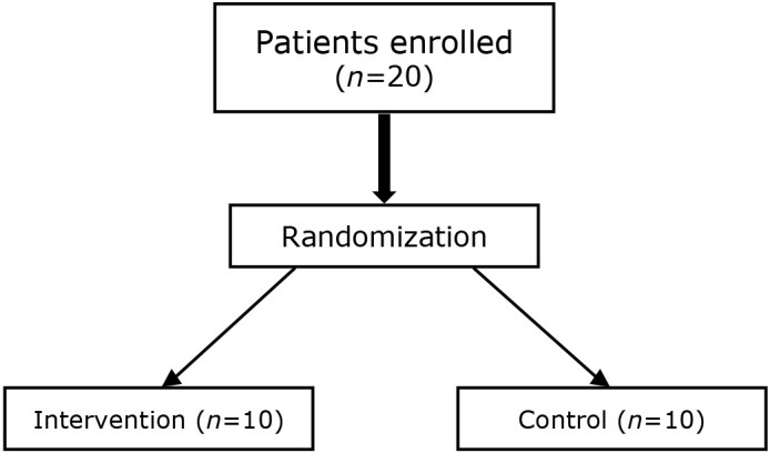
Flowchart of patient enrollment.
Randomization
The patients were randomly assigned in a 1:1 ratio to undergo nutritional guidance (intervention group) or to the control group via block randomization using computerized lists. Anonymity was ensured by allocating study-specific codes instead of using personal information, such as patient name or ID, during randomization.
Definitions
For this study, zinc deficiency was defined according to the Japanese Society of Clinical Nutrition’s recently issued Japan’s Practical Guideline for Zinc Deficiency 2018 as follows: (a) one or more symptoms of zinc deficiency or low serum alkaline phosphatase, (b) ruling out of other diseases, (c) low serum zinc level, and (d) alleviation of symptoms upon zinc administration. Serum zinc levels of <60 μg/dl and 60–80 μg/dl were considered to indicate zinc deficiency and marginal zinc deficiency, respectively.(17)
Disease remission was defined according to the UCEIS. Travis et al.,(18,19) who proposed the UCEIS, did not define effectiveness based on therapeutic evaluation or endoscopic remission (i.e., mucosal healing). In contrast, they noted that a UCEIS score of 0 was the best definition of endoscopic remission. Similarly, they concluded that a decrease in the Mayo Endoscopic Subscore by at least 1 or the UCEIS score by at least 2 was an appropriate definition of endoscopic response.(20) The UCEIS was evaluated by two expert endoscopists.
A CAI score of ≥6 and an endoscopy score of ≥4 were defined as clinically active disease, whereas a CAI score of ≤4 was defined as clinical remission.(21)
Histological assessment of disease activity was performed using the GHS. As there is no consensus on the definition of remission yet, we defined remission as a GHS score of 2 or less and set remission score as 2 or less.(22,23)
Outcome measurements
The primary outcomes included the rate of remission at 24 weeks after the initiation of nutritional guidance, as determined based on the CAI score, endoscopic activity findings (UCEIS), and GHS score. Secondary outcomes included patient weight, body mass index (BMI), energy intake, protein intake, lipid intake, n-3 fatty acid intake, zinc intake, blood zinc level (fasting), percentage of patients with blood zinc levels of >80 μg/dl, blood albumin level, white blood cell count, and C-reactive protein level.
Statistical analyses
Categorical data are presented as numbers and percentages (%) and were analyzed using Fisher’s exact test. Continuous data are presented as means ± SD. Repeated-measures analysis of variance models with between-subject (group) and within-subject (time) factors and interactions were performed. The factors of treatment group, subject, and time were included in the model. Interactions between group and time (among time) were calculated for each variable. Between-group comparisons for continuous variables were performed using unpaired t tests. Within-group comparisons for continuous variables were performed using paired t tests. Bonferroni correction was used to account for the multiplicity of comparisons between the groups. A two-sided p value of ≤0.05 was considered statistically significant. Statistical analyses were performed using SPSS for Windows ver. 26.0 (IBM Japan, Tokyo, Japan).
Results
Ten patients each were allocated to the intervention and control groups. There was no significant difference in patient background between the intervention and control groups. The Partial Mayo Score (Mayo Endoscopic Subscore) was approximately 1 in both groups. The UCEIS score was approximately 2, indicating that mucosal healing had not occurred. The CAI score was >4 in both groups (intervention group: 7.7 ± 3.4, control group: 5.8 ± 0.9; p = 0.105), indicating that clinical remission had not been achieved. GHS was >2 in both groups (intervention group: 2.6 ± 3.4, control group: 2.4 ± 0.5; p = 0.398), indicating that histological remission had not been achieved (Table 3).
At 12 and 24 weeks, the nutritional guidance intervention significantly increased body weight and BMI [body weight (baseline, 12 weeks, 24 weeks): 48.8 ± 6.7 kg → 52.4 ± 6.8 kg → 53.2 ± 7.5 kg vs 54.3 ± 8.5 kg → 55.3 ± 9.3 kg → 55.4 ± 9.5 kg in the control group, p = 0.032; BMI (baseline, 12 weeks, 24 weeks): 18.7 ± 1.9 kg/m2 → 20.2 ± 1.5 kg/m2 → 20.4 ± 1.5 kg/m2 vs 21.0 ± 3.5 kg/m2 → 21.3 ± 3.1 kg/m2 → 21.4 ± 3.3 kg/m2 in the control group, p = 0.014] relative to that observed at admission, respectively. There was also a significant difference in zinc intake between the intervention and control groups [intervention (baseline, 12 weeks, 24 weeks): 7.0 ± 2.1 g → 12.1 ± 6.3 g → 11.9 ± 6.1 g vs control: 7.6 ± 1.8 g → 8.3 ± 2.0 g → 8.6 ± 1.8 g, p = 0.049], and the blood zinc level was significantly higher in the intervention group than in the control group [intervention (baseline, 12 weeks, 24 weeks): 72.1 ± 17.8 μg/dl → 73.6 ± 10.3 μg/dl → 92.1 ± 23.2 μg/dl vs control: 72.0 ± 14.5 μg/dl → 75.5 ± 9.4 μg/dl → 71.4 ± 10.9 μg/dl, p = 0.008]. Although there was no difference in fat intake, n-3 fatty acid intake was significantly greater at 12 and 24 weeks than at admission [12 weeks: 1.5 ± 0.8 g vs 2.0 ± 0.7 g (p = 0.010) and 24 weeks: 2.3 ± 0.6 g (p = 0.002)] (Table 4).
Table 4.
Changes in study variables in each group
| n | T1. On admission |
T2. 12 weeks later |
T3. 24 weeks later |
p value | p value (group comparison) |
p value (intragroup comparison) |
||||||
|---|---|---|---|---|---|---|---|---|---|---|---|---|
| rmANOVA | T1. | T2. | T3. | T1. vs T2. | T1. vs T3. | |||||||
| Bodyweight (kg) | 0.032 | 0.097 | 0.433 | 0.561 | ||||||||
| Intervention | 10 | 48.8 ± 6.7 | 52.4 ± 6.8 | 53.2 ± 7.5 | 0.013 | 0.015 | ||||||
| Control | 10 | 54.3 ± 8.5 | 55.3 ± 9.3 | 55.4 ± 9.5 | 0.749 | 0.55 | ||||||
| BMI | 0.014 | 0.086 | 0.325 | 0.414 | ||||||||
| Intervention | 10 | 18.7 ± 1.9 | 20.2 ± 1.5 | 20.4 ± 1.5 | 0.01 | 0.011 | ||||||
| Control | 10 | 21.0 ± 3.5 | 21.3 ± 3.1 | 21.4 ± 3.3 | 0.854 | 0.505 | ||||||
| Energy (kcal) | 0.875 | 0.718 | 0.622 | 0.351 | ||||||||
| Intervention | 10 | 1,667.5 ± 509.4 | 1,781.0 ± 327.1 | 1,796.6 ± 342.9 | 0.896 | 0.708 | ||||||
| Control | 10 | 1,740.0 ± 361.7 | 1,871.6 ± 467.8 | 1,970.0 ± 458.6 | 0.83 | 0.419 | ||||||
| Protein (g) | 0.682 | 0.656 | 0.15 | 0.399 | ||||||||
| Intervention | 10 | 57.2 ± 20.5 | 60.1 ± 10.2 | 63.1 ± 12.7 | >0.999 | 0.551 | ||||||
| Control | 10 | 61.0 ± 17.5 | 69.8 ± 17.8 | 68.3 ± 13.8 | 0.351 | 0.494 | ||||||
| Fat (g) | 0.814 | 0.745 | 0.324 | 0.142 | ||||||||
| Intervention | 10 | 50.3 ± 20.5 | 54.5 ± 15.7 | 53.2 ± 11.5 | >0.999 | >0.999 | ||||||
| Control | 10 | 53.4 ± 21.6 | 61.5 ± 15.4 | 61.6 ± 12.9 | 0.408 | 0.539 | ||||||
| n-3 fatty acids (g) | 0.007 | 0.124 | 0.87 | 0.17 | ||||||||
| Intervention | 10 | 1.5 ± 0.8 | 2.0 ± 0.7 | 2.3 ± 0.6 | 0.01 | 0.002 | ||||||
| Control | 10 | 2.0 ± 0.6 | 2.1 ± 0.8 | 1.9 ± 0.6 | >0.999 | >0.999 | ||||||
| Zinc intake (g) | 0.049 | 0.492 | 0.086 | 0.112 | ||||||||
| Intervention | 10 | 7.0 ± 2.1 | 12.1 ± 6.3 | 11.9 ± 6.1 | 0.057 | 0.066 | ||||||
| Control | 10 | 7.6 ± 1.8 | 8.3 ± 2.0 | 8.6 ± 1.8 | 0.753 | 0.392 | ||||||
| Alb (g/dl) | 0.508 | 0.622 | 0.194 | 0.461 | ||||||||
| Intervention | 10 | 3.7 ± 0.6 | 4.1 ± 0.3 | 4.2 ± 0.2 | 0.16 | 0.09 | ||||||
| Control | 10 | 3.6 ± 0.9 | 4.3 ± 0.4 | 4.3 ± 0.3 | 0.067 | 0.068 | ||||||
| WBC (×103) | 0.596 | 0.874 | 0.607 | 0.195 | ||||||||
| Intervention | 10 | 7.3 ± 2.7 | 5.4 ± 1.7 | 5.4 ± 1.0 | 0.074 | 0.139 | ||||||
| Control | 10 | 7.5 ± 2.1 | 5.0 ± 1.8 | 6.3 ± 1.7 | 0.127 | 0.45 | ||||||
| CRP (mg/dl) | 0.34 | 0.298 | 0.419 | 0.339 | ||||||||
| Intervention | 10 | 1.19 ± 2.3 | 0.11 ± 0.0 | 0.12 ± 0.1 | 0.337 | 0.346 | ||||||
| Control | 10 | 3.5 ± 6.5 | 0.1 ± 0.1 | 0.4 ± 0.9 | 0.272 | 0.355 | ||||||
| Zinc (mg/dl) | 0.008 | 0.989 | 0.672 | 0.02 | ||||||||
| Intervention | 10 | 72.1 ± 17.8 | 73.6 ± 10.3 | 92.1 ± 23.2 | >0.999 | 0.073 | ||||||
| Control | 10 | 72.0 ± 14.5 | 75.5 ± 9.4 | 71.4 ± 10.9 | 0.917 | >0.999 | ||||||
| UCEIS score | 0.07 | 0.18 | 0.264 | 0.306 | ||||||||
| Intervention | 10 | 2.5 ± 0.7 | 1.7 ± 0.7 | 1.6 ± 0.7 | 0.021 | 0.02 | ||||||
| Control | 10 | 2.1 ± 0.6 | 2.0 ± 0.5 | 1.9 ± 0.6 | >0.999 | 0.887 | ||||||
| CAI score | ≤0.001 | 0.105 | ≤0.001 | ≤0.001 | ||||||||
| Intervention | 10 | 7.7 ± 3.4 | 5.0 ± 0.7 | 4.0 ± 0.0 | 0.077 | 0.015 | ||||||
| Control | 10 | 5.8 ± 0.9 | 6.4 ± 0.7 | 6.9 ± 0.6 | 0.01 | 0.002 | ||||||
| GHS | 0.209 | 0.525 | 0.478 | 0.172 | ||||||||
| Intervention | 10 | 2.6 ± 0.5 | 2.0 ± 0.8 | 1.0 ± 0.7 | 0.048 | 0.0008 | ||||||
| Control | 10 | 2.4 ± 0.5 | 2.2 ± 0.4 | 1.6 ± 1.0 | 0.894 | 0.071 | ||||||
Alb, albumin; BMI, body mass index; CAI, Clinical Activity Index; CRP, C-reactive protein; GHS, Geboes Histopathology Score; UCEIS, Ulcerative Colitis Endoscopic Index of Severity; WBC, white blood cell. p value, rmANOVA; repeated-measures ANOVA [between groups and among times (T1, T2, T3)]; group comparison, unpaired t test; intragroup comparison, paired t test (Bonferroni correction). Data are represented as mean ± SD.
In addition, endoscopic evaluation (UCEIS) revealed significant improvements in the endoscopic mucosa (by approximately 1 point) in the intervention group at both 12 and 24 weeks (2.5 ± 0.7 to 1.6 ± 0.7, p = 0.020). The CAI decreased by 3.7 points in the intervention group at 24 weeks (7.7 ± 3.4 to 4.0 ± 0.0 p = 0.015), indicating clinical remission. CAI was significantly higher in the control group.
The GHS score significantly decreased in the intervention group after 24 weeks (2.6 ± 0.5 to 1.0 ± 0.7 p = 0.0008; Table 4).
The percentage of patients in clinical remission (i.e., CAI score ≤4) was 0% in the control group and 90% in the intervention group at the 24-week assessment (p<0.001). However, there was no significant difference in the percentage of patients whose blood zinc levels were maintained above 80 μg/dl (not zinc deficient). No exacerbation of symptoms was observed in either group, and no patient required intensive treatment (Table 5).
Table 5a.
Percentage of patients in remission (CAI score ≤4) in each group
| n | T1 (at admission) |
T2 (12 weeks) |
T3 (24 weeks) |
|
|---|---|---|---|---|
| Remission (CAI score ≤4) | ||||
| Intervention | 10 | 0 (0.0) | 2 (16.7) | 9 (90.0) |
| Control | 10 | 0 (0.0) | 0 (0.0) | 0 (0.0) |
| p value | 0.474 | <0.001 | ||
Data are represented as numbers and percentages. p value: Fisher’s exact test. CAI, Clinical Activity Index.
Table 5b.
Percentage of patients with sufficient blood zinc level (zinc >80 μg/dl) in each group
| n | T1 (on admission) |
T2 (12 weeks) |
T3 (24 weeks) |
|
|---|---|---|---|---|
| Zinc >80 μg/dl | ||||
| Intervention | 10 | 5 (50.0) | 5 (30.0) | 5 (50.0) |
| Control | 10 | 3 (30.0) | 2 (20.0) | 4 (40.0) |
| p value | 0.65 | >0.999 | >0.999 | |
p value, Fisher’s exact test. Data are represented as numbers and percentages.
Discussion
In this study, we investigated whether nutritional guidance that promotes a diet rich in zinc combined with n-3 fatty acids could induce clinical remission in patients with mild active UC. The CAI and UCEIS scores were significantly lower in the intervention group than in the control group, with a significantly higher proportion of patients achieving clinical remission. Furthermore, the intervention group exhibited significant weight gain at 12 and 24 weeks, as well as significant increase in the blood zinc levels at 24 weeks. In this study, we treated only mild UC to avoid influencing the outcomes of any therapeutic intervention other than nutritional guidance. The treatment regimen, besides nutritional guidance, remained unchanged in all patients.
Although it is unclear to what extent the improvement in blood zinc levels in this study contributed to the improvement in the pathophysiology of UC, we believe that addition of zinc loading to the existing treatment for mild UC is expected to have an add-on effect on treatment.
There was no difference in total calorie, fat, and protein intake between both groups, even after nutritional guidance. However, n-3 fatty acid and zinc content was significantly higher in the intervention group, suggesting that the improvement in CAI was due to n-3 fatty acid and zinc intake.
Abnormal composition of the intestinal microbiota, known as dysbiosis, may be involved in the pathogenesis of IBD.(24) Moreover, compared with healthy individuals, patients with IBD exhibit a decrease in the diversity of intestinal microbiota.(25) Apart from monogenic IBD, microbial and other environmental exposures are more crucial in the development and progression of IBD than genetic susceptibility to the disease.(26) Diet is among the major environmental factors contributing to the epidemiological surge in IBD and mental stress.(27) In patients with UC, nutritional guidance, including that which promotes a low-fat diet, has not been considered a major contributor to the maintenance of remission. During the active disease phase, however, nutritional disorders do occur, including a loss of body weight (by 18–62%) and hypoalbuminemia (by 26–50%).(28)
Major nutrients are absorbed in the small intestine, which is not involved in most UC cases. Therefore, it is not clear whether dietary factors are pathogenic or exacerbating factors. However, diets high in fruits, vegetables, and n-3 fatty acids and low in n-6 fatty acids have been associated with a lower risk of developing both CD and UC.(29) Additionally, animal studies have shown that medium-chain and long-chain fatty acids exert different effects on the antigen-presenting cells in the intestinal tract.(30) Furthermore, trans fatty acids exacerbate intestinal mucosal inflammation.(31) Although Hou et al.(14) commented on the inconsistency in the data regarding the effects of an n-fatty acid-enriched diet on intestinal inflammation and induction of remission, their results indicated that nutritional guidance might reduce the risk of developing UC.
The intake of n-6 fatty acids, such as arachidonic acid, is reported to exacerbate inflammation and adversely affect the body; hence, a good balance should be maintained by including more food rich in n-3 fatty acids than in n-6 fatty acids in the modern diet. Although the intervention group had a higher intake of n-3 fatty acid-rich foods, fatty acid composition in the blood was not measured in this study. Thus, precise evaluation of the effectiveness of n-3 fatty acid-rich foods was not possible.
Siva et al.(32) reported that 86 of 223 (38.6%) patients with UC and 326 of 773 (42.2%) with CD had low zinc levels. In addition, patients with IBD associated with hypozincemia had a higher risk of hospitalization, surgery, and complications than patients with normal serum zinc levels.(33) One factor that may have contributed to this observation is the lack of essential nutrients due to fast food consumption and dietary restrictions. Considering the above findings, maintaining the blood zinc level may help to prevent exacerbations of UC.
In this study, nutritional guidance was provided to encourage zinc intake and the adoption of a Japanese diet rich in n-3 fatty acids because consuming zinc-rich food can lead to excessive fat intake and insufficient intake of n-3 fatty acids and dietary fiber. Zinc intake increased after starting nutritional guidance. However, there was a significant difference in zinc intake between the two groups, indicating that it may be important for these patients to comply with nutritional guidance and a Japanese diet. The actual concentration of zinc in the blood increased in the intervention group, whereas it remained almost unchanged in the control group.
In the intervention group, the clinical remission rate significantly increased, whereas endoscopic activity significantly decreased. Although the clinical remission (CAI ≤4) rate was significantly higher in the intervention group, the treatment was not standardized; therefore, this effect cannot be proven to be exclusively due to zinc loading plus the Japanese diet intervention. However, no new treatments were added in this study, and there were no significant differences in the treatment at entry, indicating that nutritional guidance encouraging the addition of zinc and conversion to a Japanese diet may be effective for inducing remission.
A previous study reported that zinc deficiency exacerbates colonic inflammation via activation of the IL-23/Th17 axis and that zinc intake is expected to increase the remission induction rate by suppressing these cytokines.(33) In the current study, the baseline blood level of zinc ranged from 80 to 130 μg/dl, and there was a significant difference in the blood zinc level between the intervention and control groups. Indeed, the zinc level exceeded 80 μg/dl and was significantly higher in the intervention group than in the control group, which may have led to the improvements in the UCEIS and CAI scores.
This study has some limitations, including its single-center design. This study did not investigate the intestinal microbiota of each patient. It also did not account for the patient’s current treatment regimen or disease duration. In addition, there was no blinding, and the sample size was small because it was a pilot study.
In conclusion, the current pilot data indicate that promotion of both zinc intake and a Japanese diet rich in n-3 fatty acids may help induce clinical remission in patients with mild active UC. Nonetheless, large-scale randomized controlled trials are required to validate our findings.
Author Contributions
Manuscript drafting, KM; study design, YT, HI; endoscopists, KM, YT, RS, HO, and HI; nutritionists, YI; statistical analysis, YT; supervision, HN and HI. The final version of the manuscript was read and approved by all authors.
Acknowledgments
We would like to thank our study participants for their invaluable contributions to this project.
Abbreviations
- BMI
body mass index
- CAI
Clinical Activity Index
- CD
Crohn’s disease
- DHA
docosahexaenoic acid
- EPA
eicosapentaenoic acid
- GHS
Geboes Histopathology Score
- IBD
inflammatory bowel disease
- UC
ulcerative colitis
- UCEIS
Ulcerative Colitis Endoscopic Index of Severity
Data Availability
All data generated or analyzed during this study are included in this article. Further enquiries can be directed to the corresponding author.
Conflicts of Interest
No potential conflicts of interest were disclosed.
References
- 1.Imaeda H, Miura S, Serizawa H, et al. Influence of fatty acid absorption on bidirectional release of immunoglobulin A into intestinal lumen and intestinal lymph in rats. Immunol Lett 1993; 38: 253–258. [DOI] [PubMed] [Google Scholar]
- 2.Strisciuglio C, Cenni S, Serra MR, et al. Effectiveness of Mediterranean diet’s adherence in children with inflammatory bowel diseases. Nutrients 2020; 12: 3206. [DOI] [PMC free article] [PubMed] [Google Scholar]
- 3.Keshteli AH, van den Brand FF, Madsen KL, et al. Dietary and metabolomic determinants of relapse in ulcerative colitis patients: a pilot prospective cohort study. World J Gastroenterol 2017; 23: 3890–3899. [DOI] [PMC free article] [PubMed] [Google Scholar]
- 4.Lewis JD, Abreu MT. Diet as a trigger or therapy for inflammatory bowel diseases. Gastroenterology 2017; 152: 398–414.e6. [DOI] [PubMed] [Google Scholar]
- 5.Yamamoto T, Nakahigashi M, Saniabadi AR. Review article: diet and inflammatory bowel disease—epidemiology and treatment. Aliment Pharmacol Ther 2009; 30: 99–112. [DOI] [PubMed] [Google Scholar]
- 6.Filippi J, Al-Jaouni R, Wiroth JB, Hébuterne X, Schneider SM. Nutritional deficiencies in patients with Crohn’s disease in remission. Inflam Bowel Dis 2006; 12: 185–191. [DOI] [PubMed] [Google Scholar]
- 7.Geerling BJ, Badart-Smook A, Stockbrügger RW, Brummer RJ. Comprehensive nutritional status in patients with long-standing Crohn disease currently in remission. Am J Clin Nutr 1998; 67: 919–926. [DOI] [PubMed] [Google Scholar]
- 8.Schneider T, Caviezel D, Ayata CK, Kiss C, Niess JH, Hruz P. The copper/zinc ratio correlates with markers of disease activity in patients with inflammatory bowel disease. Crohns Colitis 360 2020; 2: otaa001. [DOI] [PMC free article] [PubMed] [Google Scholar]
- 9.Honzawa Y, Nakase H, Matsuura M, Chiba T. Clinical significance of serum diamine oxidase activity in inflammatory bowel disease: importance of evaluation of small intestinal permeability. Inflam Bowel Dis 2011; 17: E23–E25. [DOI] [PubMed] [Google Scholar]
- 10.Finamore A, Massimi M, Conti Devirgiliis L, Mengheri E. Zinc deficiency induces membrane barrier damage and increases neutrophil transmigration in Caco-2 cells. J Nutr 2008; 138: 1664–1670. [DOI] [PubMed] [Google Scholar]
- 11.Cragg RA, Phillips SR, Piper JM, et al. Homeostatic regulation of zinc transporters in the human small intestine by dietary zinc supplementation. Gut 2005; 54: 469–478. [DOI] [PMC free article] [PubMed] [Google Scholar]
- 12.Kobayashi Y, Ohfuji S, Kondo K, et al. Association between dietary iron and zinc intake and development of ulcerative colitis: a case-control study in Japan. J Gastroenterol Hepatol 2019; 34: 1703–1710. [DOI] [PubMed] [Google Scholar]
- 13.Katayama K, Saito M, Kawaguchi T, et al. Effect of zinc on liver cirrhosis with hyperammonemia: a preliminary randomized, placebo-controlled double-blind trial. Nutrition 2014; 30: 1409–1414. [DOI] [PubMed] [Google Scholar]
- 14.Hou JK, Abraham B, El-Serag H. Dietary intake and risk of developing inflammatory bowel disease: a systematic review of the literature. Am J Gastroenterol 2011; 106: 563–573. [DOI] [PubMed] [Google Scholar]
- 15.Ananthakrishnan AN, Khalili H, Konijeti GG, et al. Long-term intake of dietary fat and risk of ulcerative colitis and Crohn’s disease. Gut 2014; 63: 776–784. [DOI] [PMC free article] [PubMed] [Google Scholar]
- 16.IBD in EPIC Study Investigators; Tjonneland A, Overvad K, et al. Linoleic acid, a dietary n-6 polyunsaturated fatty acid, and the aetiology of ulcerative colitis: a nested case-control study within a European prospective cohort study. Gut 2009; 58: 1606–1611. [DOI] [PubMed] [Google Scholar]
- 17.Kodama H, Tanaka M, Naito Y, Katayama K, Moriyama M. Japan’s practical guidelines for zinc deficiency with a particular focus on taste disorders, inflammatory bowel disease, and liver cirrhosis. Int J Mol Sci 2020; 21: 2941. [DOI] [PMC free article] [PubMed] [Google Scholar]
- 18.Travis SP, Schnell D, Krzeski P, et al. Developing an instrument to assess the endoscopic severity of ulcerative colitis: the Ulcerative Colitis Endoscopic Index of Severity (UCEIS). Gut 2012; 61: 535–542. [DOI] [PMC free article] [PubMed] [Google Scholar]
- 19.Travis SP, Schnell D, Krzeski P, et al. Reliability and initial validation of the ulcerative colitis endoscopic index of severity. Gastroenterology 2013; 145: 987–995. [DOI] [PubMed] [Google Scholar]
- 20.Vuitton L, Peyrin-Biroulet L, Colombel JF, et al. Defining endoscopic response and remission in ulcerative colitis clinical trials: an international consensus. Aliment Pharmacol Ther 2017; 45: 801–813. [DOI] [PubMed] [Google Scholar]
- 21.Rachmilewitz D. Coated mesalazine (5-aminosalicylic acid) versus sulphasalazine in the treatment of active ulcerative colitis: a randomised trial. BMJ 1989; 298: 82–86. [DOI] [PMC free article] [PubMed] [Google Scholar]
- 22.Sandborn WJ, Feagan BG, Wolf DC, et al. Ozanimod induction and maintenance treatment for ulcerative colitis. N Engl J Med 2016; 374: 1754–1762. [DOI] [PubMed] [Google Scholar]
- 23.Bessissow T, Lemmens B, Ferrante M, et al. Prognostic value of serologic and histologic markers on clinical relapse in ulcerative colitis patients with mucosal healing. Am J Gastroenterol 2012; 107: 1684–1692. [DOI] [PubMed] [Google Scholar]
- 24.van Nood E, Vrieze A, Nieuwdorp M, et al. Duodenal infusion of donor feces for recurrent Clostridium difficile. N Engl J Med 2013; 368: 407–415. [DOI] [PubMed] [Google Scholar]
- 25.Frank DN, St Amand AL, Feldman RA, Boedeker EC, Harpaz N, Pace NR. Molecular-phylogenetic characterization of microbial community imbalances in human inflammatory bowel diseases. Proc Natl Acad Sci U S A 2007; 104: 13780–13785. [DOI] [PMC free article] [PubMed] [Google Scholar]
- 26.Jostins L, Ripke S, Weersma RK, et al. Host-microbe interactions have shaped the genetic architecture of inflammatory bowel disease. Nature 2012; 491: 119–124. [DOI] [PMC free article] [PubMed] [Google Scholar]
- 27.Lee D, Albenberg L, Compher C, et al. Diet in the pathogenesis and treatment of inflammatory bowel diseases. Gastroenterology 2015; 148: 1087–1106. [DOI] [PMC free article] [PubMed] [Google Scholar]
- 28.Bischoff SC, Escher J, Hébuterne X, et al. ESPEN practical guideline: clinical nutrition in inflammatory bowel disease. Clin Nutr 2020; 39: 632–653. [DOI] [PubMed] [Google Scholar]
- 29.Tsuzuki Y, Miyazaki J, Matsuzaki K, et al. Differential modulation in the functions of intestinal dendritic cells by long- and medium-chain fatty acids. J Gastroenterol 2006; 41: 209–216. [DOI] [PubMed] [Google Scholar]
- 30.Okada Y, Tsuzuki Y, Ueda T, et al. Trans fatty acids in diets act as a precipitating factor for gut inflammation? J Gastroenterol Hepatol 2013; 28 Suppl 4: 29–32. [DOI] [PubMed] [Google Scholar]
- 31.Sigall-Boneh R, Levine A, Lomer M, et al. Research gaps in diet and nutrition in inflammatory bowel disease. A topical review by D-ECCO Working Group [Dietitians of ECCO]. J Crohns Colitis 2017; 11: 1407–1419. [DOI] [PubMed] [Google Scholar]
- 32.Siva S, Rubin DT, Gulotta G, Wroblewski K, Pekow J. Zinc deficiency is associated with poor clinical outcomes in patients with inflammatory bowel disease. Inflam Bowel Dis 2017; 23: 152–157. [DOI] [PMC free article] [PubMed] [Google Scholar]
- 33.Higashimura Y, Takagi T, Naito Y, et al. Zinc deficiency activates the IL-23/Th17 axis to aggravate experimental colitis in mice. J Crohns Colitis 2020; 14: 856–866. [DOI] [PubMed] [Google Scholar]
Associated Data
This section collects any data citations, data availability statements, or supplementary materials included in this article.
Data Availability Statement
All data generated or analyzed during this study are included in this article. Further enquiries can be directed to the corresponding author.