PLOS ONE logoLink to PLOS ONE
. 2023 Feb 6;18(2):e0281375. doi: 10.1371/journal.pone.0281375

The ratio of adaptive to innate immune cells differs between genders and associates with improved prognosis and response to immunotherapy

Johanne Ahrenfeldt 1,2,3,*, Ditte S Christensen 1,2,4, Andreas B Østergaard 2, Judit Kisistók 1,2,3, Mateo Sokač 1,2,3, Nicolai J Birkbak 1,2,3,*
Editor: Albert Rübben5
PMCID: PMC9901741  PMID: 36745657

Abstract

Immunotherapy has revolutionised cancer treatment. However, not all cancer patients benefit, and current stratification strategies based primarily on PD1 status and mutation burden are far from perfect. We hypothesised that high activation of an innate response relative to the adaptive response may prevent proper tumour neoantigen identification and decrease the specific anticancer response, both in the presence and absence of immunotherapy. To investigate this, we obtained transcriptomic data from three large publicly available cancer datasets, the Cancer Genome Atlas (TCGA), the Hartwig Medical Foundation (HMF), and a recently published cohort of metastatic bladder cancer patients treated with immunotherapy. To analyse immune infiltration into bulk tumours, we developed an RNAseq-based model based on previously published definitions to estimate the overall level of infiltrating innate and adaptive immune cells from bulk tumour RNAseq data. From these, the adaptive-to-innate immune ratio (A/I ratio) was defined. A meta-analysis of 32 cancer types from TCGA overall showed improved overall survival in patients with an A/I ratio above median (Hazard ratio (HR) females 0.73, HR males 0.86, P < 0.05). Of particular interest, we found that the association was different for males and females for eight cancer types, demonstrating a gender bias in the relative balance of the infiltration of innate and adaptive immune cells. For patients with metastatic disease, we found that responders to immunotherapy had a significantly higher A/I ratio than non-responders in HMF (P = 0.036) and a significantly higher ratio in complete responders in a separate metastatic bladder cancer dataset (P = 0.022). Overall, the adaptive-to-innate immune ratio seems to define separate states of immune activation, likely linked to fundamental immunological reactions to cancer. This ratio was associated with improved prognosis and improved response to immunotherapy, demonstrating potential relevance to patient stratification. Furthermore, by demonstrating a significant difference between males and females that associates with response, we highlight an important gender bias which likely has direct clinical relevance.

Introduction

Metastatic disease is the leading cause of cancer-related death. Metastasis is the process where cancer cells from a primary site colonise to distant organs [1], and is usually considered the terminal step in the evolution of lethal cancer. Primary cancer can in most cases be surgically removed, when it is categorised as local disease. In these cases, the patients often have a good prognosis. It has been hypothesised that the ability to metastasise is not inherent to primary cancers, but must be acquired during cancer evolution [2]. We lack understanding of how and when during cancer evolution the primary tumour achieves metastatic potential. In order to improve the survival of cancer patients this knowledge is of critical importance. Studies during the last decade have suggested that the non-cancer cells within the tumour microenvironment play an important role in the development of metastatic disease and response to immunotherapy [35].

The immune system has a critical role in cancer. It utilises T-cells to clear cancer cells harbouring novel cancer neoantigens, but it also provides some of the necessary mechanisms for developing cancer, e.g., by hijacking the inflammatory system to promote tumour growth by secretions of pro-survival, pro-migration and anti-detection factors [2].

It is now well-known that the activation of T-cells and the expression of immunoinhibitory checkpoints such as CTL-antigen (CTLA-4) and Programmed Cell Protein 1 (PD-1) play an important role in the anti-tumour response of the immune system. These molecules are expressed on T-cells, but when they bind to their ligands on antigen-presenting cells the anti-tumour response is suppressed. Checkpoint inhibitor (CPI) immunotherapy specifically Inhibits this interaction.

The immune system’s ability to combat cancer has been utilised in immunotherapy, which has recently revolutionised the treatment of metastatic cancer. E. g. in metastatic melanoma as many as half of all patients treated by CPI therapy are long-term survivors [6], where until recently, patients rarely lived past the half-year milestone. Despite the improved survival, many patients still die from their disease, and although CPI therapies are applied across cancer types, the response rates are significantly lower outside of melanoma [7]. A great amount of research is being performed to understand the dynamics of the immune response, to identify biomarkers of immunotherapy response, and to characterise which patients are sensitive to the treatment. Inflammation is the body’s response to tissue damage, and the inflammatory environment is characterised by the presence of host leukocytes and supporting stroma around the tumour. Solid tumours grow into normal tissue and recruit diverse mesenchymal and inflammatory cells. These are found both inside the primary tumour and in the tumour vicinity, and together with the neoplastic cells they form the tumour-associated stroma referred to as the tumour microenvironment (TME) [8]. Tumour associated macrophages (TAM) are one of the most abundant cell types in the TME [9]. When activated, tumour-associated macrophages can kill cancer cells and thereby play an important role in the defence against cancer cells. However, they can also stimulate tumour cell proliferation, promote angiogenesis and favour metastatic dissemination [9]. Indeed, it has previously been described how infiltration of TAM and regulatory T-cells is associated with poor prognosis, whereas infiltration of CD8+ T-cells is associated with improved outcome [10].

The immune system can roughly be divided into two major branches, the innate and the adaptive. The innate immune system is our first line of defence, but it is non-specific and its primary role is to initiate inflammation when recognizing foreign pathogens, and to use phagocytosis to engulf foreign molecules and cells, and then present antigens from these to the cells of the adaptive immune system that can activate a specific immune response [11]. The adaptive immune system contains cells that undergo recombination to create unique receptors which bind to foreign peptides or peptides not usually presented by normal, healthy cells [12].

Activation of the innate and adaptive Immune system varies between genders. Females have been shown to induce a higher production of IFN-α after stimulation with toll-like receptor 7 from plasmacytoid dendritic cells, which leads to a stronger secondary activation of CD8+ T cells. Female hormones cause delay of neutrophil apoptosis. Conversely, monocytes from males produce higher levels of pro-inflammatory cytokines after LPS stimulation. Macrophage polarisation differs between males and females and during placental development the decidual NK cells are involved in tissue remodelling [13]. Combined, these differences are associated with a higher male mortality rate from infectious diseases, while in females a relatively higher incidence of autoimmune diseases and a better response to vaccines are observed [13, 14]. There is also a difference in the cancer incidence between genders, a global study found that males have a significantly higher incidence of cancer at 32 of 35 studied sites of the body compared to females [15].

Therefore, to investigate the prognostic impact of tumour infiltration by adaptive and innate immune cells and how this may vary by gender, we divided immune cells into adaptive and innate categories, and calculated the ratio of adaptive-to-innate immune infiltration for each patient. For this study, Dendritic cells, Macrophages, Mast cells, Neutrophils, Natural killer cells and Natural killer CD56dim cells were all analysed as part of the innate immune system. Likewise, CD8 T-cells, B-cells, CD45, Cytotoxic cells, T-cells, Th1-cells and T-regulatory cells were all analysed as part of the adaptive immune system [11, 12].

This allowed us to investigate outcome and therapy response in relation to a simple ratio obtained from immune cells in the tumour microenvironment. Based on this analysis, we found that a higher level of infiltration of adaptive relative to innate immune cells associated with improved outcome and improved response to immunotherapy.

Methods

Data acquisition and preprocessing

Clinical information from 11,162 sequenced tumour samples was acquired from The Cancer Genome Atlas [16]. Gene expression data which had been uniformly normalised for all samples was acquired from the UCSC Xena database [17]. Cancers involving immune cells and tissues related to the immune system were omitted from the analysis (LAML, DLBC and THYM). Cancer type abbreviations are found in Table 1. Information regarding MSI status [18] in colon cancer was used to split the COAD patients into COAD MSI, COAD MSS and COAD, the latter for the patients where the information was not available.

Table 1. Cancer type abbreviations from the Cancer Genome Atlas.

Abbreviation  Cancer type 
LAML Acute Myeloid Leukemia
ACC Adrenocortical carcinoma
BLCA Bladder Urothelial Carcinoma
LGG Brain Lower Grade Glioma
BRCA Breast invasive carcinoma
CESC Cervical squamous cell carcinoma and endocervical adenocarcinoma
CHOL Cholangiocarcinoma
COAD Colon adenocarcinoma Microsatellite Unknown
COAD MSI Colon adenocarcinoma Microsatellite Instability
COAD MSS Colon adenocarcinoma Microsatellite Stable
ESCA Esophageal carcinoma
GBM Glioblastoma multiforme
HNSC Head and Neck squamous cell carcinoma
KICH Kidney Chromophobe
KIRC Kidney renal clear cell carcinoma
KIRP Kidney renal papillary cell carcinoma
LIHC Liver hepatocellular carcinoma
LUAD Lung adenocarcinoma
LUSC Lung squamous cell carcinoma
DLBC Lymphoid Neoplasm Diffuse Large B-cell Lymphoma
MESO Mesothelioma
MISC Miscellaneous
OV Ovarian serous cystadenocarcinoma
PAAD Pancreatic adenocarcinoma
PCPG Pheochromocytoma and Paraganglioma
PRAD Prostate adenocarcinoma
READ Rectum adenocarcinoma
SARC Sarcoma
SKCM Skin Cutaneous Melanoma
STAD Stomach adenocarcinoma
TGCT Testicular Germ Cell Tumors
THCA Thyroid carcinoma
THYM Thymoma
UCS Uterine Carcinosarcoma
UCEC Uterine Corpus Endometrial Carcinoma
UVM Uveal Melanoma

Gene expression data and sample information from 1406 cancer cell line samples from the Cancer Cell Line Encyclopedia was acquired from DepMap [19].

Clinical information and normalised gene expression data summarised to genes using transcript per million (TPM) from 1759 metastatic tumour samples was acquired from the Hartwig Medical Foundation (HMF) [20]. Clinical information and raw RNAseq data from 348 metastatic tumour samples from bladder cancer patients treated with immunotherapy was acquired from the European Genome-Phenome Archive EGAS00001002556 [21]. The raw RNAseq data was aligned against the human genome hg38 using STAR [22] version 2.7.2 and processed to generate transcript per million (TPM) expression values using Kallisto [23] version 0.46.2. For the two cohorts of Checkpoint Inhibitor treated patients, only patients with complete response (CR), partial response (PR) and progressive disease (PD) were used in the analysis of response. Patients with stable disease (SD) were not included in the analysis, as the interpretation of SD is not clearly defined as good or poor outcome. Indeed, it can be both a sign that the therapy works and contains tumour growth, or it can be a sign that the therapy has no effect but the tumour size remains unchanged due to stagnated growth.

Tumour infiltrating leukocytes

Tumour immune cell decomposition was performed using the score defined by Danaher and colleagues [24] based on whole tumour RNAseq data, implemented as described [25]. We used a defined list of genes from Danaher [24] to define the expression of immune cell types, and the mean of the cell types described in the paper was then used as the total TIL score [25]. The immune cell scores and the following TIL score was calculated based on the genes found in S1 Table.

Adaptive innate ratio

To calculate the A/I ratio, the immune cells were divided into adaptive immune cells and innate immune cells, based on which overall compartment they belong to in the immune system. Adaptive immune cell types were defined as: CD8 T-cells, B-cells, CD45, Cytotoxic cells, T-cells, Th1-cells and T-regulatory cells. Innate immune cell types were defined as: Dendritic cells, Macrophages, Mast cells, Neutrophils, Natural killer cells and Natural killer CD56dim cells. A linear scaling of the expression values for each cell type was performed as follows: first the values were reverse log-transformed, and then the values within each cell type were linearly scaled to values between 0 and 1, with this equation:

scaled_celltypen=celltypencelltypemincelltypemaxcelltypemin Eq 1

and then a score for each group (adaptive and innate) was calculated per sample as the mean scaled value for the cell types within the group, whereafter the A/I ratio was determined by dividing the adaptive with the innate score.

Ranked expression of immune genes

For each sample all genes were assigned a rank based on their expression level. The lowest expression got the rank 1, the second lowest 2 etc. If more genes had the same value, the rank was averaged. The immune genes were extracted, and for each cancer type, a mean rank for each gene was calculated.

Statistical analyses

All data analysis was performed in R version 3.6.3 [26], using tidyverse [27], survminer [28], survival [29], scales [30], ggbeeswarm [31] and Publish [32]. Survival analyses were performed by Cox proportional hazard regression [33] and Kaplan-Meier curves. Significance testing of differences between groups was performed by Wilcoxon test, unless otherwise mentioned. Fisher’s exact test was used to determine if the number of responders was higher in a subset of the data. All p-values are two-sided.

Results

Composition of the tumour immune cell microenvironment associates with outcome

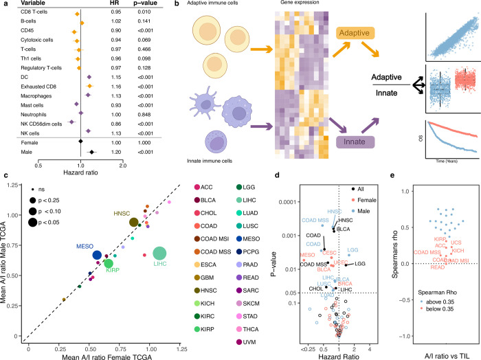
To investigate the relevance of the immune cell composition in the tumour microenvironment relative to disease progression, we performed immune cell decomposition of 8,024 tumours across 25 cancer types from the Cancer Genome Atlas (TCGA). We then fitted a multivariable Cox proportional hazard model to the progression free interval, including all immune cell types and gender, age and tumour stage as covariates (Fig 1A omitting age, stage and gender from the visualisation. Full results with all covariates listed in S2 Table). Of 14 immune cell types, 8 showed a significant association with outcome, four with improved survival, four with worse survival. Overall, we observed that adaptive immune cells associated with a lower risk of relapse (CD8 T-cells, CD45, T-cells, Th1 cells, Treg), while innate immune cells were associated with a higher risk of relapse (Dendritic cells, Macrophages, Natural killer cells) (Fig 1B). When we performed the same analysis including cancer types as covariates, the same overall pattern was observed with regard to the direction of association of the individual immune cell types, although unsurprisingly cancer type was by far the most significant covariates relative to outcome reflecting established cancer-type specific prognosis (S1A Fig). To further evaluate how the two compartments of the immune system associate with patient outcome in opposite directions, we stratified the cell types based on which of the two major immune components they belonged to and calculated a value for each component, adaptive and innate. We then performed a multivariate model including the adaptive and innate values together with age, stage and gender. This was done separately pan-cancer and with cancer type as covariates. We found that a high adaptive component was significantly associated with improved outcome (pan-cancer: HR = 0.016, P = 9.20x10⁻⁷, cancer informed: HR = 0.071, P = 0.0014,) whereas a high innate component was associated with poor outcome, although only significant in pan-cancer (pan-cancer: HR = 80.9, P = 1.94x10⁻⁶, cancer informed: HR = 1.042, P = 0.57), potentially indicating that the innate component may be of less relevance to outcome within cancer types. To further investigate the observation that high expression of adaptive immune genes and low expression of innate immune genes is associated with improved outcome, we calculated the ratio of Adaptive to Innate immune cells (A/I ratio, see methods, Fig 1B). We then compared the A/I ratio within cancer types, across the TCGA cancer cohort. While we observed a large variation in the A/I ratio, ranging from a mean of 0.25 in GBM to a median ratio of 1.0 in PRAD (S2 Fig). When we explored gender-specific differences, we observed largely similar ratio values between males and females (Fig 1C), with a few notable exceptions. The A/I ratio particularly shows large gender-specific variation in LIHC, where the median for female patients was more than 25% greater than in male patients, and in ACC and BLCA where the median for male patients was 15% and 14% greater than for female patients. To confirm that the expression of immune related genes was in fact originating from the TME and not from the cancer cells we explored the expression of the individual immune genes in the Cancer Cell Line Encyclopedia (CCLE) [19], a dataset of cancer cell lines (thus devoid of any infiltrating immune cells). Here we observed that the ranked expression of the immune genes was low for all cancer cell lines, except for cell lines originating from leukaemia and lymphoma, both cancers of the immune system. When we compared the ranked expression of cancer cell lines to TCGA tumour samples of matched tissue, we observed significantly higher ranks for all tumours (S3 Fig), indicating that the observed immune signal did indeed originate from infiltrating immune cells, and not from cancer cells expressing immune-related genes.

Fig 1. Calculating the adaptive/immune ratio.

Fig 1

a) A forest plot showing the hazard ratio for progression of cancer for the expression of each of the cell types in the TIL calculation. Yellow marks adaptive immune cells, purple marks the innate immune cells. b) A schematic showing how we calculate the A/I ratio. c) The mean A/I ratio for 29 cancer types in the TCGA cohort. The female mean ratio on the x-axis and male ratio is in the y axis. The size of the dot represents the p-value for the comparison of the mean. d) Univariate cox proportional hazard regression for the A/I ratio against progression free interval (PFI) across cancer types and genders for TCGA. e) Correlation of the A/I ratio and the TIL score across all cancer types in TCGA.

A higher ratio of adaptive to innate immune infiltration associates with improved prognosis

To investigate the impact of the A/I ratio on prognosis across cancer types, we performed univariate Cox regression against progression-free survival. As the A/I ratio varied across and within cancer types and also by gender, the hazard ratio (HR) was calculated within each cancer type for all patients and separately by gender. We observed that a higher A/I ratio significantly associated with improved outcome in 12 cancer types (BLCA, BRCA, CESC, CHOL, COAD, COAD MSS, HNSC, LIHC, LUAD, LUSC, MESO & UCEC), supporting the known role of the adaptive immune system in combating cancer [34]. Interestingly, for COAD, COAD MSS, HNSC, LIHC, LUAD and LUSC only males showed a significant association, while for MESO, only females. Lower A/I ratio only associated with improved outcome in male LGG patients (Fig 1D, S1B Fig). Across the cohort of cancer patients, we observed that the A/I ratio showed a good correlation to the total level of TIL infiltration (mean spearman rho = 0.38, Fig 1E). To investigate if the prognostic relevance of the A/I ratio was independent of the total TIL infiltration level, we performed multivariate Cox hazard regression, where we included the A/I ratio and TIL score, along with gender, age and tumour stage. We observed that the A/I ratio was highly significant and associated with improved outcome (A/I ratio: HR = 0.77, P < 2x10-16), while the total TIL score was associated with poorer outcome (TIL HR = 1.03, P = 0.0137). When we included cancer type as a covariate in the model, both terms were significantly associated with improved outcome (AI ratio: HR = 0.92, P = 0.000988, TIL: HR = 0.94, P = 0.000792), indicating that both the specific ratio of adaptive to innate immune cells and the total amount of immune cells are independently associated with outcome.

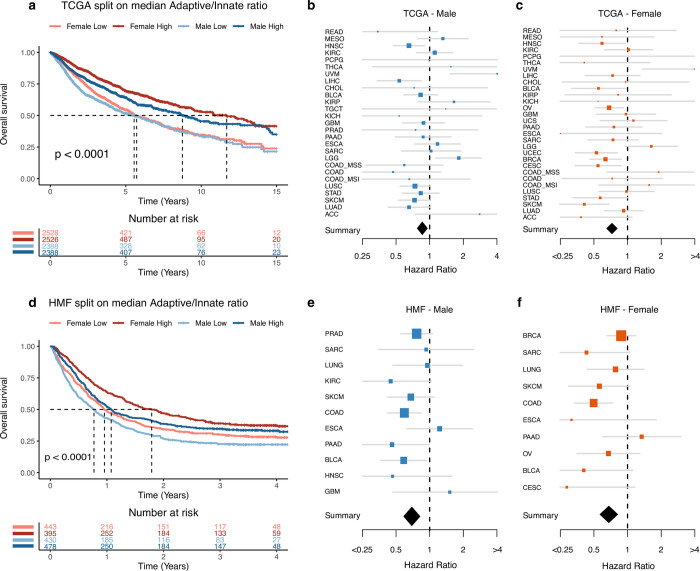
When we performed survival analysis on the combined TCGA cohort including all patients, we found that both female and male patients with an A/I ratio above median had significantly improved overall survival relative to patients with an A/I ratio below median (Fig 2A). We performed the same analysis on the individual cancer types, and found that 7/30 cancer types (BRCA, CESC, HNSC, LICH, OV, SKCM and UCEC) showed significantly improved outcome with an A/I ratio above median, while 2/30 (LGG and UVM) showed the opposite (S4 Fig). Based on these results, we performed a meta-analysis which take all cancer types into account, on male and female patients separately. Here, we observed that an A/I ratio above median associated with improved outcome in both male and female patients, but with a stronger association in females (HR females 0.73, HR males 0.86, P < 0.05, Fig 2B and 2C). To test if the observed gender difference in survival could be explained by differences in the immune systems between male and females, we compared the outcome between males with an A/I ratio above the female median A/I ratio, and females with an A/I ratio above median, for each cancer type. We used this data to do a survival analysis, and found that across cancer types, there were only two where survival was significantly improved in females (ESCA: P = 0.04, THCA: P = 0.011, S5 Fig). This suggests that some of the gender difference in outcome may be explained by the increased A/I ratio in females. To investigate if the differences in survival were solely based on gender, we performed two cox proportional hazard models, one analysing survival relative to gender, and one analysing survival relative to gender and the A/I ratio. Both models had age, stage and cancer type as covariates. We then compared the performance of the models using a likelihood ratio test. Based on this analysis, we found that the model including the A/I ratio term significantly out-performed the simpler model including only gender (P = 4.45x10⁻⁹).

Fig 2. Survival based on A/I ratio.

Fig 2

a) A Kaplan-Meier curve showing the 15-year overall survival for patients in the TCGA cohort, the patients are divided by gender and by median A/I ratio. b) A forest plot showing the univariate cox proportional hazard regression for the A/I ratio against overall survival, for the male patients in each of the TCGA cancer types. The diamond represents the hazard ratio from the meta-analysis across all cancer types. c) A forest plot showing the univariate cox proportional hazard regression for the A/I ratio against overall survival, for the female patients in each of the TCGA cancer types. The diamond represents the hazard ratio from the meta-analysis across all cancer types. d) A Kaplan-Meier curve showing the 4-year overall survival for metastatic cancer patients in the HMF cohort, the patients are divided by gender and by median A/I ratio. e) A forest plot showing the univariate cox proportional hazard regression for the A/I ratio against overall survival, for the male patients in each of the cancer types in the HMF cohort. The diamond represents the hazard ratio from the meta-analysis across all cancer types. f) A forest plot showing the univariate cox proportional hazard regression for the A/I ratio against overall survival, for the female patients in each of the cancer types in the HMF cohort. The diamond represents the hazard ratio from the meta-analysis across all cancer types.

We next asked if higher levels of innate immune infiltration may support cancer metastasis. To investigate this, we analysed the A/I ratio in 1,759 metastatic tumours from 22 different cancer types from the Hartwig Medical Foundation (HMF) dataset of metastatic cancers [20]. Initially, we performed a survival analysis on the metastatic HMF cohort and found that both male and female patients had improved overall survival if their A/I ratio was above median (Fig 2D). We performed the same analysis on the individual cancer types, and found that 2/11 cancer types (BLCA and COAD) showed significantly improved prognosis with an A/I ratio above median, while no cancer types showed the opposite (S6 Fig). When we performed a meta-analysis on male and female patients, respectively, we again found that an A/I ratio above median associated with improved outcome in both male and female patients (HR females 0.68, HR males 0.69, P < 0.05, Fig 2E and 2F).

To further investigate the observed gender differences, we specifically investigated outcome by gender. Within both cohorts we observed a gender difference in survival; female patients had significantly improved overall survival in both TCGA (P < 0.0001, S7A Fig) and HMF (P < 0.0017, S7B Fig).

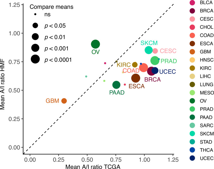
Next, we compared the A/I level between cancer types in HMF and TCGA. Of the 21 comparable cancer types, 9 had a significantly higher A/I ratio in TCGA (BRCA, CESC, COAD, ESCA, KIRC, PAAD, PRAD, SKCM and UCEC) while 2 had a significantly higher A/I ratio in HMF (GBM and OV) (Fig 3). The increased level in GBM may be related to the brain’s immune privileged status, preventing immune cells from infiltrating, potentially suggesting that a high A/I ratio may be one of the factors that contribute to a lower frequency of patients progressing to metastatic disease.

Fig 3. Comparing the A/I ratio between primary and metastatic tumours.

Fig 3

The mean A/I ratio for 20 overlapping cancer types in the TCGA and HMF cohorts. The mean ratio of primary tumours from TCGA on the x-axis and mean ratio of metastatic tumours from the HMF cohort on the y-axis.

Taken together, we have found that in both the TCGA and the HMF cohorts, which includes tumours from patients with different cancer types treated with different protocols, patients with a higher A/I ratio showed significantly improved outcomes.

A higher ratio of adaptive to innate immune infiltration associates with improved response to immunotherapy

The immune system plays a significant role in controlling cancer growth, and during anticancer treatment the adaptive immune system can be activated by the application of immunotherapy. To investigate how the balance of the adaptive to the innate immune response affects response to immunotherapy response, we determined the A/I ratio in 412 metastatic tumours treated with checkpoint inhibitor immunotherapy (CPI). These included 177 tumours from 6 cancer types in the HMF dataset [20], and 235 metastatic bladder cancer tumours from the Mariathasan dataset, treated with anti-PDL1 (atezolizumab) [21]. To determine if the previously observed gender difference in cancer prognosis also affects survival within the two cohorts of CPI treated patients, we performed a survival analysis on gender. For both cohorts we found no significant difference in survival of male and female patients treated by CPI (Mariathasan: P = 0.18, S7C Fig, HMF BLCA: P = 0.081, S7D Fig, HMF LUNG: P = 0.17, S7E Fig, HMF SKCM: P = 0.72, S7F Fig), indicating that drug-induced activation of the adaptive immune response may out-weigh any gender-specific differences in the immune response. To rule out any potential sampling biases, we investigated the percentages of patients from each cancertype that received immunotherapy, and found that in the HMF cohort, an equal percentage of male and female patients have received CPI for each of the cancer types (BLCA total: 78% male, CPI: 81% male. LUNG total: 45% male, CPI: 49% male. SKCM total: 60% male, CPI: 59% male), and the percentage of responders within each gender is similar within the HMF cohort. And between them there is an approximately equal distribution of each response category. The same is true for the metastatic bladder cancer cohort where we also find an equal distribution between males and females in the response groups. Taken together this indicates that these results are not affected by gender sampling bias.

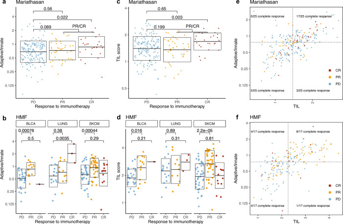
To investigate if a higher A/I ratio associates with improved response to immunotherapy, we compared the A/I ratio to response across both cohorts. In the Mariathasan bladder cancer cohort, patients with complete response (CR) to immunotherapy had a higher A/I ratio than partial responders (PR) and a significantly higher A/I ratio than patients with progressive disease (PD) (P = 0.022, Fig 4A). When each response group is split on gender, we only observe a difference in the A/I ratio in the male CR patients (P = 0.047, S8A Fig). However, this group is represented by only four patients, and should therefore be interpreted with caution. No difference in response rate was observed based on gender. In the HMF dataset including multiple cancer types, both the patients with CR and PR have a significantly higher ratio than the patients with PD (CR: P = 0.036, PR: P = 2.6x10⁻⁶, CR+PR: 2.6x10⁻⁶). There was no significant difference in the A/I ratio between CR and PR patients (Fig 4B). There were no significant differences on the A/I ratio within the response groups, when divided by gender (S8B Fig). When we restricted the HMF cohort to BLCA patients, for comparability to the Mariathasan dataset, we observed only one complete responder, and a significantly higher A/I ratio in PR patients compared to patients with PD (P = 0.00076). For lung cancer we observe a significantly higher ratio for CR compared to PR (P = 0.0385) and PD (P = 0.0035), and for Melanoma we observe no difference between CR and PR, but a significantly higher ratio in PR compared to PD (0.00044) (S8C Fig). We observed no significant gender differences within the cancer types and response groups following CPI therapy (alternative S8D Fig), indicating that gender differences in pre-treatment A/I ratio may be of limited relevance to response to immunotherapy.

Fig 4. Response to immunotherapy.

Fig 4

a) A/I ratio vs response to immunotherapy categories for the metastatic bladder cancer patients in the Mariathasan cohort. b) A/I ratio vs response to immunotherapy categories for the CPI treated metastatic cancer patients from the HMF cohort, for each of the three major cancer types. c) TIL score vs response to immunotherapy categories for the metastatic bladder cancer patients in the Mariathasan cohort. d) TIL score vs response to immunotherapy categories for the CPI treated metastatic cancer patients from the HMF cohort, for each of the three major cancer types. e) A/I ratio vs TIL score for metastatic bladder cancer patients in the Mariathasan cohort, colored by response category. 17/25 (68%) of patients with CR had both an A/I ratio and a TIL score above median (P = 0.0006). f) A/I ratio vs TIL score for the CPI treated metastatic cancer patients from the HMF cohort, colored by response category. 51/84 (61%) of all responders (CR and PR) there both have a high A/I ratio and a high TIL score (P = 1.4x10^-7).

A/I ratio associates directly with immune infiltration and predicts response to checkpoint immunotherapy

A high A/I ratio does not in itself represent a high level of immune infiltration but may be driven by a large adaptive component of a relatively small level of infiltrating immune cells. To investigate whether the A/I ratio is indeed reflective of the immune activity that takes place within the tumour, we compared it to tumour immune phenotypes scored by immunohistochemistry, defined as inflamed, excluded and desert depending on the degree of immune infiltration [21]. We found that for both male and female patients the inflamed patients have a significantly higher A/I ratio (male: P = 1.3×10⁻⁸, female: P = 0.018) and for the male patients the inflamed cohort also had a significantly higher A/I ratio than the excluded cohort (P = 0.03) (S9A Fig). This demonstrates that the A/I ratio associates directly with higher levels of immune cell infiltration, indicating that the main component of increasing TILs is associated with adaptive immune cells. We also observed that the overall level of TIL infiltration showed a significant association with outcome (S9B Fig). On a cancer specific level, five cancer types showed improved survival with a TIL score above median (S10 Fig). Next, we investigated if the combination of a high A/I ratio and a high TIL score may show improved association with immunotherapy response. We compared the TIL score of each of the response categories and found that like the A/I score, a high TIL score was associated with response in both cohorts (Mariathasan, CR: P = 0.003, Fig 4C. HMF, PR+CR: P = 4.8x10⁻⁶, Fig 4D). We defined high and low A/I ratio and high and low TIL infiltration based on the median value. In the Mariathasan cohort, 17/25 (68%) of patients with a complete response to immunotherapy had both an A/I ratio and a TIL score above median (P = 0.0006, Fig 4E). In the HMF CPI treated cohort 51/84 (61%) of patients with a response to immunotherapy (CR+PR) both have an A/I ratio and TIL score above median (P = 1.4x10⁻⁷, Fig 4F). When HMF was divided by cancer type, we observed the same tendency, there was a significantly higher number of responders with an A/I ratio and TIL score above median (BLCA: P = 0.003, LUNG: P = 0.15, SKCM: P = 0.006, S9C–S9E Fig).

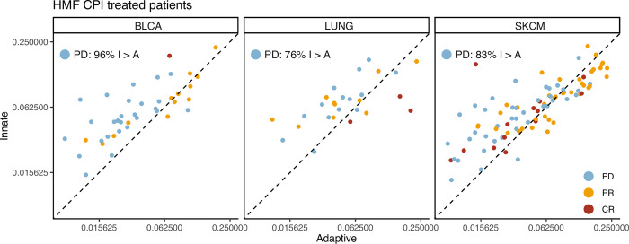
To further elucidate the relationship between expression of the adaptive and innate immune system in the tumour microenvironment of the CPI treated patients with progressive disease, we investigated the adaptive vs. the innate expression scores directly. Here, we observed that for both the HMF cohort and the Mariathasan bladder cancer, a very large proportion of patients with progressive disease showed higher innate relative to adaptive scores (BLCA: 96%, LUNG: 76%, SKCM: 83%, Fig 5. Mariathasan BLCA: 79%, S11 Fig). This supports the use of a ratio as an efficient tool to identify patients with relatively increased adaptive-to-innate expression, and potentially improved therapy response.

Fig 5. Adaptive vs Innate expression in CPI treated patients.

Fig 5

Scatterplots showing the adaptive immune expression vs. the innate immune expression in each patient for the CPI treated HMF cohort. The points are coloured by their response to immunotherapy and the plot is stratified by cancer types.

Discussion

Our findings show that large infiltration of adaptive immune cells relative to innate immune cells leads to a better prognosis for primary cancer and to improved immunotherapy response. This is consistent with literature demonstrating an immune regulatory role of innate immune cells [35]. It has previously been reported that macrophages expressing CD163 in the tumour microenvironment can lead to a poor prognosis for cancer patients [3638]. In our work, CD163 is one of the genes used to define macrophages, an element of the innate immune system. CD163+ macrophages have also recently been found to play a role in maintaining suppression in anti-PDL1 resistant melanoma in an experimental setting [39]. This supports our findings that 83% of patients with metastatic melanoma with a poor response to CPI have a lower A/I ratio, indicating that they have a relatively high expression of the innate immune system (Fig 5).

Consistent with literature we observe improved female outcomes relative to male [40, 41]. Interestingly this difference is no longer significant when we focus on patients treated by immunotherapy. While we recognise the CPI treated cohorts may be underpowered, we observed no trend supporting improved female-to-male response in either cohort. Within the HMF cohort, the percentage of responders within each gender is similar and an equal percentage of males and females have received immunotherapy. The same is true for the metastatic bladder cancer cohort where we also find an equal distribution between males and females in the response groups. To test if the gender difference could be explained by the difference in the immune system, we compared the outcome between men with an A/I ratio above the female median A/I ratio, and females with an A/I ratio above median, for each cancer type and found that across cancer types, there was no longer a significant difference in survival. This indicates that basic differences in the balance between adaptive and innate immune activation between genders may be a significant driver of differences in response rates and cancer outcome between men and women.

Currently the standard for stratifying metastatic patients into immunotherapy is to use PDL1 expression. However, some patients with lower levels of PDL1 expression still benefit from the treatment. TMB has been proposed as a predictor for response, and has been used in clinical trials, and while not all tumours with high TMB show response to immunotherapy, an association between TMB and immunotherapy response has been established [42]. TMB is a proxy measure of cancer neoantigens, which is a target of the immune system [43]. This makes neoantigens an obvious candidate for response biomarkers to immunotherapy. However, for the immune system to target cancer cells through neoantigens, T-cells must be present in the environment. We believe the A/I ratio may be relevant to more accurately identify patients who are likely to benefit from CPI therapy. As the results in this study are based on publicly available pan-cancer data from heterogeneous cohorts, independent experimental validation would be necessary to further validate using a ratio of adaptive to innate immune cells for patient stratification.

To ensure that the immune gene expression signal that defines the A/I ratio does indeed originate from the immune cells in the TME and not from the cancer cells themselves, we compared the ranked gene expression from the tumour samples from TCGA, which contain both cancer cells and cells from the surrounding tissue, to the cancer cell line samples from the CCLE project [19], that only contain cancer cells. We found that the expression of immune genes was consistently much lower in the cancer cell lines, median in the bottom 25%, relative to the tumour samples, median in the top 50%. Based on these results, we are confident that the immune signal we observed in our analysis originates from the infiltrating immune cells found within the TME.

Other studies have used a similar approach to identify elements from the immune system, which could be used to make a prediction model for cancer outcome. In a study from 2019, 11 immune regulating genes were used to calculate a risk score. With this, the authors were able to predict 5 year survival with an AUC value of 0.634 for cervical cancer [44]. While the expression of the genes included in their analysis did not correlate to the expression of the genes used in our analysis, it shows that immune related genes carry prognostic information. It has also been reported that interferon-γ, and interleukin-6 and -10 in advanced melanoma were significantly higher in the patients with response to nivolumab treatment [45]. Immune cells have also been reported to function as predictors for response; NK cells, CD4+ and CD8+ [46]. A study reports that the baseline circulating myeloid-derived suppressor cells (MDSC) correlate with outcome of ipilimumab treatment [47], they find that a low frequency correlates to improved outcome. Another study reported that a subsection of these cells, Polymorphonuclear myeloid-derived suppressor cells, is associated with bad prognosis and resistance to immune checkpoint inhibitor therapy in patients with metastatic melanoma [48]. These studies support that the immune system is the place to look if one wants to find possible predictors for response to immunotherapy.

While we have found a highly significant association between the A/I ratio and patient outcome, both in pan-cancer analysis and in a meta-analysis across cancer types, the A/I ratio was not found to be significantly associated for all cancer types individually. A part of this may be explained by limited power in some cancer types, where there are not enough patients within each group to reach statistical significance, despite a supporting trend. However, undoubtedly cancer-type specific differences also play a role. It is all but certain that the A/I ratio, despite being a systemic marker rather than a cancer-specific marker, is not prognostically relevant for all cancer types and all therapy types. However, to answer this question more accurately, further studies on larger cohorts are required.

With our study we present a more complex interaction between different compartments of the immune system and cancer cells. This presents a case for a more holistic approach to cancer biomarker development, where not only individual molecules, cell types or cancer genomic aberrations are measured and correlated to outcome, but where also the patient’s ability to mount an effective immune response to cancer is considered. A key marker here may be the relative infiltration of adaptive to innate immune cells, that may inform on whether the body recognises and is capable of fighting off cancer cells, given appropriate therapy. An advantage of our ratio-based approach is that with a ratio the values are normalised for each individual independently. Thus, while the total activation of the immune system in the tumour microenvironment of individual patients may vary greatly, the ratio may still be informative on an individual level and remain comparable across a cohort of patients.

Overall, our study supports a model where a strong activation of the adaptive immune response relative to the innate immune response in the tumour microenvironment is beneficial to patient outcome. Furthermore, our study provides a potential link between increased cancer-associated mortality among males and a relatively lower ratio of adaptive-to-innate immune cells in the tumour microenvironment. This is consistent with literature indicating gender-specific immune differences, and suggests that biomarker development and drug response predictions must consider gender both in the design and implementation phase. While we found that gender differences were reduced or eliminated when CPI were administered, this must be confirmed in further studies.

Supporting information

S1 Fig. Survival across cancer types in TCGA.

a) A forest plot showing the hazard ratio from a multivariate cox proportional hazard regression for progression of cancer for the expression of each of the cell types in the TIL calculation, gender, age, stage and cancer types as covariates. b) A forest plot showing the hazard ratio from a univariate cox proportional hazard regression for progression of cancer. A univariate model was done for each cancer type and for both genders within the cancer type individually.

(PDF)

S2 Fig. A/I ratio across TCGA cancer types.

The A/I ratio for 29 cancer types in the TCGA cohort. The cancertypes are ordered by median A/I ratio, female and male patients are represented by red and blue dots respectively, the median for each gender in each cancer type is represented by a horizontal line.

(PDF)

S3 Fig. Ranked immune gene expression.

The mean ranked expression per cancer type of the 67 immune related genes that the celltype scores are calculated from, for both the cancer cell line data from the CCLE project, and from the tumour samples in TCGA. Low rank = low expression.

(PDF)

S4 Fig. Survival vs. A/I ratio for TCGA cancer types.

Kaplan-Meier curves showing the 15-year survival for each cancer type within the TCGA cohort, the patients are stratified by gender and A/I ratio.

(PDF)

S5 Fig. Survival for TCGA cancer types.

Kaplan-Meier curves showing the 10-year survival for TCGA patients with an A/I score above the female median, Male vs. Females. A p-value for the difference in survival is available for each cancer type.

(PDF)

S6 Fig. Survival vs. A/I ratio for HMF cancer types.

Kaplan-Meier curves showing the five-year survival for each cancer type within the HMF cohort, the patients are stratified by gender and A/I ratio.

(PDF)

S7 Fig. Survival stratified on gender.

a) Kaplan-Meier curve of 10,158 patients from the TCGA dataset. b) Kaplan-Meier curve of 1746 patients from the HMF dataset. c) Kaplan-Meier curve of 348 patients from the Mariathasan bladder cancer dataset, all CPI treated. d) Kaplan-Meier curve of CPI treated patients from the HMF BLCA dataset. e) Kaplan-Meier curve of CPI treated patients from the HMF LUNG dataset. f) Kaplan-Meier curve of CPI treated patients from the HMF SKCM dataset.

(PDF)

S8 Fig. Response stratified on gender.

a) Gender stratified response to immunotherapy for the Mariathasan dataset. P-value for difference between gender for each category. b) Gender stratified response to immunotherapy for the HMF dataset, separated by cancer type. P-value for difference between gender for each category.

(PDF)

S9 Fig

a) The A/I ratio vs. the immune phenotype for the Mariathasan dataset, separated by gender. P-values are for comparisons between inflamed vs. desert and inflamed vs. excluded. b) Kaplan-Meier curve for survival of TCGA patients with a high or low TIL score. c-e) A/I ratio vs TIL score for the CPI treated patients separated by cancer type, coloured by response category. In the respective cancer types (BLCA: P = 0.003, LUNG: P = 0.15, SKCM: P = 0.006).

(PDF)

S10 Fig. Survival vs. TIL score for TCGA cancer types.

Kaplan-Meier curves showing the 15-year survival for each cancer type within the TCGA cohort, the patients are stratified by gender and TIL score.

(PDF)

S11 Fig. Adaptive vs Innate expression in CPI treated patients.

Scatterplots showing the adaptive immune expression vs. the innate immune expression in each patient for the Mariathasan cohort. The points are coloured by their response to immunotherapy.

(PDF)

S1 Table. Genes used to define the cell types.

A list of the 67 genes used in the gene expression analysis to define the expression of the cell types used for further analyses [24].

(TXT)

S2 Table. Result of multivariable Cox proportional hazard model.

Full table of results from the multivariable Cox proportional hazard model from Fig 1A with all covariates.

(TXT)

Acknowledgments

The results shown here are in part based upon data generated by the TCGA Research Network: https://www.cancer.gov/tcga. This publication and the underlying research are partly facilitated by Hartwig Medical Foundation and the Center for Personalized Cancer Treatment (CPCT) which have generated, analysed and made available data for this research.

Data Availability

Data are available from the Hartwig Medical Foundation and an application to access the data can be sent here https://www.hartwigmedicalfoundation.nl/en/data/data-acces-request/ for researchers who meet the criteria for access to confidential data. The TCGA data can be accessed here: https://www.cancer.gov/about-nci/organization/ccg/research/structural-genomics/tcga The dataset we used is the TCGA project.

Funding Statement

NJB is a fellow of the Lundbeck Foundation (R272-2017-4040), and acknowledges funding from Aarhus University Research Foundation (AUFF-E-2018-7-14), and the Novo Nordisk Foundation (NNF21OC0071483). The funders had no role in study design, data collection and analysis, decision to publish, or preparation of the manuscript.

References

  • 1.Turajlic S. and Swanton C., “Metastasis as an evolutionary process,” Science, vol. 352, no. 6282, pp. 169–175, Apr. 2016. doi: 10.1126/science.aaf2784 [DOI] [PubMed] [Google Scholar]
  • 2.Hanahan D. and Weinberg R. A., “Hallmarks of cancer: the next generation,” Cell, vol. 144, no. 5, pp. 646–674, Mar. 2011. doi: 10.1016/j.cell.2011.02.013 [DOI] [PubMed] [Google Scholar]
  • 3.Jiménez-Sánchez A. et al. , “Heterogeneous Tumor-Immune Microenvironments among Differentially Growing Metastases in an Ovarian Cancer Patient,” Cell, vol. 170, no. 5, pp. 927–938.e20, Aug. 2017. doi: 10.1016/j.cell.2017.07.025 [DOI] [PMC free article] [PubMed] [Google Scholar]
  • 4.De Mattos-Arruda L. et al. , “The Genomic and Immune Landscapes of Lethal Metastatic Breast Cancer,” Cell Rep., vol. 27, no. 9, pp. 2690–2708.e10, May 2019. doi: 10.1016/j.celrep.2019.04.098 [DOI] [PMC free article] [PubMed] [Google Scholar]
  • 5.Angelova M. et al. , “Evolution of Metastases in Space and Time under Immune Selection,” Cell, vol. 175, no. 3, pp. 751–765.e16, Oct. 2018. doi: 10.1016/j.cell.2018.09.018 [DOI] [PubMed] [Google Scholar]
  • 6.Larkin J. et al. , “Five-Year Survival with Combined Nivolumab and Ipilimumab in Advanced Melanoma,” N. Engl. J. Med., vol. 381, no. 16, pp. 1535–1546, Oct. 2019. doi: 10.1056/NEJMoa1910836 [DOI] [PubMed] [Google Scholar]
  • 7.Wang S., He Z., Wang X., Li H., and Liu X.-S., “Antigen presentation and tumor immunogenicity in cancer immunotherapy response prediction,” Elife, vol. 8, Nov. 2019, doi: 10.7554/eLife.49020 [DOI] [PMC free article] [PubMed] [Google Scholar]
  • 8.McAllister S. S. and Weinberg R. A., “The tumour-induced systemic environment as a critical regulator of cancer progression and metastasis,” Nat. Cell Biol., vol. 16, no. 8, pp. 717–727, Aug. 2014. doi: 10.1038/ncb3015 [DOI] [PMC free article] [PubMed] [Google Scholar]
  • 9.Singh N., Baby D., Rajguru J. P., Patil P. B., Thakkannavar S. S., and Pujari V. B., “Inflammation and cancer,” Ann. Afr. Med., vol. 18, no. 3, pp. 121–126, Jul. 2019. doi: 10.4103/aam.aam_56_18 [DOI] [PMC free article] [PubMed] [Google Scholar]
  • 10.Gentles A. J. et al. , “The prognostic landscape of genes and infiltrating immune cells across human cancers,” Nat. Med., vol. 21, no. 8, pp. 938–945, Aug. 2015. doi: 10.1038/nm.3909 [DOI] [PMC free article] [PubMed] [Google Scholar]
  • 11.Turvey S. E. and Broide D. H., “Innate immunity,” J. Allergy Clin. Immunol., vol. 125, no. 2 Suppl 2, pp. S24–32, Feb. 2010. doi: 10.1016/j.jaci.2009.07.016 [DOI] [PMC free article] [PubMed] [Google Scholar]
  • 12.Dempsey P. W., Vaidya S. A., and Cheng G., “The art of war: Innate and adaptive immune responses,” Cell. Mol. Life Sci., vol. 60, no. 12, pp. 2604–2621, Dec. 2003. doi: 10.1007/s00018-003-3180-y [DOI] [PMC free article] [PubMed] [Google Scholar]
  • 13.Jaillon S., Berthenet K., and Garlanda C., “Sexual Dimorphism in Innate Immunity,” Clin. Rev. Allergy Immunol., vol. 56, no. 3, pp. 308–321, Jun. 2019. doi: 10.1007/s12016-017-8648-x [DOI] [PubMed] [Google Scholar]
  • 14.Klein S. L. and Flanagan K. L., “Sex differences in immune responses,” Nat. Rev. Immunol., vol. 16, no. 10, pp. 626–638, Oct. 2016. doi: 10.1038/nri.2016.90 [DOI] [PubMed] [Google Scholar]
  • 15.Edgren G., Liang L., Adami H.-O., and Chang E. T., “Enigmatic sex disparities in cancer incidence,” Eur. J. Epidemiol., vol. 27, no. 3, pp. 187–196, Mar. 2012. doi: 10.1007/s10654-011-9647-5 [DOI] [PubMed] [Google Scholar]
  • 16.Ellrott K. et al. , “Scalable Open Science Approach for Mutation Calling of Tumor Exomes Using Multiple Genomic Pipelines,” Cell Syst, vol. 6, no. 3, pp. 271–281.e7, Mar. 2018. doi: 10.1016/j.cels.2018.03.002 [DOI] [PMC free article] [PubMed] [Google Scholar]
  • 17.Goldman M. J. et al. , “Visualizing and interpreting cancer genomics data via the Xena platform,” Nat. Biotechnol., vol. 38, no. 6, pp. 675–678, Jun. 2020. doi: 10.1038/s41587-020-0546-8 [DOI] [PMC free article] [PubMed] [Google Scholar]
  • 18.Cortes-Ciriano I., Lee S., Park W.-Y., Kim T.-M., and Park P. J., “A molecular portrait of microsatellite instability across multiple cancers,” Nat. Commun., vol. 8, p. 15180, Jun. 2017. doi: 10.1038/ncomms15180 [DOI] [PMC free article] [PubMed] [Google Scholar]
  • 19.DepMap, “DepMap 22Q2 Public. figshare. Dataset,” 2022, doi: 10.6084/m9.figshare.19700056.v2 [DOI] [Google Scholar]
  • 20.Priestley P. et al. , “Pan-cancer whole-genome analyses of metastatic solid tumours,” Nature, vol. 575, no. 7781, pp. 210–216, Nov. 2019. doi: 10.1038/s41586-019-1689-y [DOI] [PMC free article] [PubMed] [Google Scholar]
  • 21.Mariathasan S. et al. , “TGFβ attenuates tumour response to PD-L1 blockade by contributing to exclusion of T cells,” Nature, vol. 554, no. 7693, pp. 544–548, Feb. 2018. [DOI] [PMC free article] [PubMed] [Google Scholar]
  • 22.Dobin A. et al. , “STAR: ultrafast universal RNA-seq aligner,” Bioinformatics, vol. 29, no. 1, pp. 15–21, Jan. 2013. doi: 10.1093/bioinformatics/bts635 [DOI] [PMC free article] [PubMed] [Google Scholar]
  • 23.Bray N. L., Pimentel H., Melsted P., and Pachter L., “Near-optimal probabilistic RNA-seq quantification,” Nat. Biotechnol., vol. 34, no. 5, pp. 525–527, May 2016. doi: 10.1038/nbt.3519 [DOI] [PubMed] [Google Scholar]
  • 24.Danaher P. et al. , “Gene expression markers of Tumor Infiltrating Leukocytes,” J Immunother Cancer, vol. 5, p. 18, Feb. 2017. doi: 10.1186/s40425-017-0215-8 [DOI] [PMC free article] [PubMed] [Google Scholar]
  • 25.Rosenthal R. et al. , “Neoantigen-directed immune escape in lung cancer evolution,” Nature, vol. 567, no. 7749, pp. 479–485, Mar. 2019. doi: 10.1038/s41586-019-1032-7 [DOI] [PMC free article] [PubMed] [Google Scholar]
  • 26.R Core Team, “R: A Language and Environment for Statistical Computing.” R Foundation for Statistical Computing, Vienna, Austria, 2020. [Online]. Available: https://www.R-project.org/ [Google Scholar]
  • 27.Wickham H. et al. , “Welcome to the tidyverse,” J. Open Source Softw., vol. 4, no. 43, p. 1686, Nov. 2019. [Google Scholar]
  • 28.Kassambara A., Kosinski M., and Biecek P., “survminer: Drawing Survival Curves using ‘ggplot2.’” 2021. [Online]. Available: https://CRAN.R-project.org/package=survminer [Google Scholar]
  • 29.Therneau T. M. and Grambsch P. M., Modeling Survival Data: Extending the Cox Model. Springer Science & Business Media, 2000. [Google Scholar]
  • 30.Hadley W. and Seidel D., “scales: Scale functions for visualization.” GitHub; San Francisco:, 2019. [Online]. Available: https://CRAN.R-project.org/package=scales [Google Scholar]
  • 31.Clarke and Sherrill-Mix, “Ggbeeswarm: Categorical scatter (violin point) plots,” R package version 0.6. 0.
  • 32.Gerds T. A. and Ozenne B., “Publish: Format Output of Various Routines in a Suitable Way for Reports and Publication.” 2021. [Online]. Available: https://CRAN.R-project.org/package=Publish [Google Scholar]
  • 33.Cox D. R., “Regression models and life-tables,” J. R. Stat. Soc., vol. 34, no. 2, pp. 187–202, Jan. 1972. [Google Scholar]
  • 34.Spurrell E. L. and Lockley M., “Adaptive immunity in cancer immunology and therapeutics,” Ecancermedicalscience, vol. 8, p. 441, Jul. 2014. doi: 10.3332/ecancer.2014.441 [DOI] [PMC free article] [PubMed] [Google Scholar]
  • 35.Iwasaki A. and Medzhitov R., “Control of adaptive immunity by the innate immune system,” Nat. Immunol., vol. 16, no. 4, pp. 343–353, Apr. 2015. doi: 10.1038/ni.3123 [DOI] [PMC free article] [PubMed] [Google Scholar]
  • 36.Jung K. Y. et al. , “Cancers with Higher Density of Tumor-Associated Macrophages Were Associated with Poor Survival Rates,” J Pathol Transl Med, vol. 49, no. 4, pp. 318–324, Jul. 2015. doi: 10.4132/jptm.2015.06.01 [DOI] [PMC free article] [PubMed] [Google Scholar]
  • 37.Komohara Y., Jinushi M., and Takeya M., “Clinical significance of macrophage heterogeneity in human malignant tumors,” Cancer Sci., vol. 105, no. 1, pp. 1–8, Jan. 2014. doi: 10.1111/cas.12314 [DOI] [PMC free article] [PubMed] [Google Scholar]
  • 38.Park J.-Y. et al. , “Polarized CD163+ tumor-associated macrophages are associated with increased angiogenesis and CXCL12 expression in gastric cancer,” Clin. Res. Hepatol. Gastroenterol., vol. 40, no. 3, pp. 357–365, Jun. 2016. doi: 10.1016/j.clinre.2015.09.005 [DOI] [PubMed] [Google Scholar]
  • 39.Etzerodt A. et al. , “Tissue-resident macrophages in omentum promote metastatic spread of ovarian cancer,” J. Exp. Med., vol. 217, no. 4, Apr. 2020, doi: 10.1084/jem.20191869 [DOI] [PMC free article] [PubMed] [Google Scholar]
  • 40.Joosse A. et al. , “Sex is an independent prognostic indicator for survival and relapse/progression-free survival in metastasized stage III to IV melanoma: a pooled analysis of five European organisation for research and treatment of cancer randomized controlled trials,” J. Clin. Oncol., vol. 31, no. 18, pp. 2337–2346, Jun. 2013. doi: 10.1200/JCO.2012.44.5031 [DOI] [PubMed] [Google Scholar]
  • 41.Dunford A. et al. , “Tumor-suppressor genes that escape from X-inactivation contribute to cancer sex bias,” Nat. Genet., vol. 49, no. 1, pp. 10–16, Jan. 2017. doi: 10.1038/ng.3726 [DOI] [PMC free article] [PubMed] [Google Scholar]
  • 42.Chan T. A. et al. , “Development of tumor mutation burden as an immunotherapy biomarker: utility for the oncology clinic,” Ann. Oncol., vol. 30, no. 1, pp. 44–56, Jan. 2019. doi: 10.1093/annonc/mdy495 [DOI] [PMC free article] [PubMed] [Google Scholar]
  • 43.McGranahan N. et al. , “Clonal neoantigens elicit T cell immunoreactivity and sensitivity to immune checkpoint blockade,” Science, vol. 351, no. 6280, pp. 1463–1469, Mar. 2016. doi: 10.1126/science.aaf1490 [DOI] [PMC free article] [PubMed] [Google Scholar]
  • 44.Yang S. et al. , “Identification of a prognostic immune signature for cervical cancer to predict survival and response to immune checkpoint inhibitors,” Oncoimmunology, vol. 8, no. 12, p. e1659094, Oct. 2019. doi: 10.1080/2162402X.2019.1659094 [DOI] [PMC free article] [PubMed] [Google Scholar]
  • 45.Yamazaki N. et al. , “Cytokine biomarkers to predict antitumor responses to nivolumab suggested in a phase 2 study for advanced melanoma,” Cancer Sci., vol. 108, no. 5, pp. 1022–1031, May 2017. doi: 10.1111/cas.13226 [DOI] [PMC free article] [PubMed] [Google Scholar]
  • 46.Larkin J. et al. , “Combined Nivolumab and Ipilimumab or Monotherapy in Untreated Melanoma,” N. Engl. J. Med., vol. 373, no. 1, pp. 23–34, Jul. 2015. doi: 10.1056/NEJMoa1504030 [DOI] [PMC free article] [PubMed] [Google Scholar]
  • 47.Meyer C. et al. , “Frequencies of circulating MDSC correlate with clinical outcome of melanoma patients treated with ipilimumab,” Cancer Immunol. Immunother., vol. 63, no. 3, pp. 247–257, Mar. 2014. doi: 10.1007/s00262-013-1508-5 [DOI] [PMC free article] [PubMed] [Google Scholar]
  • 48.Gondois-Rey F. et al. , “Identification of an Immature Subset of PMN-MDSC Correlated to Response to Checkpoint Inhibitor Therapy in Patients with Metastatic Melanoma,” Cancers, vol. 13, no. 6, Mar. 2021, doi: 10.3390/cancers13061362 [DOI] [PMC free article] [PubMed] [Google Scholar]

Decision Letter 0

Albert Rübben

Transfer Alert

This paper was transferred from another journal. As a result, its full editorial history (including decision letters, peer reviews and author responses) may not be present.

23 Jun 2022

PONE-D-22-08886The ratio of adaptive to innate immune cells differs between genders and associates with improved prognosis and response to immunotherapyPLOS ONE

Dear Dr. Ahrenfeldt,

Thank you for submitting your manuscript to PLOS ONE. After careful consideration, we feel that it has merit but does not fully meet PLOS ONE’s publication criteria as it currently stands. Therefore, we invite you to submit a revised version of the manuscript that addresses the points raised during the review process.We have based the decision on just one external reviewer. The methodology of the study might only be evaluated by very few scientist working on exactly the same field which has limited the choice of potential reviewers and which has led a significant delay in processing the manuscript. I apologize for it.

Please submit your revised manuscript by Aug 07 2022 11:59PM. If you will need more time than this to complete your revisions, please reply to this message or contact the journal office at plosone@plos.org. When you're ready to submit your revision, log on to https://www.editorialmanager.com/pone/ and select the 'Submissions Needing Revision' folder to locate your manuscript file.

Please include the following items when submitting your revised manuscript:

  • A rebuttal letter that responds to each point raised by the academic editor and reviewer(s). You should upload this letter as a separate file labeled 'Response to Reviewers'.

  • A marked-up copy of your manuscript that highlights changes made to the original version. You should upload this as a separate file labeled 'Revised Manuscript with Track Changes'.

  • An unmarked version of your revised paper without tracked changes. You should upload this as a separate file labeled 'Manuscript'.

If you would like to make changes to your financial disclosure, please include your updated statement in your cover letter. Guidelines for resubmitting your figure files are available below the reviewer comments at the end of this letter.

If applicable, we recommend that you deposit your laboratory protocols in protocols.io to enhance the reproducibility of your results. Protocols.io assigns your protocol its own identifier (DOI) so that it can be cited independently in the future. For instructions see: https://journals.plos.org/plosone/s/submission-guidelines#loc-laboratory-protocols. Additionally, PLOS ONE offers an option for publishing peer-reviewed Lab Protocol articles, which describe protocols hosted on protocols.io. Read more information on sharing protocols at https://plos.org/protocols?utm_medium=editorial-email&utm_source=authorletters&utm_campaign=protocols.

We look forward to receiving your revised manuscript.

Kind regards,

Albert Rübben, Ass. Prof., M.D., Ph.D.

Academic Editor

PLOS ONE

Journal Requirements:

When submitting your revision, we need you to address these additional requirements.

1. Please ensure that your manuscript meets PLOS ONE's style requirements, including those for file naming. The PLOS ONE style templates can be found at

https://journals.plos.org/plosone/s/file?id=wjVg/PLOSOne_formatting_sample_main_body.pdf  and

https://journals.plos.org/plosone/s/file?id=ba62/PLOSOne_formatting_sample_title_authors_affiliations.pdf

2. In your ethics statement in the manuscript and in the online submission form, please provide additional information about the patient records used in your retrospective study. Specifically, please ensure that you have discussed whether all data were fully anonymized before you accessed them.

3. Thank you for stating the following in the Acknowledgments Section of your manuscript:

“The results shown here are in part based upon data generated by the TCGA Research Network: https://www.cancer.gov/tcga. This publication and the underlying research are partly facilitated by Hartwig Medical Foundation and the Center for Personalized Cancer Treatment (CPCT) which have generated, analysed and made available data for this research. NJB is a fellow of the Lundbeck Foundation (R272-2017-4040), and acknowledges funding from Aarhus University Research Foundation (AUFF-E-2018-7-14), and the Novo Nordisk Foundation (NNF21OC0071483).”

We note that you have provided additional information within the Acknowledgements Section that is not currently declared in your Funding Statement. Please note that funding information should not appear in the Acknowledgments section or other areas of your manuscript. We will only publish funding information present in the Funding Statement section of the online submission form.

Please remove any funding-related text from the manuscript and let us know how you would like to update your Funding Statement. Currently, your Funding Statement reads as follows:

“NJB is a fellow of the Lundbeck Foundation (R272-2017-4040), and acknowledges funding from Aarhus University Research Foundation (AUFF-E-2018-7-14), and the Novo Nordisk Foundation (NNF21OC0071483).

The funders had no role in study design, data collection and analysis, decision to publish, or preparation of the manuscript.”

Please include your amended statements within your cover letter; we will change the online submission form on your behalf.

4. We note that you have stated that you will provide repository information for your data at acceptance. Should your manuscript be accepted for publication, we will hold it until you provide the relevant accession numbers or DOIs necessary to access your data. If you wish to make changes to your Data Availability statement, please describe these changes in your cover letter and we will update your Data Availability statement to reflect the information you provide.

Additional Editor Comments:

Although it remains unclear whether the analysis of the RNAseq data might really allow determining the host immune response within a cancer, the sole fact that a pattern of protein expression associated with immune functions demonstrated correlation with the outcome of immunotherapy is of significant importance.

The authors should enter the abbreviation list for the different cancer types within the manuscript and not only in the supplement.

It could be helpful to include a table with the main genes which allow differentiation between innate and adaptive immunity.

In addition, the authors should provide information for all analyzed cancer types on gene expression of proteins involved in immune functions that might be expressed by the cancer itself (for example based on cell culture experiments) and not by infiltrating immune cells (such as IL6 in malignant melanoma), as this might constitute a confounding factor.

The possible expression of these proteins by cancer cells should be discussed within the discussion section.

[Note: HTML markup is below. Please do not edit.]

Reviewers' comments:

Reviewer's Responses to Questions

Comments to the Author

1. Is the manuscript technically sound, and do the data support the conclusions?

The manuscript must describe a technically sound piece of scientific research with data that supports the conclusions. Experiments must have been conducted rigorously, with appropriate controls, replication, and sample sizes. The conclusions must be drawn appropriately based on the data presented.

Reviewer #1: No

**********

2. Has the statistical analysis been performed appropriately and rigorously?

Reviewer #1: No

**********

3. Have the authors made all data underlying the findings in their manuscript fully available?

The PLOS Data policy requires authors to make all data underlying the findings described in their manuscript fully available without restriction, with rare exception (please refer to the Data Availability Statement in the manuscript PDF file). The data should be provided as part of the manuscript or its supporting information, or deposited to a public repository. For example, in addition to summary statistics, the data points behind means, medians and variance measures should be available. If there are restrictions on publicly sharing data—e.g. participant privacy or use of data from a third party—those must be specified.

Reviewer #1: Yes

**********

4. Is the manuscript presented in an intelligible fashion and written in standard English?

PLOS ONE does not copyedit accepted manuscripts, so the language in submitted articles must be clear, correct, and unambiguous. Any typographical or grammatical errors should be corrected at revision, so please note any specific errors here.

Reviewer #1: Yes

**********

5. Review Comments to the Author

Please use the space provided to explain your answers to the questions above. You may also include additional comments for the author, including concerns about dual publication, research ethics, or publication ethics. (Please upload your review as an attachment if it exceeds 20,000 characters)

Reviewer #1: The authors investigate an important question in immune-oncology: does the balance between innate and adaptive immunity hold important prognostic/predictive information? They investigate this questions in 3 publicly-available datasets, including TCGA. The approach of looking at adaptive /immune ratio is interesting and worthwhile, and the analyses are rich. However, there are flaws in the statistical analyses that make many of the paper’s key results unsupported. If the analyses are corrected, presumably causing many key results to change, then this manuscript could be a worthy addition to the literature.

Major concern 1:

Many of the study’s key statistical models do not consider cancer type, leaving them subject to confounding and erroneous results. These models must be corrected for the paper’s conclusions to be believed. The minor concerns below expand on this topic.

Major concern 2:

The standard model of immune-oncology would hold that, “adaptive immune cells are helpful for survival.” With its focus on adaptive/innate ratio, this paper claims that innate cells hold additional information. But this claim is never directly evaluated. (There is a multivariate analysis of A/I ratio and total TILs, but I believe total TILs includes both innate and adaptive populations.) To see whether innate cells have any prognostic information to add, multivariate models should be run with both halves of the A/I score. Then we could see whether adaptive cells are the whole story, or whether innate cells have a countervailing effect. Unless this analysis is done, I’m left to wonder whether A/I ratio is really only meaningful insofar as it provides an oblique readout of total adaptive immunity.

More minor concerns are below:

The approach of contrasting innate and adaptive scores is interesting and sound. (Not a concern.)

Line 72: “macrophages are the most abundant” – add a reference please.

The text needs some light editing for spelling, grammar, and conciseness.

Line 106: only PD, PR and CR were used. Were Stable Disease cases omitted then? If so, why? Without a justification, this looks like cherry-picking the data.

117: “a linear scaling was performed” – please give the full details of this operation, and the motivation. Because the cell type scores are log-scale, some scaling approaches might lead to strange interpretations, while simply mean-centering would retain the log-scale interpretation of the scores.

Figure 1a: Fitting a single model to all cancer types doesn’t make much sense. In particular, I’m worried that cancer type acts as a powerful confounder in the analysis – e.g. melanoma is both highly infiltrated and deadly, confounding the relationship between immune abundance and survival. Secondly, it seems hard to justify the assumption that immune cells have the same survival implications in each cancer type. (For example, in glioblastoma, I believe high immune abundance predicts poor survival). To address these concerns, please run this analysis separately for each cancer type and report those results instead. And ideally, BRCA would be split into HR+ vs. TNBC, and COAD would be split into MSI-high vs. MSS.

Figure 1a: in addition, please expand your regressions to adjust for other variables relevant to survival, e.g. tumor grade, stage, sex, age, etc…

The TCGA datasets DLBC and LAML (and possibly THYM) are tricky for immune decomposition, as these immune-driven cancers express many immune marker genes. I recommend removing them to ensure they don’t contaminate the more reliable results from solid tumors.

Please define how “total TIL infiltration” is calculated.

Figure 1d: a forest plot might show these results to better effect. For example, is the COAD hazard ratio for males significantly different than it is for females? A forest plot would show this, while the volcano plot cannot.

Figure 1e: can you label the low outlier cancer type? That seems interesting.

“…the specific ratio of adaptive 161 to innate immune cells is more relevant to disease outcome than total TIL infiltration.” This claim is both important and novel, and so it deserves to be backed up more thoroughly.

Very important: the regression of survival on A/I and TILs should be run separately for each cancer type; otherwise cancer type is a confounder.

Figure 2a: same concern: a model combining all cancer types together is probably invalid. Please re-run this model separately for each cancer type.

Lines 167-171: This argument ignores how different cancer types have different incidence in males and females. E.g. breast cancer, the biggest TCGA dataset, occurs mainly in females. Thus the argument does not support the conclusion that “This suggests that some of the 171 gender difference in survival can be predominantly explained by the increased A/I ratio in females.” Instead, you could model survival vs. sex, then again vs. sex + A/I ratio. The change in sex’s effect size between the models would get at how much of sex effects are explained by A/I. (As with all the other models, this analysis would have to be performed per cancer type.)

Line 178, “This suggests that a high A/I ratio may lead to a lower frequency of patients progressing to 179 metastatic disease.” That’s making too strong a conclusion from the available data – there are all sorts of reasons different study populations could differ. Please remove this claim or support it more rigorously.

Figure 2b: please clarify which cohort this analysis is from.

Figure 2b: please stratify by cancer type

Figure S5b: please stratify by cancer type

Line 279-281: “This suggests that immunotherapy may be relatively more effective in males than females, 280 with males achieving greater benefit from immunotherapy compared to chemotherapy or targeted 281 therapies.” This is phrased too strongly, unless you can back it up with some statistics (including confidence intervals).

**********

6. PLOS authors have the option to publish the peer review history of their article (what does this mean?). If published, this will include your full peer review and any attached files.

If you choose “no”, your identity will remain anonymous but your review may still be made public.

Do you want your identity to be public for this peer review? For information about this choice, including consent withdrawal, please see our Privacy Policy.

Reviewer #1: No

**********

[NOTE: If reviewer comments were submitted as an attachment file, they will be attached to this email and accessible via the submission site. Please log into your account, locate the manuscript record, and check for the action link "View Attachments". If this link does not appear, there are no attachment files.]

While revising your submission, please upload your figure files to the Preflight Analysis and Conversion Engine (PACE) digital diagnostic tool, https://pacev2.apexcovantage.com/. PACE helps ensure that figures meet PLOS requirements. To use PACE, you must first register as a user. Registration is free. Then, login and navigate to the UPLOAD tab, where you will find detailed instructions on how to use the tool. If you encounter any issues or have any questions when using PACE, please email PLOS at figures@plos.org. Please note that Supporting Information files do not need this step.

PLoS One. 2023 Feb 6;18(2):e0281375. doi: 10.1371/journal.pone.0281375.r002

Author response to Decision Letter 0


14 Sep 2022

Dear reviewer,

First of all, we would like to thank you for taking the time to revise our paper. We greatly appreciate this. We have found your comments useful, and followed them to make the paper even better. Below is a point by point list of our responses to each of them.

Best Regards,

Nicolai J. Birkbak & Johanne Ahrenfeldt

Reviewer #1: The authors investigate an important question in immune-oncology: does the balance between innate and adaptive immunity hold important prognostic/predictive information? They investigate this questions in 3 publicly-available datasets, including TCGA. The approach of looking at adaptive /immune ratio is interesting and worthwhile, and the analyses are rich. However, there are flaws in the statistical analyses that make many of the paper’s key results unsupported. If the analyses are corrected, presumably causing many key results to change, then this manuscript could be a worthy addition to the literature.

Major concern 1:

Many of the study’s key statistical models do not consider cancer type, leaving them subject to confounding and erroneous results. These models must be corrected for the paper’s conclusions to be believed. The minor concerns below expand on this topic.

We apologise for this oversight. We have now performed all models on each cancer type individually or with the cancer types as covariates (particularly note the updated supplementary figure 1). We are happy to report that this has not in any major way changed our results, in most cases the A/I ratio remains significantly associated with outcome. In some cancer types, the association is not significant or less significant, but here the trend mostly remains, and some of this is likely a power issue.

Major concern 2:

The standard model of immune-oncology would hold that, “adaptive immune cells are helpful for survival.” With its focus on adaptive/innate ratio, this paper claims that innate cells hold additional information. But this claim is never directly evaluated. (There is a multivariate analysis of A/I ratio and total TILs, but I believe total TILs includes both innate and adaptive populations.) To see whether innate cells have any prognostic information to add, multivariate models should be run with both halves of the A/I score. Then we could see whether adaptive cells are the whole story, or whether innate cells have a countervailing effect. Unless this analysis is done, I’m left to wonder whether A/I ratio is really only meaningful insofar as it provides an oblique readout of total adaptive immunity.

We appreciate this insightful comment from the reviewer. This is indeed a very good point. Based on this suggestion, we have made a multivariate model where we test for both adaptive and innate immune cells as covariates This has been done with and without cancer types as covariates in the model. Omitting cancer types, we see a very striking result, where both innate and adaptive immune cells are significantly associated with outcome in opposite directions (Adaptive HR = 0.016, P = 9.20*10^-7, Innate HR = 80.9, P= 1.94*10^-6). Including cancer types in the model, we observe the same signal, albeit weaker (Adaptive HR = 0.071, P = 0.0014, Innate HR = 1.042, P= 0.57).

We have included this analysis in the manuscript:

“To evaluate if the two compartments of the immune system do indeed pull in opposite directions when it comes to patient outcome, we used a multivariate model, where the innate and the adaptive values were covariates together with age, stage and gender. This was done both pan-cancer and including the cancer types as covariates. We find that a high adaptive component is significantly associated better survival (pan-cancer: HR = 0.016, P = 9.20*10-7, cancer informed: HR = 0.071, P = 0.0014,) where as a high innate component is associated with an poor prognosis, although only significantly in pan-cancer (pan-cancer: HR = 80.9, P= 1.94*10-6, cancer informed: HR = 1.042, P= 0.57).”

More minor concerns are below:

The approach of contrasting innate and adaptive scores is interesting and sound. (Not a concern.)

Line 72: “macrophages are the most abundant” – add a reference please.

Our apologies, the reference for this was inserted further below. We have now moved the reference up to the end of this sentence. Furthermore the sentence was changed to “Tumour associated macrophages are one of the most abundant cells in the TME (Singh et al. 2019)”

The text needs some light editing for spelling, grammar, and conciseness.

Line 106: only PD, PR and CR were used. Were Stable Disease cases omitted then? If so, why? Without a justification, this looks like cherry-picking the data.

Thank you for pointing this out. Not including our justification was an oversight. We have now added our reasons behind this decision. Stable disease is specifically not included in the analysis, as it is not clearly associated with either response or disease progression. Patients with stable disease may have extended survival can be considered as experiencing clinical benefit from treatment, even if the tumour does not shrink following therapy. To make this clear in the manuscript, we have added the following to the methods:

“Patients with stable disease (SD) were not included in the analysis, as the interpretation of SD is not clearly defined as good or poor outcome. Indeed, it can be both a sign that the therapy works and contains tumour growth, or it can be a sign that the therapy has no effect but the tumour size remains unchanged due to stagnated growth”

117: “a linear scaling was performed” – please give the full details of this operation, and the motivation. Because the cell type scores are log-scale, some scaling approaches might lead to strange interpretations, while simply mean-centering would retain the log-scale interpretation of the scores.

We wanted to have all of the values between 0 and 1, for easy interpretation of the calculation. This makes the value for all cell types comparable, and we can then sum them up and use their mean. All values were un-logged prior to scaling. For each cell type the maximum and minimum value was found, and the scaled value was calculated as scaled = (unscaled - minimum ) / (maximum - minimum).

We have changed the text in the methods to describe this in more detail:

“A linear scaling of the expression values for each cell type was performed as follows, first the values were reverse log-transformed, and then the values within each cell type were linearly scaled to values between 0 and 1, with this equation: scaled_celltypen = (celltypen - celltypemin ) / (celltypemax - celltypemin), and then a mean scaled expression a score for each group (adaptive and innate) was calculated per sample as the mean scaled value for the cell types within the group per sample, whereafter the A/I ratio was determined by dividing the adaptive with the innate score.”

Figure 1a: Fitting a single model to all cancer types doesn’t make much sense. In particular, I’m worried that cancer type acts as a powerful confounder in the analysis – e.g. melanoma is both highly infiltrated and deadly, confounding the relationship between immune abundance and survival. Secondly, it seems hard to justify the assumption that immune cells have the same survival implications in each cancer type. (For example, in glioblastoma, I believe high immune abundance predicts poor survival). To address these concerns, please run this analysis separately for each cancer type and report those results instead. And ideally, BRCA would be split into HR+ vs. TNBC, and COAD would be split into MSI-high vs. MSS.

When we modelled on single cancer types, or added cancer types as a covariate, most cell types were not individually significant, occasionally except for Macrophages, Mast cells and NK cells. Thus, to leverage the power of multiple cell types with correlated abundance, we decided to investigate the ratio of adaptive to innate immune cells. To make this more clear, we have now added a forest plot from the model with cancer types as covariates to Supplementary figure 1a, and expanded on our reasoning behind the AI-ratio in the explanatory text:

“We then fitted a multivariable Cox proportional hazard model to the progression free interval, including all immune cell types and gender, age and tumour stage as covariates (Figure 1A omitting age, stage and gender from the visualisation. Full results with all covariates listed in Table S2). Of 14 immune cell types, 8 showed a significant association with outcome, four with improved survival, four with worse survival. Overall, we observed that adaptive immune cells associated with a lower risk of relapse (CD8 T-cells, CD45, T-cells, Th1 cells, Treg), while innate immune cells was associated with a higher risk of relapse (Dendritic cells, Macrophages, Natural killer cells) (Figure 1a). When we performed the same analysis including cancer types as covariates, the same overall pattern was observed (Figure S1a). To further evaluate how the two compartments of the immune system associate with patient outcome in opposite directions, we divided the cell types based on which of the two major immune components they belong to and calculated a value for each component. We then performed a multivariate model including the innate and the adaptive values together with age, stage and gender. This was done separately pan-cancer and with cancer type as covariates. We found that a high adaptive component is significantly associated with improved survival (pan-cancer: HR = 0.016, P = 9.20*10-7, cancer informed: HR = 0.071, P = 0.0014,) whereas a high innate component is associated with an poor prognosis, although only significantly in pan-cancer (pan-cancer: HR = 80.9, P= 1.94*10-6, cancer informed: HR = 1.042, P= 0.57).”

Additionally, while we agree that splitting BRCA and COAD into subtypes would be optimal, this also reduces the power of the cohorts.

Figure 1a: in addition, please expand your regressions to adjust for other variables relevant to survival, e.g. tumor grade, stage, sex, age, etc…

The model used to produce figure 1a, do already contain age, sex and stage. To improve clarity, we have added more detail to the text where we explain this:

“We then fitted a multivariable Cox proportional hazard model to the progression free interval, including all immune cell types and gender, age and tumour stage as covariates (Figure 1A omitting age, stage and gender from the visualisation. Full results with all covariates listed in supplementary table 2)

The TCGA datasets DLBC and LAML (and possibly THYM) are tricky for immune decomposition, as these immune-driven cancers express many immune marker genes. I recommend removing them to ensure they don’t contaminate the more reliable results from solid tumors.

We thank the reviewer for this insightful comment. We agree that is a valid point, and have removed these three cancer types from the analysis.

Please define how “total TIL infiltration” is calculated.

We apologise for the lack of clarity. We have expanded the text describing how the total TIL infiltration is calculated in the methods section, pasted here below:

“Tumour immune cell decomposition was performed using the score defined by Danaher and colleagues21 based on whole tumour RNAseq data, implemented as described22. We used a defined list of genes from Danaher(Danaher et al. 2017) to define the expression of immune cell types, and the mean of the cell types described in the paper was then used as the total TIL score”

Figure 1d: a forest plot might show these results to better effect. For example, is the COAD hazard ratio for males significantly different than it is for females? A forest plot would show this, while the volcano plot cannot.

We thank the reviewer for this suggestion. While we specifically chose a volcano plot as we believe it is a more illustrative method to show the effect of gender, we have now also made a forest plot version. Both methods of course show the same results, but we are respectfully of the opinion that the volcano plot is more illustrative. Nevertheless, we have included the forest plot as Supplementary Figure 1b.

Figure 1e: can you label the low outlier cancer type? That seems interesting.

We agree this is of interest and thank the reviewer for the suggestion. We have added labels to the outliers.

“…the specific ratio of adaptive 161 to innate immune cells is more relevant to disease outcome than total TIL infiltration.” This claim is both important and novel, and so it deserves to be backed up more thoroughly.

Very important: the regression of survival on A/I and TILs should be run separately for each cancer type; otherwise cancer type is a confounder.

We thank the reviewer for the nice comments and the suggestion, we have performed the analysis, and we have changed the paragraph in the paper so reflect the added analyses:

“While the total TIL score was significant in univariate analysis (HR = 0.93, p = 2.95x10^-07), it was not significant in the multivariate analysis (TIL HR = 1.032, P = 0.06114). However when we run this model with cancetype as a covariate, both terms are associated with significantly better outcome (AI ratio: HR = 0.92 , P = 0.000917, TIL: HR = 0.94 , P = 0.002245), indicating that the specific ratio of adaptive to innate immune cells is slightly more relevant to disease outcome than total TIL infiltration, but not for all cancer types.”

Figure 2a: same concern: a model combining all cancer types together is probably invalid. Please re-run this model separately for each cancer type.

Thank you for this comment, we agree that the cancer types are important to incorporate in the analysis. We have now performed the analysis, the results are in a supplementary figure 4.

Lines 167-171: This argument ignores how different cancer types have different incidence in males and females. E.g. breast cancer, the biggest TCGA dataset, occurs mainly in females. Thus the argument does not support the conclusion that “This suggests that some of the 171 gender difference in survival can be predominantly explained by the increased A/I ratio in females.” Instead, you could model survival vs. sex, then again vs. sex + A/I ratio. The change in sex’s effect size between the models would get at how much of sex effects are explained by A/I. (As with all the other models, this analysis would have to be performed per cancer type.)

This model has been performed for each cancer type individually, and cancer types where there are mainly female patients have been excluded from this analysis, so Breast, Cervix and Ovarian cancer are not part of this analysis. Neither is Prostate cancer, as there are no females to compare with.

We have followed the suggestion to model survival vs. gender and compared it to survival vs. gender + A/I ratio. And we did this with the cancer types as covariates. The results show that the model with our A/I ratio is a significantly better model than only gender. This has been added to the results:

“To investigate if the differences in survival were solely based on gender, we performed two cox proportional hazard models, one analysing survival relative to gender, and one analysing survival relative to gender and the A/I ratio. We then compared the performance of the models using a likelihood ratio test. Based on this analysis, we found that the model including the A/I ratio term significantly out-performed the simpler model including only gender (P = 4.45 * 10-9).”

Line 178, “This suggests that a high A/I ratio may lead to a lower frequency of patients progressing to 179 metastatic disease.” That’s making too strong a conclusion from the available data – there are all sorts of reasons different study populations could differ. Please remove this claim or support it more rigorously.

We apologise the strong language, we have rephrased this section as:

“Taken together, this suggests that a high A/I ratio may be one of the factors that contribute to a lower frequency of patients progressing to metastatic disease.”

Figure 2b: please clarify which cohort this analysis is from.

We apologise for the oversight, we have improved the text describing Figure 2 as:

“Next, we performed a survival analysis on the metastatic HMF cohort and found that both male and female patients had improved overall survival if their A/I ratio was above median (figure 2b)”

Additionally, the cohort is mentioned on the figure itself (“HMF”), and in the figure legend

Figure 2b: please stratify by cancer type

Apologies, we have made the figure for each individual cancertype, these have been added as supplementary figure 6.

Figure S5b: please stratify by cancer type

We have stratified based on cancertype, and added this as supplementary figure 10.

Line 279-281: “This suggests that immunotherapy may be relatively more effective in males than females, 280 with males achieving greater benefit from immunotherapy compared to chemotherapy or targeted 281 therapies.” This is phrased too strongly, unless you can back it up with some statistics (including confidence intervals).

Thank you for pointing this out, we have tried and failed to phrase it less strongly or to find a way to show this statistically, but instead we have ended up removing this sentence, as we do not have sufficient data to back it up.

Additional Editor Comments:

Although it remains unclear whether the analysis of the RNAseq data might really allow determining the host immune response within a cancer, the sole fact that a pattern of protein expression associated with immune functions demonstrated correlation with the outcome of immunotherapy is of significant importance.

The authors should enter the abbreviation list for the different cancer types within the manuscript and not only in the supplement.

We have added a list of abbreviations as Table 1.

It could be helpful to include a table with the main genes which allow differentiation between innate and adaptive immunity.

Thank you for this input, we have added the genes as Supplementary Table 1.

In addition, the authors should provide information for all analyzed cancer types on gene expression of proteins involved in immune functions that might be expressed by the cancer itself (for example based on cell culture experiments) and not by infiltrating immune cells (such as IL6 in malignant melanoma), as this might constitute a confounding factor.

The possible expression of these proteins by cancer cells should be discussed within the discussion section.

Thank you for this very good point, we agree that this does add a layer of nuance to the discussion. We therefore used data from the CCLE (Cancer Cell Line Expression) project downloaded from DepMap (DepMap, Broad (2022): DepMap 22Q2 Public. figshare. Dataset. https://doi.org/10.6084/m9.figshare.19700056.v2) to evaluate the expression of immune genes. For each sample the genes were ranked based on expression values (low rank = low expression), and for each cancer type, we calculated the mean rank score per gene. We did the same for TCGA, and in supplementary figure 3 we now show the values for the cancer types where data from both datasets were available.

We have added the following to the results:

“To confirm that the expression of immune related genes are in fact originating from the TME and not from the cancer cells we explored the expression of the individual immune genes in the Cancer Cell Line Encyclopedia (CCLE)29, a dataset of cancer cell lines (thus devoid of any infiltrating immune cells). Here we observed that the ranked expression of the immune genes were low for all cancer cell lines, except for cell lines originating from leukaemia and lymphoma, both cancers of the immune system. When we compared the ranked expression of cancer cell lines to TCGA tumour samples of matched tissue, we observed significantly higher ranks for all tumours (Figure S3), indicating that the observed immune signal is indeed originating from infiltrating immune cells, and is not from cancer cells expressing immune-related genes.”

And the following to the discussion:

“To ensure that the immune gene expression signal we see from the A/I ratio does indeed come from the immune cells in the TME and not from the cancer itself, we compared the ranked gene expression from the tumour samples from TCGA, which contain both cancer cells and cells from the surrounding tissue, to the cancer cell line samples from the CCLE project29, that only contain cancer cells. We found that the expression of immune genes are consistently much lower in the cancer cells, median in the bottom 25 %, than in the mixed cells, median in the top 50 %. Based on these results, we are confident that the immune signal we observe in our data analysis originates from the infiltrating immune cells found within the TME.”

Attachment

Submitted filename: Response_to_Reviewers.docx

Decision Letter 1

Albert Rübben

17 Oct 2022

PONE-D-22-08886R1The ratio of adaptive to innate immune cells differs between genders and associates with improved prognosis and response to immunotherapyPLOS ONE

Dear Dr. Ahrenfeldt,

Thank you for submitting your manuscript to PLOS ONE. After careful consideration, we feel that it has merit but does not fully meet PLOS ONE’s publication criteria as it currently stands. Therefore, we invite you to submit a revised version of the manuscript that addresses the points raised during the review process.

Please submit your revised manuscript by Dec 01 2022 11:59PM. If you will need more time than this to complete your revisions, please reply to this message or contact the journal office at plosone@plos.org. When you're ready to submit your revision, log on to https://www.editorialmanager.com/pone/ and select the 'Submissions Needing Revision' folder to locate your manuscript file.

Please include the following items when submitting your revised manuscript:

  • A rebuttal letter that responds to each point raised by the academic editor and reviewer(s). You should upload this letter as a separate file labeled 'Response to Reviewers'.

  • A marked-up copy of your manuscript that highlights changes made to the original version. You should upload this as a separate file labeled 'Revised Manuscript with Track Changes'.

  • An unmarked version of your revised paper without tracked changes. You should upload this as a separate file labeled 'Manuscript'.

If you would like to make changes to your financial disclosure, please include your updated statement in your cover letter. Guidelines for resubmitting your figure files are available below the reviewer comments at the end of this letter.

If applicable, we recommend that you deposit your laboratory protocols in protocols.io to enhance the reproducibility of your results. Protocols.io assigns your protocol its own identifier (DOI) so that it can be cited independently in the future. For instructions see: https://journals.plos.org/plosone/s/submission-guidelines#loc-laboratory-protocols. Additionally, PLOS ONE offers an option for publishing peer-reviewed Lab Protocol articles, which describe protocols hosted on protocols.io. Read more information on sharing protocols at https://plos.org/protocols?utm_medium=editorial-email&utm_source=authorletters&utm_campaign=protocols.

We look forward to receiving your revised manuscript.

Kind regards,

Albert Rübben, Ass. Prof., M.D., Ph.D.

Academic Editor

PLOS ONE

[Note: HTML markup is below. Please do not edit.]

Reviewers' comments:

Reviewer's Responses to Questions

Comments to the Author

1. If the authors have adequately addressed your comments raised in a previous round of review and you feel that this manuscript is now acceptable for publication, you may indicate that here to bypass the “Comments to the Author” section, enter your conflict of interest statement in the “Confidential to Editor” section, and submit your "Accept" recommendation.

Reviewer #1: (No Response)

**********

2. Is the manuscript technically sound, and do the data support the conclusions?

The manuscript must describe a technically sound piece of scientific research with data that supports the conclusions. Experiments must have been conducted rigorously, with appropriate controls, replication, and sample sizes. The conclusions must be drawn appropriately based on the data presented.

Reviewer #1: No

**********

3. Has the statistical analysis been performed appropriately and rigorously?

Reviewer #1: No

**********

4. Have the authors made all data underlying the findings in their manuscript fully available?

The PLOS Data policy requires authors to make all data underlying the findings described in their manuscript fully available without restriction, with rare exception (please refer to the Data Availability Statement in the manuscript PDF file). The data should be provided as part of the manuscript or its supporting information, or deposited to a public repository. For example, in addition to summary statistics, the data points behind means, medians and variance measures should be available. If there are restrictions on publicly sharing data—e.g. participant privacy or use of data from a third party—those must be specified.

Reviewer #1: (No Response)

**********

5. Is the manuscript presented in an intelligible fashion and written in standard English?

PLOS ONE does not copyedit accepted manuscripts, so the language in submitted articles must be clear, correct, and unambiguous. Any typographical or grammatical errors should be corrected at revision, so please note any specific errors here.

Reviewer #1: Yes

**********

6. Review Comments to the Author

Please use the space provided to explain your answers to the questions above. You may also include additional comments for the author, including concerns about dual publication, research ethics, or publication ethics. (Please upload your review as an attachment if it exceeds 20,000 characters)

Reviewer #1: Thank you for addressing my comments; I agree the manuscript is stronger now. It’s got many interesting results, presented well.

Before publication, I have to insist: the manuscript should not include any results from pan-cancer analyses ignoring cancer type. Because of the very strong confounding by tumor type, the results of these analyses are misleading. The detailed comments call out results of this nature. To be clear: I consider all these detailed comments to be essential and will not support publication unless they are addressed.

Detailed comments:

- Thanks for clarifying the scaling approach. I have no concerns about the described approach.

- This claim in the abstract is from a pan-cancer / cancer-type-blind analysis and must be replaced: “Pan-cancer analysis of primary tumour samples from TCGA showed improved progression free survival in 30 patients with an A/I ratio above median (P < 0.0001).”

- This claim in the abstract is from a pan-cancer / cancer-type-blind analysis and must be replaced: For patients with metastatic disease, we found that 33 responders to immunotherapy have a significantly higher A/I ratio than non-responders in HMF (P = 0.036).

- Figure 1a still reports results ignoring cancer type. Please remove these results entirely and replace them with the results of Figure S1.

- The COAD samples still haven’t been split into MSI high and MSS. Because these subtypes are profoundly immunologically different, ignoring them makes it hard to interpret your results for this cancer type. E.g. I see males and females have very different results for COAD in Figure S1A, but I don’t know if that’s just confounding due to MSI status. The TCGA clinical metadata will have this field. I know that stratifying further reduces sample size, but if the alternative to an underpowered analysis is a confounded analysis, then the underpowered analysis is the right choice. This same comment applies to TNBC BRCA, but the effect these is less dramatic.

- “We found that a high adaptive component is significantly associated with improved survival (pan-cancer: HR = 0.016, P = 9.20*10-7 173 , cancer informed: HR = 0.071, P = 0.0014,) whereas a high 174 innate component is associated with an poor prognosis, although only significantly in pan-cancer (pancancer: HR = 80.9, P= 1.94*10-6 175 , cancer informed: HR = 1.042, P= 0.57)” -> Please remove the result from the unadjusted analysis.

- Thank you for the analysis including AI ratio and total TILs. I think this statement is not supported by the printed results: “indicating that the specific ratio of adaptive to innate immune cells is slightly more relevant to disease outcome than total TIL infiltration, but not for all cancer types”. Please either remove it or give a p-value for the difference between the 0.92 and 0.94 hazard ratios.

- “We observed that the AI ratio remained highly significant (AI ratio: HR = 0.77 , P < 2x10^-16). While the total TIL score was significant in univariate analysis (HR = 0.93, p = 2.95x10⁻⁷ 215 ), it was not 216 significant in the multivariate analysis (TIL HR = 1.03, P = 0.061).” -> Please remove any reference to pan-cancer analyses ignoring tumor type.

- Figure 2a: please remove, since it ignores cancer type. Moving the results of S5 to a main figure would be appropriate (or at least a summary of them, e.g. a forest plot).

- Figure 2b: please remove, since it ignores cancer type.

- “To investigate if the differences in survival were solely based on gender, we performed two cox proportional hazard models, one analysing survival relative to gender, and one analysing survival relative to gender and the A/I ratio. We then compared the performance of the models using a likelihood ratio test. Based on this analysis, we found that the model including the A/I ratio term significantly out-performed the simpler model including only gender (P = 4.45 * 10-9 237 )” -> I’m reading this as ignoring cancer type. Please remove, or at a minimum adjust for cancer type.

- Figure S7: Same as above: please remove or stratify by cancer type. (I believe the Mariathasan dataset is all bladder cancer; if so, then its results can be kept.)

- Line 280: “For both cohorts we found no significant difference in 281 survival of male and and female patients treated by CPI” -> the HMF results should be stratified by cancer type.

- Figure 3b,d,f: please remove or stratify by cancer type.

- “This supports our findings that metastatic melanoma with a poor response to CPI have a lower A/I ratio, indicating that they have a relatively high expression of the innate immune system.” -> This is a rather indirect inference. You should either remove this statement or back it up with an analysis of the innate immune score in the relevant samples.

- It looks like your analyses found that innate abundance wasn’t informative when considered alongside adaptive abundance. At a minimum, please discuss the implications of this in the Discussion. Should we just be looking at adaptive abundance alone, or does your work provide additional reasons to look at A/I ratio?

**********

7. PLOS authors have the option to publish the peer review history of their article (what does this mean?). If published, this will include your full peer review and any attached files.

If you choose “no”, your identity will remain anonymous but your review may still be made public.

Do you want your identity to be public for this peer review? For information about this choice, including consent withdrawal, please see our Privacy Policy.

Reviewer #1: No

**********

[NOTE: If reviewer comments were submitted as an attachment file, they will be attached to this email and accessible via the submission site. Please log into your account, locate the manuscript record, and check for the action link "View Attachments". If this link does not appear, there are no attachment files.]

While revising your submission, please upload your figure files to the Preflight Analysis and Conversion Engine (PACE) digital diagnostic tool, https://pacev2.apexcovantage.com/. PACE helps ensure that figures meet PLOS requirements. To use PACE, you must first register as a user. Registration is free. Then, login and navigate to the UPLOAD tab, where you will find detailed instructions on how to use the tool. If you encounter any issues or have any questions when using PACE, please email PLOS at figures@plos.org. Please note that Supporting Information files do not need this step.

PLoS One. 2023 Feb 6;18(2):e0281375. doi: 10.1371/journal.pone.0281375.r004

Author response to Decision Letter 1


6 Dec 2022

Response to reviewers is uploaded as file and inserted below.

Reviewer #1: Thank you for addressing my comments; I agree the manuscript is stronger now. It’s got many interesting results, presented well.

Before publication, I have to insist: the manuscript should not include any results from pan-cancer analyses ignoring cancer type. Because of the very strong confounding by tumor type, the results of these analyses are misleading. The detailed comments call out results of this nature. To be clear: I consider all these detailed comments to be essential and will not support publication unless they are addressed.

We appreciate the reviewers excellent comments, particularly we note the reviewer agrees with us that the manuscript has been significantly improved following the initial review and with incorporation of the reviewers excellent suggestions into the text and figure layout.

However, we politely disagree with the firm notion that pan-cancer analysis is not a valid scientific approach. Cancer is inherently a genomic disease inflicted upon host cells, all sharing the *exact* same genome initially. The site of origin is critical for the clinical trajectory of the disease, but different cancer types are not fundamentally different diseases, as may be the case for diseases caused by external pathogens, viruses and bacteria. Indeed, pan-cancer analysis may reveal common characteristics in the body’s natural defenses against cancer, but also differences likely dictated by the tissue microenvironments and cellular programming depending on the tissue of origin.

Indeed, pan-cancer analysis is not novel. In the scientific literature, pan-cancer analysis is commonly found already. Eg., in this manuscript by Combes and colleagues, published in Cell earlier this year, the authors deployed a similar type of analysis, as shown in figure 3b:

https://www.sciencedirect.com/science/article/pii/S0092867421014264

And for further examples:

Figure 3a:

https://cancerci.biomedcentral.com/articles/10.1186/s12935-021-02266-3

Figur 8;

https://www.nature.com/articles/s41389-019-0121-7

Figur 1:

https://www.mdpi.com/2072-6694/11/10/1562/htm

We accept that the reviewer may disagree on this approach, however in our humble opinion that does not invalidate the analysis but can be presented as a weakness. To alleviate this concern, we have now included both pan-cancer and cancer-type specific analysis throughout the manuscript, and a specific section in the discussion:

“While we have found a highly significant association between the A/I ratio and patient outcome , both in pan-cancer analysis and in a meta-analysis across cancer types, the A/I ratio was not found to be significantly associated for all cancer types individually. A part of this may be explained by limited power in some cancer types, where there are not enough patients within each group to reach statistical significance, despite a supporting trend. However, undoubtedly cancer-type specific differences also play a role. It is all but certain that the A/I ratio, despite being a systemic marker rather than a cancer-specific marker, is not prognostically relevant for all cancer types and all therapy types. However, to answer this question properly, further studies on larger cohorts are required.

Furthermore, we have added to or changed most manuscript figures to make the cancertype specific results more visible:

Figure 1, updated with COAD MSS & MSI subtypes

Figure 2, completely redesigned with new panels b, c, e, & f to show cancer type specific results

Figure 3, previous figure 2d, reassigned to its own figure, showing cancer type specific results

Figure 4, redesigned previous figure 3, showing cancer type specific results

Figure 5, new figure, showing cancer type specific results comparing adaptive to innate scores

Supplementary figure 4, added COAD MSS and COAD MSI

Supplementary figure 5, added COAD MSS and COAD MSI

Supplementary figure 7, added cancer type to HMF

Supplementary figure 10, added COAD MSS and COAD MSI

Supplementary figure 11, new figure comparing adaptive to innate scores in Mariathasan BLCA

While the reviewer was quite firm in their arguments, we hope the above may sufficiently alleviate their concerns so that this project may now be shared with a wider audience.

All considered, we are very appreciative of the time and effort the reviewer has placed on our manuscript, and there is no question that with the extensive revisions performed during this process the resulting manuscript and the presented analysis has been thoroughly improved. We hope that with this last round of major revisions, we may convince the reviewer that the manuscript is now in a state sufficient for publication.

Detailed comments:

- Thanks for clarifying the scaling approach. I have no concerns about the described approach.

We thank the reviewer for the comment

- This claim in the abstract is from a pan-cancer / cancer-type-blind analysis and must be replaced:

“Pan-cancer analysis of primary tumour samples from TCGA showed improved progression free survival in 30 patients with an A/I ratio above median (P < 0.0001).”

We have removed this sentence from the abstract, and replaced it with a meta-analysis that considers cancer types

“A meta-analysis of 32 cancer types from TCGA overall showed improved progression free survival in patients with an A/I ratio above median (Hazard ratio (HR) females 0.73, HR males 0.86, P < 0.05)”.

- This claim in the abstract is from a pan-cancer / cancer-type-blind analysis and must be replaced: For patients with metastatic disease, we found that 33 responders to immunotherapy have a significantly higher A/I ratio than non-responders in HMF (P = 0.036).

We respectfully disagree with this comment, as here we are specifically investigating IO response within a limited group of patients. The question is relevant to ask across all samples, and the text in the main body of the manuscript goes into detail with the individual cancer types where the observation still stands. Hence, in our humble opinion, this is not misleading.

- Figure 1a still reports results ignoring cancer type. Please remove these results entirely and replace them with the results of Figure S1.

The concept behind this manuscript is that the type of immune cell infiltration matters. Figure 1A demonstrates the logical reasoning behind the project, and serves to illustrate which cell types are part of each group. The take-away message here is that some immune cell types are associated with improved outcome, others vice-versa. For full transparency, Figure S1 shows the same analysis with cancer type, here, the HRs demonstrate the same trend, though prognostically, this is unsurprisingly often not significant given the strong prognostic effect of primary cancer type, which we also clearly describe as such in the text. Overall, based on this analysis, we found that, pan-cancer, there was a tendency for the cell types to behave differently depending on which immune cell group they belonged to, compared to survival. We also found that it was different cells within the groups for different cancer types that would be significantly associated with outcome. This is illustrated very poorly by the plot including cancer types, Figure S1A, but remarkably well on the plot without them. Therefore, as this is a discovery analysis, we will argue that it is reasonable to keep the current figure, and politely disagree with the reviewers’ firm opposition to this concept. However we have altered the text to ensure the reader is aware of the purpose of the analysis and the strong association of cancer type and outcome.

“When we performed the same analysis including cancer types as covariates, the same overall pattern was observed with regard to the direction of association of the individual immune cell types, although unsurprisingly cancer type was by far the most significant covariates relative to outcome reflecting established cancer-type specific prognosis (Figure S1a).”

- The COAD samples still haven’t been split into MSI high and MSS. Because these subtypes are profoundly immunologically different, ignoring them makes it hard to interpret your results for this cancer type. E.g. I see males and females have very different results for COAD in Figure S1A, but I don’t know if that’s just confounding due to MSI status. The TCGA clinical metadata will have this field. I know that stratifying further reduces sample size, but if the alternative to an underpowered analysis is a confounded analysis, then the underpowered analysis is the right choice. This same comment applies to TNBC BRCA, but the effect these is less dramatic.

We acknowledge that the MSI status of the COAD samples is important, and we have now added the status to the TCGA patients, and rerun all the plots. What we see most clearly is that the MSS COAD patients have by far the largest gain from a high A/I ratio, and then unlabeled COAD - but still only for the male patients. For the female COAD patients none of the three groups are significant.

We have added the new results in the manuscript and added the division of the COAD patients in the methods section:

Methods:

“Cancer type abbreviations are found in Table 1. Information regarding MSI status [16] in colon cancer was used to split the COAD patients into COAD MSI, COAD MSS and COAD, the latter for the patients where the information was not available. ”

Results:

“We observed that a higher A/I ratio significantly associated with improved outcome in 12 cancer types (COAD, COAD MSS, HNSC, BLCA, CESC, MESO, UCEC, BRCA, CHOL, LIHC, LUAD & LUSC), supporting the known role of the adaptive immune system in combating cancer[32]. Interestingly, for COAD, COAD MSS, HNSC, LIHC, LUAD and LUSC only males showed a significant association, while for MESO, only females.”

- “We found that a high adaptive component is significantly associated with improved survival (pan-cancer: HR = 0.016, P = 9.20*10-7 173 , cancer informed: HR = 0.071, P = 0.0014,) whereas a high 174 innate component is associated with an poor prognosis, although only significantly in pan-cancer (pancancer: HR = 80.9, P= 1.94*10-6 175 , cancer informed: HR = 1.042, P= 0.57)” -> Please remove the result from the unadjusted analysis.

Here we clearly show the results for the cancer informed model, as well as the pan-cancer model, thus we politely insist that it is not unreasonable to keep the pan-cancer results.

- Thank you for the analysis including AI ratio and total TILs. I think this statement is not supported by the printed results: “indicating that the specific ratio of adaptive to innate immune cells is slightly more relevant to disease outcome than total TIL infiltration, but not for all cancer types”. Please either remove it or give a p-value for the difference between the 0.92 and 0.94 hazard ratios.

We apologise for our poor phrasing and thank the reviewer for bringing this to our attention. As both terms are significantly associated with outcome, we have changed the text as indicated below:

“When we included cancer type as a covariate in the model, both terms remained significantly associated improved outcome (AI ratio: HR = 0.92 , P = 0.000988, TIL: HR = 0.94 , P = 0.000792), indicating that both the specific ratio of adaptive to innate immune cells and the total amount of immune cells are independently associated with outcome.”

- “We observed that the AI ratio remained highly significant (AI ratio: HR = 0.77 , P < 2x10^-16). While the total TIL score was significant in univariate analysis (HR = 0.93, p = 2.95x10⁻⁷ 215 ), it was not 216 significant in the multivariate analysis (TIL HR = 1.03, P = 0.061).” -> Please remove any reference to pan-cancer analyses ignoring tumor type.

Here we clearly show both cancer-informed and pan-cancer, thus we politely disagree with the reviewers position.

- Figure 2a: please remove, since it ignores cancer type. Moving the results of S5 to a main figure would be appropriate (or at least a summary of them, e.g. a forest plot).

- Figure 2b: please remove, since it ignores cancer type.

We have added forest plots for both TCGA and HMF, as 2b-c and 2e-f, respectively. To these we have added a meta analysis, to show that overall an A/I ratio above median leads to an improved survival. The text regarding these plots have been changed to:

“When we performed survival analysis on the combined TCGA cohort including all patients, we found that both female and male patients with an A/I ratio above median had significantly improved overall survival relative to patients with an A/I ratio below median (Figure 2a). We performed the same analysis on the individual cancer types, and found that 7/30 cancer types (BRCA, CESC, HNSC, LICH, OV, SKCM and UCEC) showed significantly improved outcome with an A/I ratio above median, while 2/30 (LGG and UVM) showed the opposite (Figure S4). Based on these results, we performed a metaanalysis which take all cancer types into account, on male and female patients separately. Here, we observed that an A/I ratio above median associated with improved outcome in both male and female patients, but with a stronger association in females (HR females 0.73, HR males 0.86, P < 0.05, Figure 2b-c).”

“Initially, we performed a survival analysis on the metastatic HMF cohort and found that both male and female patients had improved overall survival if their A/I ratio was above median (Figure 2d). We performed the same analysis on the individual cancer types, and found that 2/11 cancer types (BLCA and COAD) showed significantly improved prognosis with an A/I ratio above median, while no cancer types showed the opposite (Figure S6). When we performed a meta-analysis on male and female patients, respectively, we again found that an A/I ratio above median associated with improved outcome in both male and female patients (HR females 0.68, HR males 0.69, P < 0.05, Figure 2e-f).”

- “To investigate if the differences in survival were solely based on gender, we performed two cox proportional hazard models, one analysing survival relative to gender, and one analysing survival relative to gender and the A/I ratio. We then compared the performance of the models using a likelihood ratio test. Based on this analysis, we found that the model including the A/I ratio term significantly out-performed the simpler model including only gender (P = 4.45 * 10-9 237 )” -> I’m reading this as ignoring cancer type. Please remove, or at a minimum adjust for cancer type.

This analysis was actually adjusted for cancer types, unfortunately this was not clearly written, this has been corrected now.

“To investigate if the differences in survival were solely based on gender, we performed two cox proportional hazard models, one analysing survival relative to gender, and one analysing survival relative to gender and the A/I ratio. Both models had age, stage and cancer type as covariates. ”

- Figure S7: Same as above: please remove or stratify by cancer type. (I believe the Mariathasan dataset is all bladder cancer; if so, then its results can be kept.)

The Mariathasan data is all bladder cancer. We have stratified the HMF results by cancertype, and inserted these as figure 7d-f. We have already included many large supplementary figures with km plots for both the TCGA and the HMF and their many different cancer types. Here, it is logical to include S7A+B in order to also illustrate the gender difference on a pan-cancer level.

“To determine if the previously observed gender difference in cancer prognosis also affects survival within the two cohorts of CPI treated patients, we performed a survival analysis on gender. For both cohorts we found no significant difference in survival of male and and female patients treated by CPI (Mariathasan: P = 0.18, Figure S7c, HMF BLCA: P = 0.081, Figure S7d, HMF LUNG: P = 0.17, Figure S7e, HMF SKCM: P = 0.72, Figure S7f), indicating that drug-induced activation of the adaptive immune response may out-weigh any gender-specific differences in the immune response.”

- Line 280: “For both cohorts we found no significant difference in 281 survival of male and and female patients treated by CPI” -> the HMF results should be stratified by cancer type.

We have stratified the HMF results by cancertype, and inserted these as figure 7d-f.

“For both cohorts we found no significant difference in survival of male and and female patients treated by CPI (Mariathasan: P = 0.18, Figure S7c, HMF BLCA: P = 0.081, Figure S7d, HMF LUNG: P = 0.17, Figure S7e, HMF SKCM: P = 0.72, Figure S7f), indicating that drug-induced activation of the adaptive immune response may out-weigh any gender-specific differences in the immune response.”

- Figure 3b,d,f: please remove or stratify by cancer type.

Figure 3 is now re-labeled Figure 4. Figure 4b and 4d have been stratified by cancer type to allow for investigation of individual cancer types. Figure 4f is a summary and is kept as is.

- “This supports our findings that metastatic melanoma with a poor response to CPI have a lower A/I ratio, indicating that they have a relatively high expression of the innate immune system.” -> This is a rather indirect inference. You should either remove this statement or back it up with an analysis of the innate immune score in the relevant samples.

Thank you for this comment, we agree that we could show this in a more direct manner, and we have therefore added a new figure to show this more clearly, Figure 5, which we have inserted below for your convenience. We have added the following text to the Result section:

“To further elucidate the relationship between expression of the adaptive and innate immune system in the tumour microenvironment of the CPI treated patients with progressive disease, we investigated the adaptive vs. the innate expression scores directly. Here, we observed that for both the HMF cohort and the Mariathasan bladder cancer, a very large proportion of patients with progressive disease showed higher innate relative to adaptive scores (BLCA: 96%, LUNG: 76 %, SKCM: 83%, Figure 5. Mariathasan BLCA: 79%, Figure S11). This supports the use of a ratio as a proper tool to identify patients with relatively increased adaptive-to-innate expression, and potentially improved therapy response”

and to the Discussion:

“This supports our findings that 83% of patients with metastatic melanoma with a poor response to CPI have a lower A/I ratio, indicating that they have a relatively high expression of the innate immune system (Figure 5).”

- It looks like your analyses found that innate abundance wasn’t informative when considered alongside adaptive abundance. At a minimum, please discuss the implications of this in the Discussion. Should we just be looking at adaptive abundance alone, or does your work provide additional reasons to look at A/I ratio?

It is our opinion that our work very convincingly shows that the ratio is better to utilize than the abundance alone. Considering innate and adaptive scores individually, there is no question that most information is found in the adaptive component. However, the ratio shows a more significant association, and on the new figure 5, we demonstrate how the higher innate abundance is associated with poor response to immunotherapy. Furthermore a ratio has the benefit that it is by definition normalised within each patient, thus we get a patient specific measurement that remains robust across patients and across cancer types.

Attachment

Submitted filename: Rebuttal_letter.pdf

Decision Letter 2

Albert Rübben

8 Jan 2023

PONE-D-22-08886R2The ratio of adaptive to innate immune cells differs between genders and associates with improved prognosis and response to immunotherapy

PLOS ONE

Dear Dr. Ahrenfeldt,

Thank you for submitting your manuscript to PLOS ONE. After careful consideration, we feel that it has merit but does not fully meet PLOS ONE’s publication criteria as it currently stands. Therefore, we invite you to submit a revised version of the manuscript that addresses the points raised during the review process.

Please submit your revised manuscript by Feb 22 2023 11:59PM. If you will need more time than this to complete your revisions, please reply to this message or contact the journal office at plosone@plos.org. When you're ready to submit your revision, log on to https://www.editorialmanager.com/pone/ and select the 'Submissions Needing Revision' folder to locate your manuscript file.

Please include the following items when submitting your revised manuscript:

  • A rebuttal letter that responds to each point raised by the academic editor and reviewer(s). You should upload this letter as a separate file labeled 'Response to Reviewers'.

  • A marked-up copy of your manuscript that highlights changes made to the original version. You should upload this as a separate file labeled 'Revised Manuscript with Track Changes'.

  • An unmarked version of your revised paper without tracked changes. You should upload this as a separate file labeled 'Manuscript'.

If you would like to make changes to your financial disclosure, please include your updated statement in your cover letter. Guidelines for resubmitting your figure files are available below the reviewer comments at the end of this letter.

If applicable, we recommend that you deposit your laboratory protocols in protocols.io to enhance the reproducibility of your results. Protocols.io assigns your protocol its own identifier (DOI) so that it can be cited independently in the future. For instructions see: https://journals.plos.org/plosone/s/submission-guidelines#loc-laboratory-protocols. Additionally, PLOS ONE offers an option for publishing peer-reviewed Lab Protocol articles, which describe protocols hosted on protocols.io. Read more information on sharing protocols at https://plos.org/protocols?utm_medium=editorial-email&utm_source=authorletters&utm_campaign=protocols.

We look forward to receiving your revised manuscript.

Kind regards,

Albert Rübben, Ass. Prof., M.D., Ph.D.

Academic Editor

PLOS ONE

Journal Requirements:

Please review your reference list to ensure that it is complete and correct. If you have cited papers that have been retracted, please include the rationale for doing so in the manuscript text, or remove these references and replace them with relevant current references. Any changes to the reference list should be mentioned in the rebuttal letter that accompanies your revised manuscript. If you need to cite a retracted article, indicate the article’s retracted status in the References list and also include a citation and full reference for the retraction notice.

Additional Editor Comments:

I would like to congratulate the authors for their important contribution to cancer immunology and bioinformatic analysis. The last round of reviews have resulted in only minor requests which can be addressed by minor corrections and changes in wording.

The bottom line is the request to emphasize the limitations of the study with regard to pan-cancer analysis and to the need of future validation of the results.

I am looking forward to receiving the final version of the manuscript.

Yours sincerely

Albert Rübben

[Note: HTML markup is below. Please do not edit.]

Reviewers' comments:

Reviewer's Responses to Questions

Comments to the Author

1. If the authors have adequately addressed your comments raised in a previous round of review and you feel that this manuscript is now acceptable for publication, you may indicate that here to bypass the “Comments to the Author” section, enter your conflict of interest statement in the “Confidential to Editor” section, and submit your "Accept" recommendation.

Reviewer #2: All comments have been addressed

Reviewer #3: All comments have been addressed

**********

2. Is the manuscript technically sound, and do the data support the conclusions?

The manuscript must describe a technically sound piece of scientific research with data that supports the conclusions. Experiments must have been conducted rigorously, with appropriate controls, replication, and sample sizes. The conclusions must be drawn appropriately based on the data presented.

Reviewer #2: Yes

Reviewer #3: Yes

**********

3. Has the statistical analysis been performed appropriately and rigorously?

Reviewer #2: Yes

Reviewer #3: Yes

**********

4. Have the authors made all data underlying the findings in their manuscript fully available?

The PLOS Data policy requires authors to make all data underlying the findings described in their manuscript fully available without restriction, with rare exception (please refer to the Data Availability Statement in the manuscript PDF file). The data should be provided as part of the manuscript or its supporting information, or deposited to a public repository. For example, in addition to summary statistics, the data points behind means, medians and variance measures should be available. If there are restrictions on publicly sharing data—e.g. participant privacy or use of data from a third party—those must be specified.

Reviewer #2: Yes

Reviewer #3: Yes

**********

5. Is the manuscript presented in an intelligible fashion and written in standard English?

PLOS ONE does not copyedit accepted manuscripts, so the language in submitted articles must be clear, correct, and unambiguous. Any typographical or grammatical errors should be corrected at revision, so please note any specific errors here.

Reviewer #2: Yes

Reviewer #3: Yes

**********

6. Review Comments to the Author

Please use the space provided to explain your answers to the questions above. You may also include additional comments for the author, including concerns about dual publication, research ethics, or publication ethics. (Please upload your review as an attachment if it exceeds 20,000 characters)

Reviewer #2: This manuscript presents a carfully performed pan-cancer analysis, showing that the the ratio of adaptive to innate immune cells differs between females and males and associates with improved prognosis and response to immunotherapy. Using RNAseq data, an adaptive-to-innate immune ratio (A/I ratio) was defined and it was found with high significance that primary tumour samples from TCGA showed improved progression free survival in patients with an A/I ratio above median.

This is a quite interesting observation in these data sets, as the present standards for stratifying metastatic patients for immunotherapy (for e.g. via PDL1 expression or TMB measurement) is far from perfect. Thus, the proposede A/I ratio may be an additional suitable tool to identify patients which are likely to benefit from immunotherapy using checkpoint inhibitors. The gender specific aspect is also interesting and should be more carefully adressed by drug developers in future studies.

I think the manuscript is suitable for PLOS1, if the authors emphasise again that the observation is so far only based on pan-cancer data and needs further indepedant experimental confirmation. But as an introduction to this field of research (adaptive-to-innate immune ratio), it is certainly very exciting for others researchers as well.

Reviewer #3: (No Response)

**********

7. PLOS authors have the option to publish the peer review history of their article (what does this mean?). If published, this will include your full peer review and any attached files.

If you choose “no”, your identity will remain anonymous but your review may still be made public.

Do you want your identity to be public for this peer review? For information about this choice, including consent withdrawal, please see our Privacy Policy.

Reviewer #2: No

Reviewer #3: No

**********

[NOTE: If reviewer comments were submitted as an attachment file, they will be attached to this email and accessible via the submission site. Please log into your account, locate the manuscript record, and check for the action link "View Attachments". If this link does not appear, there are no attachment files.]

While revising your submission, please upload your figure files to the Preflight Analysis and Conversion Engine (PACE) digital diagnostic tool, https://pacev2.apexcovantage.com/. PACE helps ensure that figures meet PLOS requirements. To use PACE, you must first register as a user. Registration is free. Then, login and navigate to the UPLOAD tab, where you will find detailed instructions on how to use the tool. If you encounter any issues or have any questions when using PACE, please email PLOS at figures@plos.org. Please note that Supporting Information files do not need this step.

Attachment

Submitted filename: Reviewer Comment for PONE-D-22-08886.docx

Attachment

Submitted filename: review.pdf

PLoS One. 2023 Feb 6;18(2):e0281375. doi: 10.1371/journal.pone.0281375.r006

Author response to Decision Letter 2


13 Jan 2023

Dear reviewers,

We strongly appreciate the time spent reviewing our manuscript. We have found your comments very useful, and we hope you will find that we have addressed them adequately. In our view, your comments have helped improve our manuscript, which we are very grateful for.

Below you will find a point-by-point reply to each comment.

Reviewer 2

This manuscript presents a carefully performed pan-cancer analysis, showing that the the ratio of adaptive to innate immune cells differs between females and males and associates with improved prognosis and response to immunotherapy. Using RNAseq data, an adaptive-to-innate immune ratio (A/I ratio) was defined and it was found with high significance that primary tumour samples from TCGA showed improved progression free survival in patients with an A/I ratio above median.

This is a quite interesting observation in these data sets, as the present standards for stratifying metastatic patients for immunotherapy (for e.g. via PDL1 expression or TMB measurement) is far from perfect. Thus, the proposed A/I ratio may be an additional suitable tool to identify patients which are likely to benefit from immunotherapy using checkpoint inhibitors. The gender specific aspect is also interesting and should be more carefully addressed in future studies.

I think the manuscript is suitable for PLOS1, if the authors emphasise again that the observation is so far only based on pan-cancer data and needs further independent experimental confirmation. But as an introduction to this field of research (adaptive-to-innate immune ratio), it is certainly very exciting for others researchers as well.

Thank you very much for your comments and for reviewing our manuscript. We have added an extra paragraph to the discussion, where we emphasize the need for experimental validation.

“As the results in this study are based on publicly available pan-cancer data from heterogeneous cohorts, independent experimental validation would be necessary to further validate using a ratio of adaptive to innate immune cells for patient stratification.”

Reviewer 3

The manuscript has already been through many revisions, and thus I think it is in good shape. It reads well and the concepts, ideas and evidence are clear. The authors offer a concise introduction to immunotherapy and provide a solid base for their rationale. Furthermore, I think that enough evidence is provided in favour of the hypothesis that the type of immune cell infiltration is important for patient stratification. While more statistical power is necessary to provide final evidence, we applaud the authors’ efforts to mine currently available data. Advances in machine learning and statistical extrapolation, combined with the increasing number of patient data could take this concept into a fully-fledged clinical tool in the not-too- distant future.

Before publication, we would kindly ask the author to address the following:

Could you please mention in the introduction the cell types that Adaptive immune cell types (CD8 T-cells, B-cells, CD45, Cytotoxic cells, T-cells, Th1-cells and T-regulatory cells) and innate immune cell types (Dendritic cells, Macrophages, Mast cells, Neutrophils, Natural killer cells and Natural killer CD56dim cells). 


Thank you for this very sensible suggestion, we humbly apologise for not already having included such a section. We agree that it improves understanding to have these mentioned in the introduction. We have therefore added the following paragraphs to the introduction, and included two new references (11 and 12).

“The immune system can roughly be divided into two major branches, the innate and the adaptive. The innate immune system is our first line of defence, but it is non-specific and its primary role is to initiate inflammation when recognizing foreign pathogens, and to use phagocytosis to engulf foreign molecules and cells, and then present antigens from these to the cells of the adaptive immune system that can activate a specific immune response[11]. The adaptive immune system contains cells that undergo recombination to create unique receptors which bind to foreign peptides or peptides not usually presented by normal, healthy cells[12].”

“For this study, Dendritic cells, Macrophages, Mast cells, Neutrophils, Natural killer cells and Natural killer CD56dim cells were all analysed as part of the innate immune system. Likewise CD8 T-cells, B-cells, CD45, Cytotoxic cells, T-cells, Th1-cells and T-regulatory cells were all analysed as part of the adaptive immune system [11], [12].”

Could you please fix the following typos and grammar:

Ln 20: ... “are” far from perfect

This has been corrected.

Ln 49: ... “plays” an ..
.

This has been corrected.

Ln 68: A great amount of research is “being” performed ...

This has been corrected.

Ln 82: please lowercase “immune system”

This has been corrected.

Ln 110: needs a new line to separate paragraphs

New lines have been added both above and below, to separate the three separate paragraphs.

Ln 135: Please isolate in a new line the equation:

scaled_celltypen = (celltypen - celltypemin ) / (celltypemax - celltypemin) (eq1)

This has been correctly formatted as an equation.

Ln 196- why are the abbreviated cancers not in alphabetical order? Is there a significance to the current order? It would be easier to look them up in Table 1 if they were.

We have changed the order to be alphabetical. They were ordered from lowest to highest p-value, the same order in which they appeared on the figure, but we agree that it is easier to look them up, when they are in alphabetical order.

Line 326- I would suggest softening the claim by using the word “useful” or “efficient” instead of “proper”.

Thank you for this suggestion, we changed the word to “efficient”, as we agree that this better expresses the meaning of the sentence.

Line 360- Same here. I would suggest to use the words “more accurately” instead of “properly”.

Thank you for this suggestion, we have changed the words to “more accurately”, as we again agree that this better expresses the meaning of the sentence.

Line 381 - Did you mean that “ , whereas here a low frequency ...”

No, that sentence was referring to the findings in the paper, and to make this clear we have changed the sentence to

“A study reports that the baseline circulating myeloid-derived suppressor cells (MDSC) correlate with outcome of ipilimumab treatment[45], they find that a low frequency correlates to improved outcome.”

Figure 3- some cancers are missing label. Could you create a legend that goes next to it for those missing a label?

We have only labelled the significantly different cancer types. A legend has been added, to show the colour of all cancers, as we agree that this improves the understanding of the figure.

Figure general- the margins of the figures are too small, and some of the figure text overlaps with text generated by the automatic review pdf print.

Thank you for noticing this! We have made all the margins wider, to avoid any overlaps.

Attachment

Submitted filename: Rebuttal_letter.pdf

Decision Letter 3

Albert Rübben

23 Jan 2023

The ratio of adaptive to innate immune cells differs between genders and associates with improved prognosis and response to immunotherapy

PONE-D-22-08886R3

Dear Dr. Ahrenfeldt,

We’re pleased to inform you that your manuscript has been judged scientifically suitable for publication and will be formally accepted for publication once it meets all outstanding technical requirements.

Within one week, you’ll receive an e-mail detailing the required amendments. When these have been addressed, you’ll receive a formal acceptance letter and your manuscript will be scheduled for publication.

An invoice for payment will follow shortly after the formal acceptance. To ensure an efficient process, please log into Editorial Manager at http://www.editorialmanager.com/pone/, click the 'Update My Information' link at the top of the page, and double check that your user information is up-to-date. If you have any billing related questions, please contact our Author Billing department directly at authorbilling@plos.org.

If your institution or institutions have a press office, please notify them about your upcoming paper to help maximize its impact. If they’ll be preparing press materials, please inform our press team as soon as possible -- no later than 48 hours after receiving the formal acceptance. Your manuscript will remain under strict press embargo until 2 pm Eastern Time on the date of publication. For more information, please contact onepress@plos.org.

Kind regards,

Albert Rübben, Ass. Prof., M.D., Ph.D.

Academic Editor

PLOS ONE

Additional Editor Comments (optional):

The authors have presented a highly interesting study with significant implications for the fields of cancer immunology and cancer bioinformatics.

Reviewers' comments:

Reviewer's Responses to Questions

Comments to the Author

1. If the authors have adequately addressed your comments raised in a previous round of review and you feel that this manuscript is now acceptable for publication, you may indicate that here to bypass the “Comments to the Author” section, enter your conflict of interest statement in the “Confidential to Editor” section, and submit your "Accept" recommendation.

Reviewer #2: All comments have been addressed

Reviewer #3: All comments have been addressed

**********

2. Is the manuscript technically sound, and do the data support the conclusions?

The manuscript must describe a technically sound piece of scientific research with data that supports the conclusions. Experiments must have been conducted rigorously, with appropriate controls, replication, and sample sizes. The conclusions must be drawn appropriately based on the data presented.

Reviewer #2: Yes

Reviewer #3: Yes

**********

3. Has the statistical analysis been performed appropriately and rigorously?

Reviewer #2: Yes

Reviewer #3: Yes

**********

4. Have the authors made all data underlying the findings in their manuscript fully available?

The PLOS Data policy requires authors to make all data underlying the findings described in their manuscript fully available without restriction, with rare exception (please refer to the Data Availability Statement in the manuscript PDF file). The data should be provided as part of the manuscript or its supporting information, or deposited to a public repository. For example, in addition to summary statistics, the data points behind means, medians and variance measures should be available. If there are restrictions on publicly sharing data—e.g. participant privacy or use of data from a third party—those must be specified.

Reviewer #2: Yes

Reviewer #3: Yes

**********

5. Is the manuscript presented in an intelligible fashion and written in standard English?

PLOS ONE does not copyedit accepted manuscripts, so the language in submitted articles must be clear, correct, and unambiguous. Any typographical or grammatical errors should be corrected at revision, so please note any specific errors here.

Reviewer #2: Yes

Reviewer #3: Yes

**********

6. Review Comments to the Author

Please use the space provided to explain your answers to the questions above. You may also include additional comments for the author, including concerns about dual publication, research ethics, or publication ethics. (Please upload your review as an attachment if it exceeds 20,000 characters)

Reviewer #2: The authors have taken my suggestions satisfactorily and the paper is now acceptable to PLOS1. xxxxxxxxxxxxxxxxxxxxxxxxxxxxxxxxxxxxxxxxxxxxxxxxxxxxxxxxxxxxxxxxxxxxxxxxxxxxxxxxxxxxxxxxxxxxxxxxxxxxxxxxxxxxxxxxxxxxxxxxxxxxxxxxxxxxxxxxxxxxxxxxxxxxxxxxxxxxxxxx

Reviewer #3: (No Response)

**********

7. PLOS authors have the option to publish the peer review history of their article (what does this mean?). If published, this will include your full peer review and any attached files.

If you choose “no”, your identity will remain anonymous but your review may still be made public.

Do you want your identity to be public for this peer review? For information about this choice, including consent withdrawal, please see our Privacy Policy.

Reviewer #2: No

Reviewer #3: No

**********

Acceptance letter

Albert Rübben

26 Jan 2023

PONE-D-22-08886R3

The ratio of adaptive to innate immune cells differs between genders and associates with improved prognosis and response to immunotherapy

Dear Dr. Ahrenfeldt:

I'm pleased to inform you that your manuscript has been deemed suitable for publication in PLOS ONE. Congratulations! Your manuscript is now with our production department.

If your institution or institutions have a press office, please let them know about your upcoming paper now to help maximize its impact. If they'll be preparing press materials, please inform our press team within the next 48 hours. Your manuscript will remain under strict press embargo until 2 pm Eastern Time on the date of publication. For more information please contact onepress@plos.org.

If we can help with anything else, please email us at plosone@plos.org.

Thank you for submitting your work to PLOS ONE and supporting open access.

Kind regards,

PLOS ONE Editorial Office Staff

on behalf of

Albert Rübben

Academic Editor

PLOS ONE

Associated Data

    This section collects any data citations, data availability statements, or supplementary materials included in this article.

    Supplementary Materials

    S1 Fig. Survival across cancer types in TCGA.

    a) A forest plot showing the hazard ratio from a multivariate cox proportional hazard regression for progression of cancer for the expression of each of the cell types in the TIL calculation, gender, age, stage and cancer types as covariates. b) A forest plot showing the hazard ratio from a univariate cox proportional hazard regression for progression of cancer. A univariate model was done for each cancer type and for both genders within the cancer type individually.

    (PDF)

    S2 Fig. A/I ratio across TCGA cancer types.

    The A/I ratio for 29 cancer types in the TCGA cohort. The cancertypes are ordered by median A/I ratio, female and male patients are represented by red and blue dots respectively, the median for each gender in each cancer type is represented by a horizontal line.

    (PDF)

    S3 Fig. Ranked immune gene expression.

    The mean ranked expression per cancer type of the 67 immune related genes that the celltype scores are calculated from, for both the cancer cell line data from the CCLE project, and from the tumour samples in TCGA. Low rank = low expression.

    (PDF)

    S4 Fig. Survival vs. A/I ratio for TCGA cancer types.

    Kaplan-Meier curves showing the 15-year survival for each cancer type within the TCGA cohort, the patients are stratified by gender and A/I ratio.

    (PDF)

    S5 Fig. Survival for TCGA cancer types.

    Kaplan-Meier curves showing the 10-year survival for TCGA patients with an A/I score above the female median, Male vs. Females. A p-value for the difference in survival is available for each cancer type.

    (PDF)

    S6 Fig. Survival vs. A/I ratio for HMF cancer types.

    Kaplan-Meier curves showing the five-year survival for each cancer type within the HMF cohort, the patients are stratified by gender and A/I ratio.

    (PDF)

    S7 Fig. Survival stratified on gender.

    a) Kaplan-Meier curve of 10,158 patients from the TCGA dataset. b) Kaplan-Meier curve of 1746 patients from the HMF dataset. c) Kaplan-Meier curve of 348 patients from the Mariathasan bladder cancer dataset, all CPI treated. d) Kaplan-Meier curve of CPI treated patients from the HMF BLCA dataset. e) Kaplan-Meier curve of CPI treated patients from the HMF LUNG dataset. f) Kaplan-Meier curve of CPI treated patients from the HMF SKCM dataset.

    (PDF)

    S8 Fig. Response stratified on gender.

    a) Gender stratified response to immunotherapy for the Mariathasan dataset. P-value for difference between gender for each category. b) Gender stratified response to immunotherapy for the HMF dataset, separated by cancer type. P-value for difference between gender for each category.

    (PDF)

    S9 Fig

    a) The A/I ratio vs. the immune phenotype for the Mariathasan dataset, separated by gender. P-values are for comparisons between inflamed vs. desert and inflamed vs. excluded. b) Kaplan-Meier curve for survival of TCGA patients with a high or low TIL score. c-e) A/I ratio vs TIL score for the CPI treated patients separated by cancer type, coloured by response category. In the respective cancer types (BLCA: P = 0.003, LUNG: P = 0.15, SKCM: P = 0.006).

    (PDF)

    S10 Fig. Survival vs. TIL score for TCGA cancer types.

    Kaplan-Meier curves showing the 15-year survival for each cancer type within the TCGA cohort, the patients are stratified by gender and TIL score.

    (PDF)

    S11 Fig. Adaptive vs Innate expression in CPI treated patients.

    Scatterplots showing the adaptive immune expression vs. the innate immune expression in each patient for the Mariathasan cohort. The points are coloured by their response to immunotherapy.

    (PDF)

    S1 Table. Genes used to define the cell types.

    A list of the 67 genes used in the gene expression analysis to define the expression of the cell types used for further analyses [24].

    (TXT)

    S2 Table. Result of multivariable Cox proportional hazard model.

    Full table of results from the multivariable Cox proportional hazard model from Fig 1A with all covariates.

    (TXT)

    Attachment

    Submitted filename: Response_to_Reviewers.docx

    Attachment

    Submitted filename: Rebuttal_letter.pdf

    Attachment

    Submitted filename: Reviewer Comment for PONE-D-22-08886.docx

    Attachment

    Submitted filename: review.pdf

    Attachment

    Submitted filename: Rebuttal_letter.pdf

    Data Availability Statement

    Data are available from the Hartwig Medical Foundation and an application to access the data can be sent here https://www.hartwigmedicalfoundation.nl/en/data/data-acces-request/ for researchers who meet the criteria for access to confidential data. The TCGA data can be accessed here: https://www.cancer.gov/about-nci/organization/ccg/research/structural-genomics/tcga The dataset we used is the TCGA project.


    Articles from PLOS ONE are provided here courtesy of PLOS

    RESOURCES