Abstract
Context.—
Stanford Pathology began stepwise subspecialty implementation of whole slide imaging (WSI) in 2018 soon after the first US Food and Drug Administration approval. In 2020, during the COVID-19 pandemic, the Centers for Medicare & Medicaid Services waived the requirement for pathologists to perform diagnostic tests in Clinical Laboratory Improvement Amendments (CLIA)– licensed facilities. This encouraged rapid implementation of WSI across all surgical pathology subspecialties.
Objective.—
To present our experience with validation and implementation of WSI at a large academic medical center encompassing a caseload of more than 50 000 cases per year.
Design.—
Validation was performed independently for 3 subspecialty services with a diagnostic concordance threshold above 95%. Analysis of user experience, staffing, infrastructure, and information technology was performed after department-wide expansion.
Results.—
Diagnostic concordance was achieved in 96% of neuropathology cases, 100% of gynecologic pathology cases, and 98% of immunohistochemistry cases. After full implementation, 8 high-capacity scanners were operational, with whole slide images generated on greater than 2000 slides per weekday, accounting for approximately 80% of histologic slides at Stanford Medicine. Multiple modifications in workflow and information technology were needed to improve performance. Within months of full implementation, most attending pathologists and trainees had adopted WSI for primary diagnosis.
Conclusions.—
WSI across all surgical subspecialities is achievable at scale at an academic medical center; however, adoption required flexibility to adjust workflows and develop tailored solutions. WSI at scale supported the health and safety of medical staff while facilitating high-quality patient care and education during COVID-19 restrictions.
Whole slide imaging (WSI) for digital review of histologic slides in surgical pathology is a revolution in the practice of our craft and has significant operational, diagnostic, and research implications.1–3 As part of Stanford Medicine’s commitment to digitally driven medicine, implementation and validation of WSI for primary diagnostics began in 2018. The initial strategy of stepwise digital conversion of subspecialties was significantly accelerated by the COVID-19 pandemic in 2020 as we leveraged the technology to optimize social distancing and workplace flexibility for our pathologists and trainees.
Faculty, staff, and trainees in surgical pathology at Stanford Medicine are predominantly located at a single, on-campus site; however, a few surgical pathologists and staff are located off-site in the San Francisco Bay Area, and the histology and immunohistochemistry laboratories are housed at an off-site clinical laboratory a few miles from campus. The yearly volume of surgical pathology cases in 2018–2019, including specimens from procedures performed at Stanford Medicine and outside consultations, was approximately 94 000 with a daily average of 751 blocks and 1588 slides.
Here we outline our experience of initial stepwise implementation and validation in selected subspecialties, the rapid expansion of the system in the spring of 2020 compelled by mandates in response to the COVID-19 pandemic, and stabilization as procedural volumes returned to prepandemic levels later in 2020 and 2021 (Figure 1). Many studies have evaluated concordance of WSI-based diagnosis compared to glass, however fewer have discussed broad scale implementation in clinical practice.4–18 To our knowledge, 2 other institutions in the United States have recently reported adoption of digital pathology at a comparable scale: Ohio State University reported a Philips IntelliSite Pathology Solution (PIPS) system implementation19 and Memorial Sloan Kettering implemented a third-party viewer system.2,12 However, limited guidance is currently available regarding data storage infrastructure for clinical applications of WSI and use of home computing hardware for remote reporting.20 As relative early adopters, we conclude by reviewing the lessons learned, hoping that this may benefit other academic surgical pathology groups considering WSI at scale.21
Figure 1.
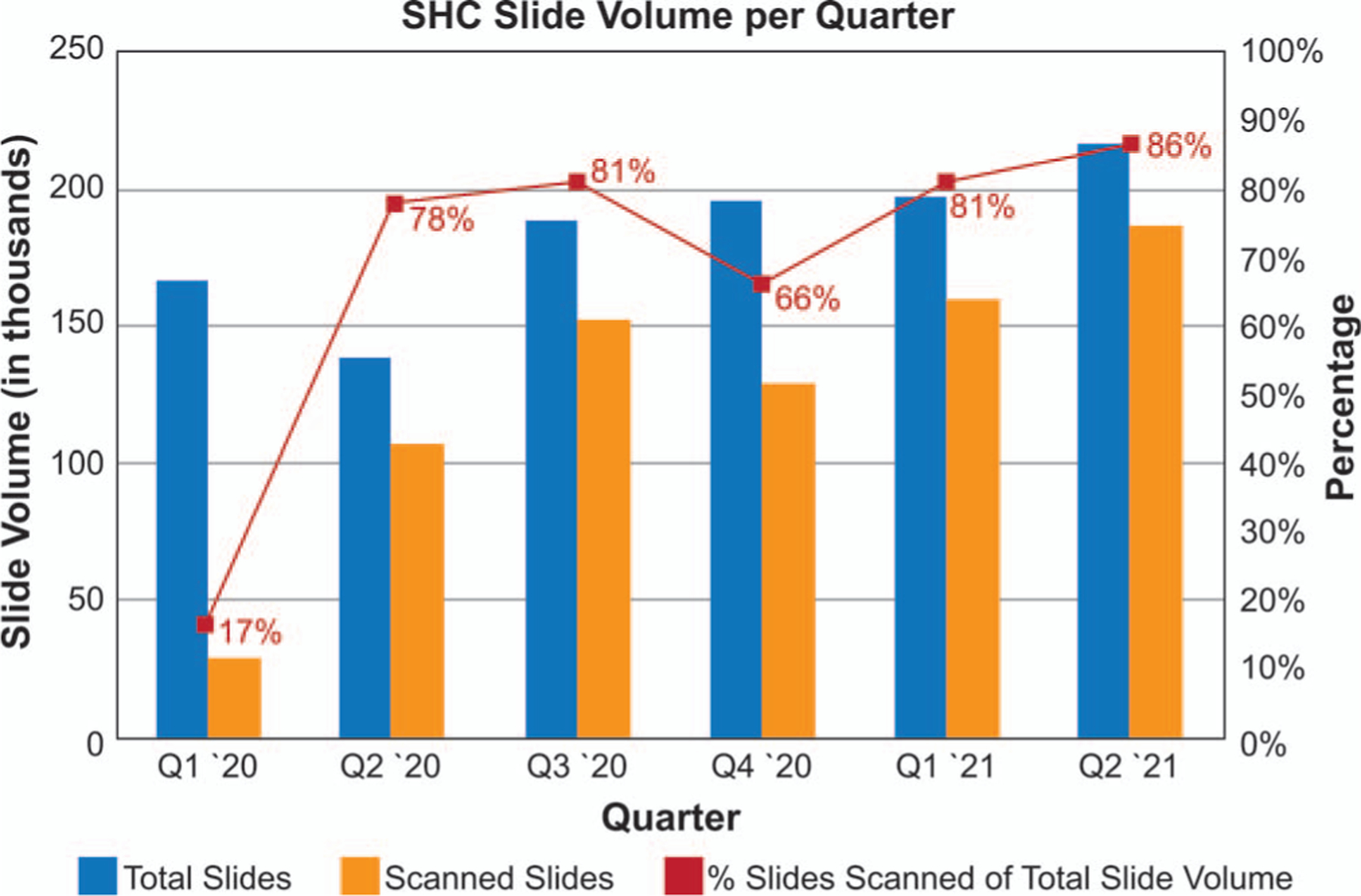
Stanford Pathology scanning volume by quarter in 2020 and 2021. Total number of slides produced by Stanford Histology Laboratory (blue bars). Total number of slides scanned (yellow bars). Percentage of total slide volume that was scanned (red line). Abbreviations: SHC, Stanford Healthcare; Q1, January through March; Q2, April through June; Q3, July through September; Q4, October through December.
METHODS
Rapid Conversion to WSI in Response to COVID-19 Pandemic
Since the first reported death in California and the United States on February 6, 2020, COVID-19 cases continued to rise, prompting a mandatory shelter-in-place order for 6 Bay Area counties (Alameda, Contra Costa, Marin, San Mateo, San Francisco, and Santa Clara, the site of Stanford University) effective March 17, 2020, limiting only persons involved in essential activities to report to work.
In alignment with shelter-in-place orders, Stanford Medicine began transitioning to essential care activities, which resulted in a substantial decrease in the surgical specimen case load for Stanford Pathology. In March 2020, the Centers for Medicare & Medicaid Services (CMS) waived the requirement for pathologists to perform diagnostic tests in Clinical Laboratory Improvement Amendments (CLIA)–licensed facilities. Stanford Pathology immediately pursued the digitization of all amenable surgical pathology cases to ensure uninterrupted care to patients and education to our trainees, while maintaining the safety of our health care workforce.
Digital Pathology Instrumentation
The (De Novo pathway) US Food and Drug Administration (FDA)–granted PIPS (IMS software version 3.3.3, Ultra Fast Scanner [UFS] with software version 1.8; Royal Philips, Amsterdam, the Netherlands) was implemented. This platform was FDA granted on the basis of its noninferiority to glass slides in a large multicenter study.16
For all our on-site clinical end users, we deployed standard HP Z4 G4 tower PCs (Hewlett Packard, Palo Alto, California) running the Microsoft Windows 7 operating system (department-wide upgrade to Windows 10 is ongoing) with dual 27-inch monitors, both with 2560 × 1440 pixel resolution: a Philips Barco PP27QHD monitor (Barco, Kortrijk, Belgium) and an HP z27n monitor (Hewlett Packard). Notably, high-resolution medical-grade monitors minimize loss of resolution along the pixel pathway and are preferred by pathologists.22–24 Use of a 4-MP medical-grade Barco display as part of the PIPS is a stipulation of the FDA. For off-site use, after an initial testing and validation period, we offered all faculty the same HP towers with the FDA-conforming Philips Barco PP27QHD monitors in their homes, with a Cisco Meraki MX68CW hardware VPN and a high-performance ASUS PCE-AC88 Wi-Fi card to further increase performance (Table 1). In both on-site and at-home settings, the pixel pathway was maintained on FDA-authorized devices.
Table 1.
Performance of the Scanning and Viewing Application Using Various Virtual Private Network Configurations
| Standard Software VPN, s | Citrix-Hosted Virtual Desktop, s | Wireless Meraki VPN, s | Wired Meraki VPN, s | |
|---|---|---|---|---|
| Average LIS first time to launch | 25 | 14 | 18 | 7 |
| Max IMS zoom/pan tiling response under load | 4.1 | 2.9 | 0.8 | 0.6 |
Abbreviations: IMS, image management system; LIS, laboratory information system; VPN, virtual private network.
Digital Pathology Validation Studies
Pathologists involved in validation received a 1-hour tutorial from vendor representatives. The validation model used was based on College of American Pathologists (CAP) guidelines for the validation of whole slide images,25 which has been successfully used to validate whole slide images at other institutions.26 These guidelines have since been updated and still support our approach to validation.27 Consistent with these guidelines, validation of a distinct application included review of at least 60 cases reflecting the spectrum and complexity of specimen types and diagnoses encountered in routine practice. The glass slide diagnosis was taken from the finalized pathology report. After a washout period of at least 2 weeks, the same attending pathologist who provided the original diagnosis interpreted the digital images without access to clinical information, to minimize unintentional unblinding. A separate pathologist compared the digital diagnosis to the original glass diagnosis and scored the cases as concordant or discordant. Discordant cases were determined to be either major or minor discordances (Supplemental Table 1; see supplemental digital content containing 2 tables). A threshold of 95% concordance was required for validation. Scanner validation studies were also performed upon installation of additional scanners in Q3 of 2020. For each additional scanner, 20 cases were scanned, and slide images reviewed for quality by a histotechnician. A pathologist then reviewed each image for concordance with the images from the first scanner and documented pass/fail for each.
Information Technology Infrastructure
The Philips IntelliSite scanning and viewing application (SVA) server was deployed in a Stanford Medicine virtual server environment. Three tiers of disk storage were initially used. In order of increasing storage capacity and decreasing price and speed, these are BLOCK, FILE, and OBJECT storage (Table 2). BLOCK is composed of a series of solid-state disks and is used for image ingestion, short-term retrieval, database access, and metadata. FILE is composed of a mix of solid state and high-speed mechanical disk drives configured to be the primary image repository for near-term access. OBJECT, a form of cloud storage with a duplicate off-site repository, was initially used for archival data that are not accessed on a continual basis. As a different communication protocol is used for access to each tier of storage, a storage virtualization product, GATEWAY, capable of communicating in all 3 protocols was initially used as an intermediary between the scanner and the 3 storage tiers. Both GATEWAY and OBJECT storage were ultimately removed owing to poor performance under high case load, and a 2-tier storage system using only BLOCK and FILE was put in place as a temporary solution.
Table 2.
Whole Slide Image Storage Solutions
| Tier | Basic Structure | Use | Estimated Performance Relative to BLOCK | Cost per Usable TB (Including Backup) |
|---|---|---|---|---|
| Tier 1 (BLOCK) | High-speed controllers, solid-state disks, fiber channel access | Initial WSI ingestion, short-term retrieval (24 h), database access and image metadata | 1.0 | $4789 |
| Tier 2 (FILE) | Mix of solid state and high-speed mechanical disk, Server Message Block access | WSI storage after the first 24 h of WSI creation up to 6 mo | 1.2 | $2368 |
| Tier 3 (OBJECT) | On site S3 “Cloud” storage | Long-term (>6 mo) storage of WSI with duplicate off-site backup | 7.0 | $199 per year |
Abbreviations: TB, terabyte; WSI, whole slide image.
Originally, an SVA server was provided by the vendor for ease of deployment during the pilot phase. As we continued the implementation of digital pathology across all applicable services, our infrastructure needs grew, necessitating the deployment of a higher-capacity configuration (Table 3).
Table 3.
Pilot Versus Full Deployment Server Characteristics
| Server Make and Model | Physical Processors (CPUs) | Virtual Processors (vCPUs) | Cores per Processor | Processor Speed, GHz | Server RAM, GB | Disk Storage (for Scanners), GB | |
|---|---|---|---|---|---|---|---|
| Vendor pilot deployment | Lenovo x3650 M5 | 2 | N/A | 6 | 2.4 | 32 | 40 |
| SHC full VM deployment | Cisco UCS-B200-M5 | 2 | 24 | 12 | 2.9 | 128 | 300+ |
Abbreviations: CPU, central processing unit; GB, gigabyte; GHz, gigahertz; N/A, not applicable; RAM, random access memory; SHC, Stanford Health Care; vCPU, virtual central processing unit; VM, virtual machine.
From a network perspective, the vendor allotted 40 MB/s (320 Mbps) per scanner against the facility network line (10 Gbps) and up to 3 MB/s (25 Mbps) per viewing end user. Per vendor recommendation, we segregated the server’s scanning and viewing network traffic across 2 network interface cards.
Workflow Changes for WSI Adoption
For most WSI-related workflows, we leveraged our laboratory information system (LIS), PowerPath (Sunquest Information Systems, Tucson, Arizona). Within the LIS, clinicians work from a subspecialty-specific worklist of pending cases. When in a given case, end users click hyperlinked slide icons in the LIS to open the entire case for viewing in the WSI viewer. By contrast, the histology laboratory quality control (QC) workflow operates outside of the LIS. Instead, for slide QC, laboratory technicians use the digital pathology application case list to navigate cases and tag slides with notes, including “Blurry Slide,” “Tissue Not Scanned,” “Image QC Failed,” or “Image QC Complete.” This populates the image with a corresponding colored flag to notify users of the QC status.
Outside Case Scanning
Rapid conversion because of COVID-19 also compelled WSI conversion of outside cases. Scanning of outside cases for diagnostic review posed unique challenges because of the variation in the quality of the slide preparation and labeling slides for automated linking to cases. As the WSI scanners used the same barcode as our LIS tracking, it was necessary that a unique Stanford Pathology label with QR code be placed on the slide, while preserving outside information such as stain type and slide number. This method needed to be flexible to the extreme diversity of labels and slides that arrive for consultation. Initially, individual stickers were manually cut and placed on outside slides. Ultimately, a novel modular adhesive label was designed in-house and contracted for production. These modular labels allowed an individualized approach to applying the Stanford-specific accession and QR code labels while preserving all case-relevant information associated with a slide (Figure 2).
Figure 2.
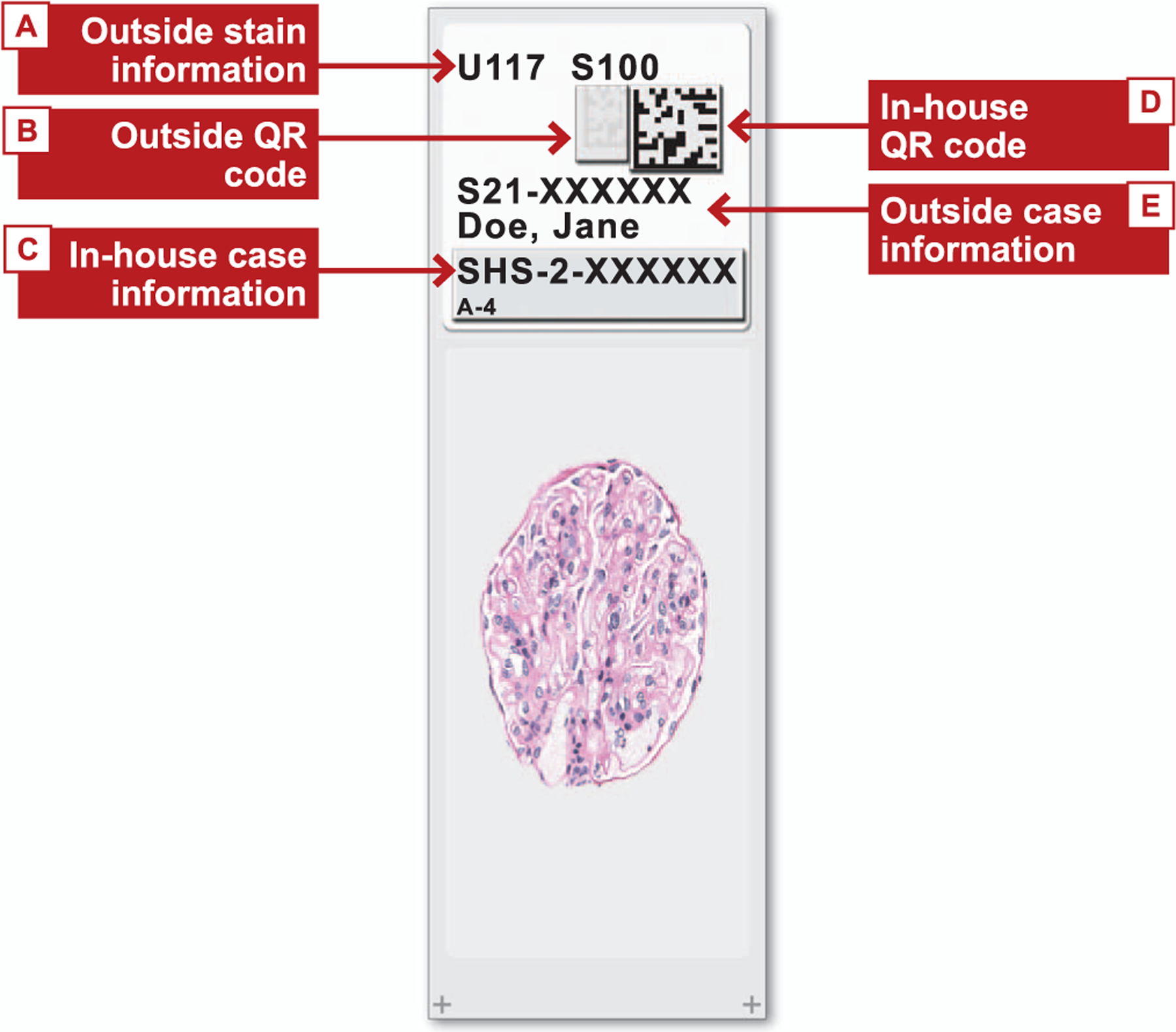
Schematic showing modular labels designed to preserve important information from outside (consult and referral) labels for slides received by Stanford Pathology from other institutions. The outside stain information is preserved (A), the originating institution’s QR code is obscured (B), an in-house case identifier code and slide number are added (C), an in-house QR code is added (D), and the original case number and patient name are preserved (E). Abbreviation: QR, quick response.
Survey of WSI Users
On July 23, 2020, a voluntary survey composed of 1 demographic, 7 multiple choice with optional short answer, 4 Likert scale, and 3 short answer questions was sent electronically to all anatomic pathologists, hematopathologists, cytopathologists, and trainees (52 attending pathologists, 42 fellows, and 35 residents). Data were analyzed anonymously after a 3-week period.
RESULTS
Validation and Stepwise Conversion of Subspecialty Services in 2018 and 2019
WSI was piloted in neuropathology because of its relatively low case volume, small number of slides per case, and high level of interest among faculty and trainees. A total of 64 hematoxylin-eosin (H&E)–stained, formalin-fixed, paraffin-embedded (FFPE) cases and 61 frozen muscle cases were reviewed. Diagnostic concordance was achieved in 62 of 64 FFPE cases (97%, 1 major discordance, 1 minor discordance) and 58 of 61 frozen muscle cases (95%, 1 major discordance, 2 minor discordances) (Supplemental Table 2).
All cases with discordant findings underwent subsequent review by additional subspeciality neuropathologists to confirm the discordance. The reasons for discordance fell into 2 broad categories: (1) critical regions with poor scan quality and (2) lack of clinical information leading to tumor misclassification. Poor scan quality caused discordance when critical regions were out of focus owing to scanner artifact or tissue folds. The absence of clinical history has been previously identified as a cause of discordance in WSI validation studies.28 While not leading to discordant diagnoses, certain special stains on frozen muscle sections and mitotic figures were noted to be more difficult to discern with WSI.
Following validation of the WSI platform for H&E-stained FFPE neuropathology cases, we evaluated additional subspecialty services as they transitioned to WSI. The second subspecialty evaluated was gynecologic pathology, with the goal of testing workflows at higher case volumes of both small specimens and larger resections. Twenty gynecologic pathology cases were validated. Diagnostic concordance was 100%.
CAP guidelines recommend that validation include confirmation that all material present on the glass slide be included in the image (Statement 11 in the CAP guideline on whole slide imaging for diagnostic purposes).25 We observed high rates of scanning failure for fragmented and scant specimens obtained from endocervical curettages and endometrial biopsies. The lower limit of size detection in WSI results in fragments smaller than 0.4 mm not being reliably scanned. We found that most (77%, 43 of 56 cases evaluated) digital scans of endocervical curettage specimen slides had tissue detection failure.29 As tumor cells can be present very focally, missing these scant tissue fragments is suboptimal for patient care. While image analysis methods have been developed to detect remote fragments on digital images,30 as has been reported for fragmented brain tumor specimens,31 our solution used a collodion bag protocol that is routinely used by cytology for cell blocks. This method allowed fragmented tissue to be aggregated to a single area and formed a distinct collodion bag rim around the fragmented tissue. With this workflow adaptation, tissue detection failure rates were reduced from 77% (43 of 56) in non–collodion bag cases to 23 of 52 in collodion bag cases (44%), representing a 42% reduction.29 While we observed a marked improvement in tissue detection with the implementation of the collodion bag protocol, the method does not completely prevent tissue detection failure. Therefore, even when using collodion bags, we advise pathologists to exercise caution for missed tissue and to maintain a low threshold for conversion to glass slide evaluation.
WSI conversion of the immunohistochemistry service provided a platform to equip and involve faculty and trainees across many subspecialties, thereby expanding stakeholder engagement. All attending pathologists were polled to recommend immunohistochemical stains that were frequently ordered, challenging to interpret, or diagnostically crucial for their subspecialty. Forty immunohistochemical stains were chosen, and 67 cases were reviewed. Importantly, cases were also chosen to include different chromogens, dynamic ranges of intensity, subcellular localizations, and methods of preparation. Whole slide images were reviewed by subspecialty experts for each case, including 24 dermatopathology whole slide images. Diagnostic concordance was achieved in 66 of 67 cases (98.5%). Our results were similar to those described by other laboratories.32 Notably, for the 1 discordant case, the digital scan was uninterpretable owing to scratches in the coverslip obscuring the diagnostic material. Although not formally assessed in our validation, in anticipation of scanning whole slide images, we also increased the hematoxylin (counter-stain) stain time on 1 of 2 of our immunohistochemical instrument platforms to assess whether negatively stained tissue would be more readily detected with a darker counterstain. In the validation study, we noted no limitations due to undetected tissue for either counterstain intensity. Our experience since the validation, however, has indicated that stains for which we are unable to increase the counterstain owing to effects on signal, such as SV40, remain problematic to scan when negative.
We achieved greater than 95% concordance between glass slides and whole slide images in each subsequent subspecialty conversion. Our phased-in approach of pilot, pressure test, and then broad roll out via the immunohistochemistry service was well received by the anatomic pathology faculty and trainees.
From our surgical pathology case volumes and an estimated practical scanning time of 2 minutes per glass slide, we had initially projected that 7 ultrafast scanners were needed for comprehensive WSI. At the time we decided to expand our scanning to all subspecialties, Stanford Pathology was actively operating 3 scanners, all of which were dedicated to in-house cases. A fourth scanner, designated for scanning consult cases, had arrived but had not been validated (Figure 3). Over the course of the next few months, we expanded to a total of 8 scanners. Six scanners were dedicated to the histology laboratory. Figure 1 shows the rapid increase in the number of total whole slide images versus the number of slides ordered during the spring and summer of 2020. The additional 2 scanners were dedicated to consult materials amounting to 13 376 cases with 87 129 slides in 2020.
Figure 3.
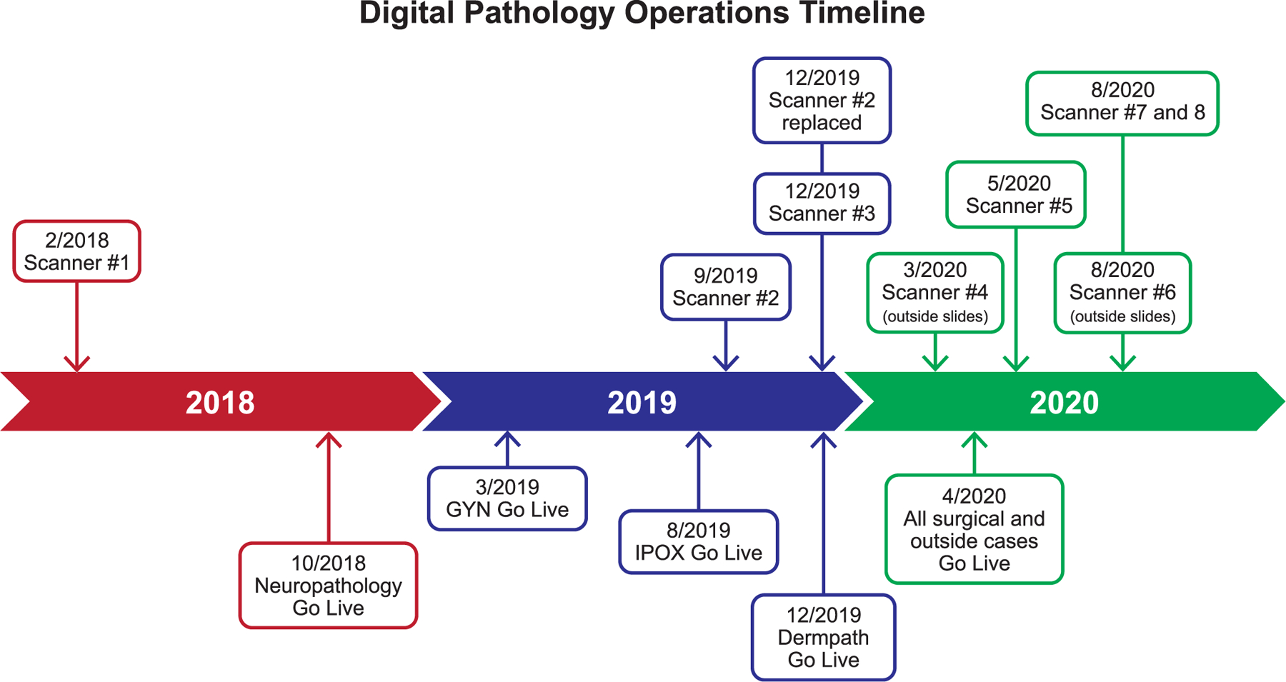
Timeline of Stanford Pathology whole slide imaging (WSI) implementation beginning February 2018. Blue boxes show points at which scanners were added. Red boxes show the dates of the initial stepwise implementation of WSI for each of 4 subspecialty services followed by broad implementation across all surgical pathology and consult services in April 2020. Abbreviations: GYN, gynecologic pathology; IPOX, immunohistochemistry.
The FDA-specified intended use states that the PIPS is not intended for use with frozen section, cytology, or non-FFPE hematopathology specimens.33 Based on this guidance, analysis of specimen-type and content, and our prior experience digitizing the gynecologic and neuropathology subspecialties, we decided that the only subspecialty cases that would not be included in digitization were cytology cases, bone marrow aspirates and smears, whole mount prostate cases, and breast resection cases. Notably, evaluation of cytology and bone marrow aspirates/smears requires the ability to adjust the plane of focus, whereas PIPS whole slide images are obtained at a single depth of focus. Whole mount slides were not included in the FDA intended use statement, and our scanners were not compatible with oversized whole mount prostate resection slides. We initially excluded breast resection cases because previous reports suggested high rates of tissue detection failure of fatty tissue sections in breast specimens.34 We later included these cases after our in-house validation was favorable with a concordance of 99% (1 discordance of 40 cases reviewed).
Staffing and hours in the histology laboratory necessitated major operational changes to adapt to WSI across surgical pathology. Slide scanning added approximately 3 hours to the overall turnaround time for availability of glass slides. We determined that the optimal ratio of histology laboratory staffing with WSI in the different shifts would be 30% of total staff in the morning shift, 20% in the midday shift, and 50% in the evening shift (3:2:5). This was a change from 4:2:4 staffing used before implementation of WSI. Scheduling half of the histotechnicians and histotechnologists in the evening shift allowed for glass slides to be available earlier and loaded onto the scanners to meet the expected turnaround time for WSI availability in the morning.
Workflows in both the gross room and histology laboratory were made to accommodate the additional time needed to perform slide scanning. Tissue processors used in the histology laboratory are routinely operated with run settings specific to a subset of specimen types. To facilitate continuous use of the available tissue processor capacity, the run settings were consolidated from 4 different run types to 3. As a result, more cassettes could be batched into each run type, reducing downtime spent awaiting space on the next applicable run.
Overall, we found that scanning of materials added approximately 3 hours to turnaround time for in-house slide availability. Initially, we added 1 laboratory technician per scanner, but over time, we found that the increased work of screening and organizing slides, cleaning/drying slides, loading, machine troubleshooting, and QC required an additional half-time laboratory technician. However, this ratio may vary at other institutions, depending on their specific workflow. At our institution, distribution of outside cases operates independently from in-house cases. Scanning added about 24 hours to the availability of glass slides for outside cases, owing to major changes in workflow, limited availability of administrative staff because of pandemic restrictions, and special labeling requirements.
Stabilization in the Second Half of 2020 and 2021
WSI was adopted broadly across all surgical subspecialty services in Q2 of 2020 to manage the challenges to clinical service and teaching caused by response to the pandemic. This innovative response during a low case volume scenario was quickly stressed by recovering case volumes in Q3 and Q4 of 2020 (Figure 1). Given the success of WSI during Q2 of 2020, faculty, trainees, and administration all strongly favored maintaining the high level of digital surgical pathology achieved and not returning to glass slides and stepwise digital conversion of subspecialty services. To maintain a high level of WSI in the face of rapidly increasing case volumes, additional scanners were installed and validated in Q3 2020 (Figure 3).
As we rapidly expanded the number of slides scanned per day, we found that viewing speed and system stability degraded significantly at high load. We also quickly reached the limit of our vendor-supplied server environment, which has a stated maximum of 4 scanners for our pilot configuration (the vendor recommends 10 GB of disk storage per scanner at a minimum; see Table 3). Stanford Medicine information technology (IT) team deployed a new owned-and-administered infrastructure built to support up to 8 concurrent scanners, based on the vendor-provided specifications. The SVA server was built by using the same FDA-granted open virtualization appliance file deployed in the initial server environment and hosted in the Stanford Medicine virtual server environment, which was subsequently validated by the vendor. Three tiers of disk storage were used for image acquisition and retention, storage, and retrieval as well as associated metadata required for their use (Table 2). A storage virtualization product, GATEWAY, served as the intermediary between tiered storage and SVA. Stanford Medicine went live with this configuration in May 2020, but as volumes increased in Q3 of 2020 performance deteriorated substantially. Testing revealed that GATEWAY was the likely culprit.
Throughout Q4 of 2020, the Pathology Imaging System was redesigned to exclude GATEWAY. We went live on the latest IT design that excluded GATEWAY in early January 2021, resulting in a performance improvement, measured as time for WSI tiles to load in the Pathology Imaging System Viewer, of approximately 200% (Figure 4). Currently, new solutions are being developed to regain the full functionality of GATEWAY, including improving research access and regaining archival and backup WSI storage while maintaining the current high level of clinical performance.
Figure 4.
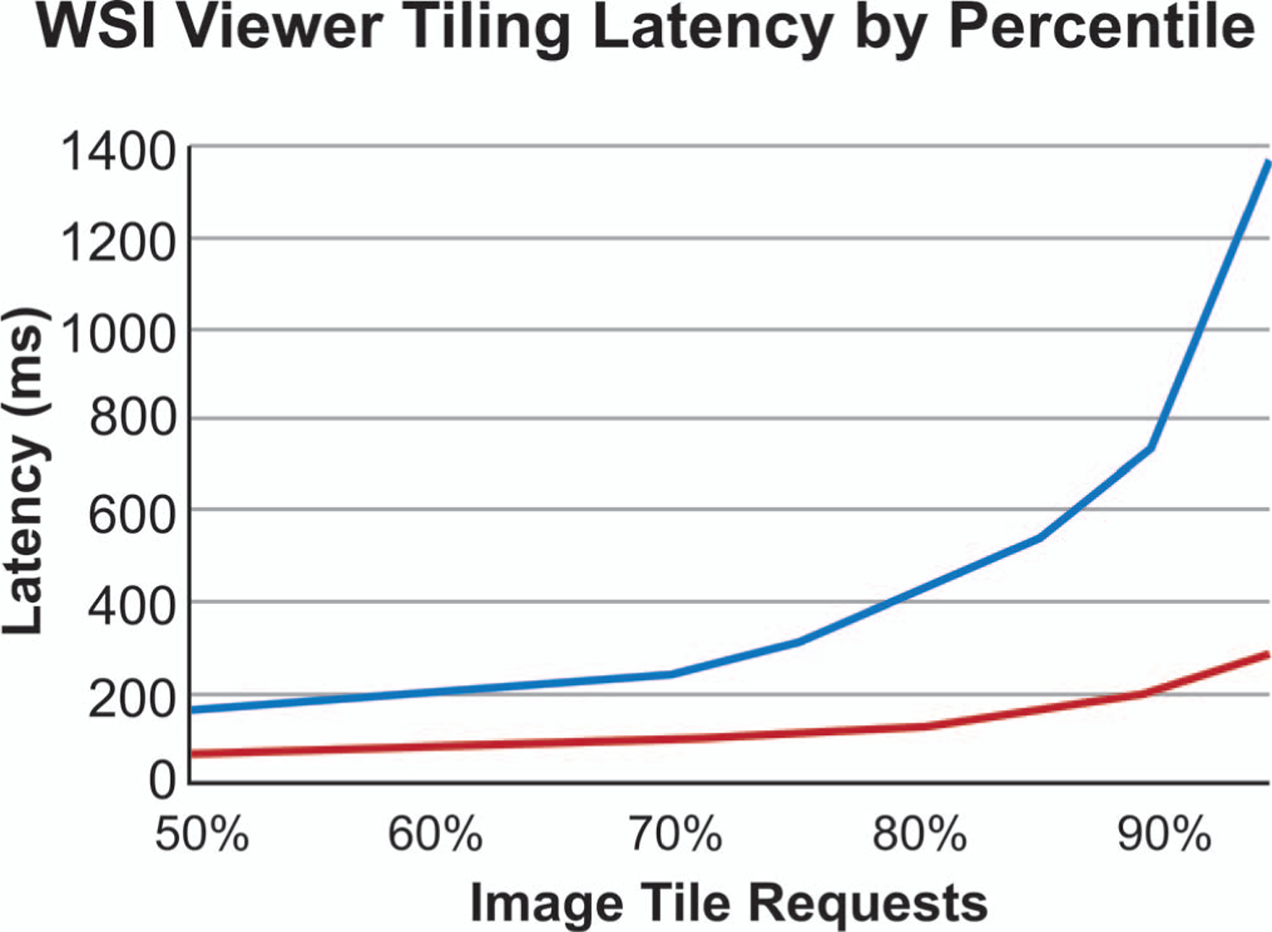
Whole slide image (WSI) viewer tiling latency by percentile. Time to load an image is shown in milliseconds per tile (grouped by percentile). Blue line shows latency with GATEWAY storage virtualization as intermediary between scanner and file storage drives. Red line shows latency after removal of GATEWAY.
Workflow Changes for Ancillary Studies
We took advantage of WSI to streamline several work-flows, which were otherwise more complex. A subset of our immunohistochemical stains use off-slide positive controls, which were reviewed daily by a single designated pathologist, as making these slides available to multiple pathologists simultaneously was logistically challenging. WSI obviated the need for a single pathologist to review all off-slide controls. Instead, the whole slide images were now available to all users on demand. Similarly, prior to WSI, when fluorescence in situ hybridization was required, the ordering pathologist or trainee would enter the required probe information in a logbook and circle the region of interest on the physical slide. These slides would be batched and sent by courier to the off-site cytogenetics laboratory. With WSI available, we transitioned to a fully digital workflow. The region of interest is circled on the whole slide images, and the order is placed in the LIS. Both are immediately accessible to the cytogenetics laboratory without the need for a courier.
Current Status and Pathologist Response
As of March 2021, Stanford Medicine is generating whole slide images from more than 80% of the slides produced by the histology laboratory (Figure 1). Consults are scanned prospectively before pathologist review, if this can be done within 2 days of receipt. Otherwise, they bypass scanning, get reviewed and finalized, and are scanned retrospectively before being returned to the consulting institution. Currently, we are scanning more than 2000 slides per day on weekdays (Figure 5).
Figure 5.
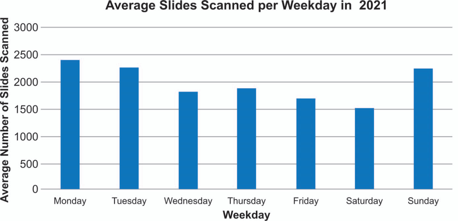
Average number of slides scanned per weekday by Stanford Pathology during Q2, April through June 2021.
In July 2020, shortly after the initial expansion of WSI, we surveyed attending pathologists, residents, and fellows in Stanford Pathology to determine the breadth of adoption of WSI diagnosis and opportunities for improvement (Figure 6; Table 4). Although the survey was available for 3 weeks, all responses were received within 48 hours of survey opening. The 32 respondents included 17 faculty members (53%), 7 fellows (21.9%), and 8 residents (25%). Most respondents (21 of 32, 66%) reported using WSI every day and only 4 respondents (13%) reported using the system less than once a week. The majority had no formal training in digital pathology (25 of 32, 78%), but 21 (66%) had reviewed an in-house developed training guide or sought IT support. Nonetheless, 24 (75%) said they felt comfortable signing out at least some cases exclusively digitally and 14 (44%) said they could sign out all their cases digitally. The majority (20 of 32, 63%) used WSI for real-time conferencing at least several times a week, with 19 (95%) opting to use a dedicated videoconference or screen share application such as Webex or Zoom as opposed to the WSI platform’s screen share feature. Our survey data showed that while nearly all respondents (31 of 32, 97%) used WSI at the hospital, 47% (15 of 32) also used the system at home, and 1 respondent used WSI at home exclusively. Additionally, the majority felt that slow image load speed (24 of 32, 75%) and/or hardware (17 of 32, 53%) had the strongest impact on their ability to perform digital diagnosis.
Figure 6.
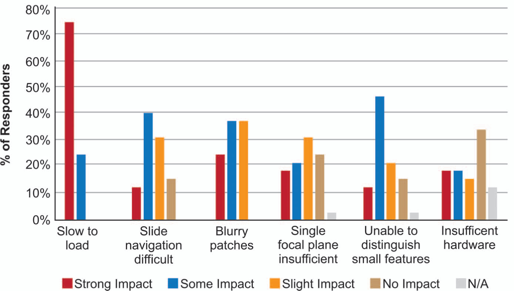
Pathologists’ perceptions of the degree to which image load time, slide navigation, blurry images, focal plane, small features, and hardware impacted their ability to perform digital diagnosis using whole slide images (attending pathologist, fellow, and resident survey responses). Abbreviation: N/A, not applicable.
Table 4.
Pathologists’ Response to Digital Pathology
| No. (%) | |
|---|---|
| Role | |
| Faculty | 17/32 (53) |
| Fellow | 7/32 (22) |
| Resident | 8/32 (25) |
| Training in digital pathology | |
| Formal demo/tutorial | 6/32 (19) |
| Review of tip sheet or IT help | 15/32 (47) |
| Trial and error | 10/32 (31) |
| Frequency of WSI use | |
| Less than once a week | 4/32 (13) |
| A few times per week | 7/32 (22) |
| Every day | 21/32 (66) |
| Location of WSI use | |
| On-site | 16/32 (50) |
| Off-site | 1/32 (3) |
| Both | 15/32 (47) |
| Comfort with digital diagnosis | |
| Somewhat (would not sign out cases) | 7/32 (22) |
| Moderate (would sign out some cases) | 10/32 (31) |
| Comfortable but prefer glass | 10/32 (31) |
| Very (sign out most cases) | 4/32 (13) |
| Frequency of real-time conferencing | |
| Daily | 6/32 (19) |
| A few times | 9/32 (28) |
| Weekly | 15/32 (47) |
| Never | 2/32 (6) |
| Collaboration method | |
| Screenshare app | 28/32 (88) |
| WSI viewer | 1/32 (3) |
| N/A | 3/32 (9) |
Abbreviations: IT, information technology; N/A, not applicable; WSI, whole slide image.
Implementing Remote Sign-Out
When CMS relaxed the requirement for remote locations to have separate CLIA licenses it opened the possibility for remote sign-out. Given this waiver, and the fact that we provided faculty with identical hardware on- and off-site, we did not separately validate remote versus on-site reporting; however, we performed an initial test of off-site SVA use during which we measured latency and subjective performance of the SVA, using various VPN configurations (Table 1). Recent reports of off-site sign-out from other institutions supports our own experience of its feasibility.35,36 The vendor recommends a minimum network speed for remote use of 32 Mbps for both download and upload as well as wired connection. In our experience with off-site use, application performance correlated strongly with network speed, which varied by location from 10 Mbps to 1þ Gbps. Additionally, we noted viewing performance improvements in the use of the SVA at network speeds of up to 1 Gbps even though 3 MB/s is the vendor’s stated maximum application file download speed.
About half of attending surgical pathologists opted for setup of workstations, and trainees were provided hospital-owned laptops for secure remote access. With the workstations at home, faculty had the flexibility to remotely sign out with trainees and review and finalize cases at home.
DISCUSSION
Pandemic restrictions led to near complete adoption of WSI by our attending pathologists and trainees mostly because of remote reviewing capability with trainees and sign-out. Although the capacity for social distancing afforded by WSI in the context of pandemic restrictions drove its pervasive adoption in surgical pathology, multiple other benefits were noted by our attending pathologists and trainees. First, obtaining the opinion of colleagues on difficult cases is an important aspect of any high-functioning surgical pathology group. In the past, individuals at our institution approached this in various ways: sending the trainee to find the attending pathologist, setting a time to meet together, leaving the slides in a colleague’s mailbox, or sending slides by a courier to a different site. All of these methods were time-consuming for the individuals involved and could take multiple days. With WSI, showing cases is as simple as sending an email with a link to the slides (annotated with areas of interest) and the question to the consultant. Second, rapid access to case material in a complex organization with multiple sites can be challenging. WSI allows immediate access to digital slides without having to search various locations for materials.10 Third, WSI facilitates efficiency and accuracy of measurements for important histologic features, such as depth of invasion and margin status. Synchronized viewing of H&E slides with corresponding immunohistochemical stains allows more efficient interpretation, especially when the cells of interest are uncommon or rare. Fourth, we are able to readily access archived images including immediate access to WSI at the time of frozen section and review of prior cases to inform the current evaluation, cytology-histology correlation, and block selection for ancillary studies and send-out testing. Fifth, all of our tumor boards went from in-person to fully online in a matter of weeks. WSI provided a much clearer picture than a video-based image of the slide on a microscope. Moreover, the availability of WSI saved more than 100 hours of administrative time per month previously required to search for the glass slides needed for 46 monthly subspecialty tumor boards. Sixth, ancillary testing in the clinical pathology laboratories, including fluorescence in situ hybridization, flow cytometry, and molecular testing for both pathogenic mutations and microorganisms, is performed on many surgical pathology cases. With the implementation of WSI, slides are now viewable by colleagues on these services in real time. This enables efficient triage and clinical decision-making with regard to appropriate test utilization and tissue selection. The final benefit already realized is archiving of consultation material. One of the benefits of working at a large academic institution is the exposure to a diverse array of consultation cases, including many rare entities. With WSI, there is preserved access to the entire case without additional labor of creating and storing recut slides and without additional risk of misplaced materials.
We also appreciated benefits to our teaching and research missions during our rapid conversion to WSI. Instructive cases easily can be flagged by subspecialty either in the LIS or the Pathology Imaging System. The maintenance of glass teaching sets for trainee education and outside presentations is a large endeavor for any academic institution. Conversion to a digitally driven platform offers ease of access by multiple users and stability of the images over generations of trainees.37 WSI provides the option of asynchronous review and sign-out for cases not reviewed together by faculty and trainee, where the attending pathologist can annotate the image for fellows and residents to review later. Additional research benefits that are beginning to accrue from WSI include greatly simplified gathering and organizing of materials for review, a very time-consuming and often frustrating manual process, readily available high-quality images for publication and presentations, and a resource for development of in-house machine learning enhancements for the practice of surgical pathology.
Overall, our experience was similar to that of others38,39 in that the confidence with interpretation was greater with glass than with digital slides, likely due in part to familiarity, but also due to WSI challenges with rare and small events or regions of poor focus. Panning and zooming on a whole slide image is physically quite different from pushing a glass slide on a microscope. Pathologists needed time to adjust to this different mode of review. As others have reported, even when the pathologist was proficient, we found that WSI review is still slower than glass slide review for an experienced pathologist.6,13 In general, users find it fairly easy to review biopsy samples but become fatigued when reviewing larger resection cases.
Adoption of WSI for clinical cases creates opportunities for enhanced intra-institutional and interinstitutional collaboration. We plan to leverage WSI to link with radiologic imaging to improve case correlation through body part matching. The development of a digital consult portal for review of outside materials from clients with various types of scanners is part of our long-term strategic plan for reducing the transport of glass slides. Finally, we expect that there soon will be rapid progress in the deployment of machine learning enhancements to aid in various aspects of screening and interpreting WSI.
CONCLUSIONS
COVID-19 highlighted the advantages of WSI for clinical diagnosis and created the impetus for its broad adoption. We saw numerous advantages to implementing WSI for our clinical, educational, and research activities. The relatively rapid transition we underwent from low to high volume slide scanning demonstrated important operational and technical considerations that should be taken into account by other groups when embarking on WSI for surgical pathology. Chief among these were the need for skilled staff, changes in workflow, and digital storage solutions appropriate to the volume of image data. Perhaps the most novel aspect of WSI during the COVID-19 pandemic was the option for remote sign-out, bringing with it additional flexibility, along with unique technical challenges. In our experience, successful adoption of WSI requires strong commitment and collaboration between all facets of the laboratory system while supporting continued high-quality patient care during challenging times.
Supplementary Material
Acknowledgments
We would like to thank Megan Troxell, MD, PhD (Department of Pathology, School of Medicine, Stanford University, Stanford, California) for her helpful comments, and Norm Cyr (School of Medicine, Stanford University, Stanford, California) for assistance with graphics.
Footnotes
The authors have no relevant financial interest in the products or companies described in this article.
Supplemental digital content is available for this article. See text for hyperlink.
References
- 1.Lujan G, Quigley JC, Hartman D, et al. Dissecting The Business Case for Adoption and Implementation of Digital Pathology: a white paper from the Digital Pathology Association. J Pathol Inform. 2021;12:17. doi: 10.4103/jpi.jpi_67_20 [DOI] [PMC free article] [PubMed] [Google Scholar]
- 2.Schüffler PJ, Geneslaw L, Yarlagadda DVK, et al. Integrated digital pathology at scale: a solution for clinical diagnostics and cancer research at a large academic medical center. J Am Med Inform Assoc. 2021;28(9):1874–1884. doi: 10.1093/jamia/ocab085 [DOI] [PMC free article] [PubMed] [Google Scholar]
- 3.Zarella MD, Bowman D, Aeffner F, et al. A Practical Guide to Whole Slide Imaging: a white paper from the Digital Pathology Association. Arch Pathol Lab Med. 2019;143(2):222–234. doi: 10.5858/arpa.2018-0343-RA [DOI] [PubMed] [Google Scholar]
- 4.Al-Janabi S, Huisman A, Nap M, Clarijs R, van Diest PJ. Whole slide images as a platform for initial diagnostics in histopathology in a medium-sized routine laboratory. J Clin Pathol. 2012;65(12):1107–1111. doi: 10.1136/jclinpath-2012-200878 [DOI] [PubMed] [Google Scholar]
- 5.Bauer TW, Schoenfield L, Slaw RJ, Yerian L, Sun Z, Henricks WH. Validation of whole slide imaging for primary diagnosis in surgical pathology. Arch Pathol Lab Med. 2013;137(4):518–524. doi: 10.5858/arpa.2011-0678-OA [DOI] [PubMed] [Google Scholar]
- 6.Borowsky AD, Glassy EF, Wallace WD, et al. Digital whole slide imaging compared with light microscopy for primary diagnosis in surgical pathology. Arch Pathol Lab Med. 2020;144(10):1245–1253. doi: 10.5858/arpa.2019-0569-OA [DOI] [PubMed] [Google Scholar]
- 7.Buck TP, Dilorio R, Havrilla L, O’Neill DG. Validation of a whole slide imaging system for primary diagnosis in surgical pathology: a community hospital experience. J Pathol Inform. 2014;5(1):43. doi: 10.4103/2153-3539. 145731 [DOI] [PMC free article] [PubMed] [Google Scholar]
- 8.Campbell WS, Lele SM, West WW, Lazenby AJ, Smith LM, Hinrichs SH. Concordance between whole-slide imaging and light microscopy for routine surgical pathology. Hum Pathol. 2012;43(10):1739–1744. doi: 10.1016/j.humpath.2011.12.023 [DOI] [PubMed] [Google Scholar]
- 9.Cheng CL, Azhar R, Sng SH, et al. Enabling digital pathology in the diagnostic setting: navigating through the implementation journey in an academic medical centre. J Clin Pathol. 2016;69(9):784–792. doi: 10.1136/jclinpath-2015-203600 [DOI] [PubMed] [Google Scholar]
- 10.Evans AJ, Salama ME, Henricks WH, Pantanowitz L. Implementation of whole slide imaging for clinical purposes: issues to consider from the perspective of early adopters. Arch Pathol Lab Med. 2017;141(7):944–959. doi: 10.5858/arpa.2016-0074-OA [DOI] [PubMed] [Google Scholar]
- 11.Gilbertson JR, Ho J, Anthony L, Jukic DM, Yagi Y, Parwani AV. Primary histologic diagnosis using automated whole slide imaging: a validation study. BMC Clin Pathol. 2006;6:4. doi: 10.1186/1472-6890-6-4 [DOI] [PMC free article] [PubMed] [Google Scholar]
- 12.Hanna MG, Reuter VE, Ardon O, et al. Validation of a digital pathology system including remote review during the COVID-19 pandemic. Mod Pathol. 2020;33(11):2115–2127. doi: 10.1038/s41379-020-0601-5 [DOI] [PMC free article] [PubMed] [Google Scholar]
- 13.Hanna MG, Reuter VE, Hameed MR, et al. Whole slide imaging equivalency and efficiency study: experience at a large academic center. Mod Pathol. 2019;32(7):916–928. doi: 10.1038/s41379-019-0205-0 [DOI] [PubMed] [Google Scholar]
- 14.Houghton JP, Ervine AJ, Kenny SL, et al. Concordance between digital pathology and light microscopy in general surgical pathology: a pilot study of 100 cases. J Clin Pathol. 2014;67(12):1052–1055. doi: 10.1136/jclinpath-2014-202491 [DOI] [PubMed] [Google Scholar]
- 15.Jukić DM, Drogowski LM, Martina J, Parwani AV. Clinical examination and validation of primary diagnosis in anatomic pathology using whole slide digital images. Arch Pathol Lab Med. 2011;135(3):372–378. doi: 10.5858/2009-0678-OA.1 [DOI] [PubMed] [Google Scholar]
- 16.Mukhopadhyay S, Feldman MD, Abels E, et al. Whole slide imaging versus microscopy for primary diagnosis in surgical pathology. Am J Surg Pathol. 2018; 42(1):39–52. doi: 10.1097/pas.0000000000000948 [DOI] [PMC free article] [PubMed] [Google Scholar]
- 17.Snead DR, Tsang YW, Meskiri A, et al. Validation of digital pathology imaging for primary histopathological diagnosis. Histopathology. 2016;68(7): 1063–1072. doi: 10.1111/his.12879 [DOI] [PubMed] [Google Scholar]
- 18.Tabata K, Mori I, Sasaki T, et al. Whole-slide imaging at primary pathological diagnosis: validation of whole-slide imaging-based primary pathological diagnosis at twelve Japanese academic institutes. Pathol Int. 2017;67(11): 547–554. doi: 10.1111/pin.12590 [DOI] [PubMed] [Google Scholar]
- 19.Lujan GM, Savage J, Shana’ah A, et al. Digital pathology initiatives and experience of a large academic institution during the coronavirus disease 2019 (COVID-19) pandemic. Arch Pathol Lab Med. 2021;145(9):1051–1061. doi: 10.5858/arpa.2020-0715-sa [DOI] [PubMed] [Google Scholar]
- 20.Williams BJ, Fraggetta F, Hanna MG, et al. The future of pathology: what can we learn from the COVID-19 pandemic? J Pathol Inform. 2020;11:15. doi: 10.4103/jpi.jpi_29_20 [DOI] [PMC free article] [PubMed] [Google Scholar]
- 21.Betmouni S Diagnostic digital pathology implementation: learning from the digital health experience. Digit Health. 2021;7:205520762110202. doi: 10.1177/20552076211020240 [DOI] [PMC free article] [PubMed] [Google Scholar]
- 22.Abel JT, Ouillette P, Williams CL, et al. Display characteristics and their impact on digital pathology: a current review of pathologists’ future “microscope”. J Pathol Inform. 2020;11:23. doi: 10.4103/jpi.jpi_38_20 [DOI] [PMC free article] [PubMed] [Google Scholar]
- 23.Clarke EL, Munnings C, Williams B, Brettle D, Treanor D. Display evaluation for primary diagnosis using digital pathology. J Med Imaging (Bellingham). 2020;7(2):027501. doi: 10.1117/1.JMI.7.2.027501 [DOI] [PMC free article] [PubMed] [Google Scholar]
- 24.Norgan AP, Suman VJ, Brown CL, Flotte TJ, Mounajjed T. Comparison of a medical-grade monitor vs commercial off-the-shelf display for mitotic figure enumeration and small object (Helicobacter pylori) detection. Am J Clin Pathol. 2018;149(2):181–185. doi: 10.1093/ajcp/aqx154 [DOI] [PubMed] [Google Scholar]
- 25.Pantanowitz L, Sinard JH, Henricks WH, et al. Validating Whole Slide Imaging for Diagnostic Purposes in Pathology: guideline from the College of American Pathologists Pathology and Laboratory Quality Center. Arch Pathol Lab Med. 2013;137(12):1710–1722. doi: 10.5858/arpa.2013-0093-cp [DOI] [PMC free article] [PubMed] [Google Scholar]
- 26.Samuelson MI, Chen SJ, Boukhar SA, et al. Rapid validation of whole-slide imaging for primary histopathology diagnosis. Am J Clin Pathol. 2021;155(5): 638–648. doi: 10.1093/ajcp/aqaa280 [DOI] [PMC free article] [PubMed] [Google Scholar]
- 27.Evans AJ, Brown RW, Bui MM, et al. Validating Whole Slide Imaging Systems for Diagnostic Purposes in Pathology: guideline update from the College of American Pathologists in collaboration with the American Society for Clinical Pathology and the Association for Pathology Informatics [published online May 18, 2021]. Arch Pathol Lab Med. doi: 10.5858/arpa.2020-0723-cp [DOI] [Google Scholar]
- 28.Bauer TW, Behling C, Miller DV, et al. Precise identification of cell and tissue features important for histopathologic diagnosis by a whole slide imaging system. J Pathol Inform. 2020;11(1):3. doi: 10.4103/jpi.jpi_47_19 [DOI] [PMC free article] [PubMed] [Google Scholar]
- 29.Jhun I, Levy D, Lim H, et al. Implementation of collodion bag protocol to improve whole-slide imaging of scant gynecologic curettage specimens. J Pathol Inform. 2021;12(1):2. doi: 10.4103/jpi.jpi_82_20 [DOI] [PMC free article] [PubMed] [Google Scholar]
- 30.Pantanowitz L, Michelow P, Hazelhurst S, et al. A digital pathology solution to resolve the tissue floater conundrum. Arch Pathol Lab Med. 2020;145(3):359–364. doi: 10.5858/arpa.2020-0034-oa [DOI] [PubMed] [Google Scholar]
- 31.Cadwell CR, Bowman S, Laszik ZG, Pekmezci M. Loss of fidelity in scanned digital images compared to glass slides of brain tumors resected using cavitron ultrasonic surgical aspirator. Brain Pathol. 2021;31(4). doi: 10.1111/bpa.12938 [DOI] [PMC free article] [PubMed] [Google Scholar]
- 32.Williams BJ, Jayewardene D, Treanor D. Digital immunohistochemistry implementation, training and validation: experience and technical notes from a large clinical laboratory. J Clin Pathol. 2019;72(5):373–378. doi: 10.1136/jclinpath-2018-205628 [DOI] [PubMed] [Google Scholar]
- 33.Food US and Administration Drug, Center for Drug Evaluation and Research. Philips IntelliSite Pathology Solution (PIPS) approval letter, April 12, 2017. Accessed July 16, 2021. https://www.accessdata.fda.gov/cdrh_docs/pdf16/DEN160056.pdf [Google Scholar]
- 34.Rabban J Tissue detection failure rates for selected gynecologic and breast specimens using an FDA approved digital pathology imaging system: practical implications for pathology workflow and patient safety. Platform presentation at: USCAP 2020; Los Angeles, CA. [Google Scholar]
- 35.Ramaswamy V, Tejaswini BN, Uthaiah SB. Remote reporting during a pandemic using digital pathology solution: Experience from a tertiary care cancer center. J Pathol Inform. 2021;12(1):20. doi: 10.4103/jpi.jpi_109_20 [DOI] [PMC free article] [PubMed] [Google Scholar]
- 36.Rao V, Kumar R, Rajaganesan S, et al. Remote reporting from home for primary diagnosis in surgical pathology: a tertiary oncology center experience during the COVID-19 pandemic. J Pathol Inform. 2021;12(1):3. doi: 10.4103/jpi.jpi_72_20 [DOI] [PMC free article] [PubMed] [Google Scholar]
- 37.Christian RJ, VanSandt M. using dynamic virtual microscopy to train pathology residents during the pandemic: perspectives on pathology education in the age of COVID-19. Acad Pathol. 2021;8:237428952110068. doi: 10.1177/23742895211006819 [DOI] [PMC free article] [PubMed] [Google Scholar]
- 38.Babawale M, Gunavardhan A, Walker J, et al. Verification and validation of digital pathology (whole slide imaging) for primary histopathological diagnosis: all Wales experience. J Pathol Inform. 2021;12(1):4. doi: 10.4103/jpi.jpi_55_20 [DOI] [PMC free article] [PubMed] [Google Scholar]
- 39.Thorstenson S, Molin J, Lundström C. Implementation of large-scale routine diagnostics using whole slide imaging in Sweden: digital pathology experiences 2006–2013. J Pathol Inform. 2014;5(1):14. doi: 10.4103/2153-3539.129452 [DOI] [PMC free article] [PubMed] [Google Scholar]
Associated Data
This section collects any data citations, data availability statements, or supplementary materials included in this article.


