



Abstract
Purpose:
The aim of this study was to present the signs, symptoms, management, and outcome of a series of cases of cluster endophthalmitis caused by a multi-drug resistant fungus, Trichosporon.
Methods:
This was a retrospective, non-randomized, consecutive interventional case series. Ten cases of postoperative endophthalmitis operated by a surgeon on three consecutive operation theater (OT) days presented 3–5 months after their surgery. All cases were microbiologically confirmed. The pathogen was found to be resistant to most antifungals, including amphotericin B. The cases had a latent period of around 45 days. Management of endophthalmitis included intravitreal injections, anterior chamber (AC) lavage, Pars Plana vitrectomy (PPV), posterior capsulotomy, IOL, and capsular bag removal. Multiple intravitreal injections were required due to recurrence of infections after initial improvement with voriconazole injections.
Results:
Structural integrity was maintained and infection-free status was achieved in all the eyes. The presenting vision ranged from 6/60 to PL (perception of light). Seven out of 10 had improvement in their final vision over the presenting vision. Final outcome of four patients had vision of 6/24 or better, 4 patients had vision in the range of 2/60 to 6/36 and 2 patients had PL.
Conclusion:
Trichosporon can cause devasting infections even in the immunocompetent, especially in association with implants and catheters. Triazoles form the mainstay of treatment of Trichosporon infection due to the high susceptibility of the organism in vitro. A regimen including voriconazole and amphotericin B may prove to be the most effective. This is the first report of an outbreak of cluster endophthalmitis caused by Trichosporon.
Keywords: Amphotericin B–resistant, cluster endophthalmitis, intravitreal injections, Trichosporon, voriconazole
Postoperative fungal endophthalmitis often presents as late onset endopthalmitis with latent period of 20 days.[1] The delay between the surgery, when the pathogen gains access to the eye, and the appearance of the first signs and symptoms of the infection depends on the virulence and replication rate of the pathogen. Chronic endophthalmitis presents as insidious intraocular inflammation mimicking granulomatous uveitis. This delays the identification of infective etiology. The capsular bag is often the reservoir of the infection and resolution requires removal of the capsular bag with the intraocular lens and intravitreal antibiotic injections.[2]
Pathogenic fungi in humans are classified as molds, yeasts, and dimorphic. Yeasts are a rare cause of endophthalmitis. However, Candida species are a common cause of disseminated nosocomial infections and endogenous endophthalmitis in the immunocompromised.[3] Rare fungal pathogens like Trichosporon have recently emerged as a significant cause of opportunistic infections.[4] Systemic infections with Trichosporon spp. are associated with neutropenic cancer patients, bed-ridden patients with indwelling intravenous lines and catheterization, or patients with implants on dialysis.[5,6] Despite the use of antifungal drugs to treat trichosporonosis, infection is often persistent and is associated with high mortality.[7] The propensity of the organism to form biofilms on the substrate of the implants provides a route to gain systemic access and cause infections. Ocular implants like the intraocular lens (IOL) may also provide a similar substrate for the fungus to adhere, multiply, and resist the host defenses to cause endophthalmitis.
Antifungals can be grouped into four classes based on their mechanism of action: azoles (fluconazole, itraconazole, voriconazole), polyenes (Amphotericin B), echinocandins (caspofungin, micafungin), and others (5-fluorocytosine). All antifungals, however, have limitations in treatment of fungal endophthalmitis like poor ocular penetration of systemic and topical drugs, innate resistance of fungal pathogens to the drugs, fungistatic action of drugs (azoles), difference in in vitro and in vivo susceptibility of the pathogen to the drug and also difference in the susceptibility of the drug among the fungal species or isolates.[8,9] Combining antifungal drugs with complimentary mechanism of action has proved to be a successful treatment strategy against several systemic fungal infections.[10]
The high in vitro susceptibility of Trichosporon spp. to the azole group of antifungals (fluconazole, voriconazole) makes regimens including the azoles the most prudent treatment approach. This case series provides important insights for the ophthalmologists to be aware and cautious of a potentially catastrophic but easily avoidable pathogen.
Methods
The cataract (manual small-incision) surgeries for the cases were performed by a single surgeon elsewhere on three days: “day 1”, “day 2”, and “day 3”, each seven days apart. The patients had presented at least a month after surgery with symptoms of pain, redness, and decrease in vision to the surgeon and were treated as having persistent postoperative inflammation. When the inflammation did not resolve completely with the treatment, the surgeon referred the patients for a second opinion and further management.
The cases presented 70–122 days after cataract surgery (mean 98.7 days). The onset of symptoms was after 36–99 days (latent period) of surgery (mean 56 days, median 46.5 days). The duration of symptoms varied from 15 days to 70 days.
The average age of the patients was 62 years, with male-to-female ratio being 4:6. All patients were from rural backgrounds. One patient had diabetes mellitus though it was well controlled by oral hypoglycemics.
All the patients had received treatment for “prolonged post operative inflammation” in the intervening periods in the form of topical antibiotic-steroid drops. Six patients had received a short course of oral steroids in the intervening period.
All patients presented with redness, pain, excessive watering, and gradual decrease in visual acuity in the operated eye.
The initial visual acuity at presentation ranged from 6/60 to hand movements (HM). Eight out of ten patients had vision equal to or less than counting fingers (CF) close to face.
All patients had congestion with corneal edema and severe anterior chamber (AC) reaction with hypopyon. The hypopyon varied from minimal (<1 mm) to 4 mm. All patients had fibrinous exudates in the AC with thick aggregates in the pupillary area. Miotic pupil with thick exudates and an occlusion pupillae type of picture [Figs. 1 and 2] was present in 6/10 cases. B-scan showed exudates in anterior vitreous in all the cases [Fig. 3]. Patients 6 and 7 had the most aggressive signs, with severe corneal edema and exudates filling almost the entire AC.
Figure 1.
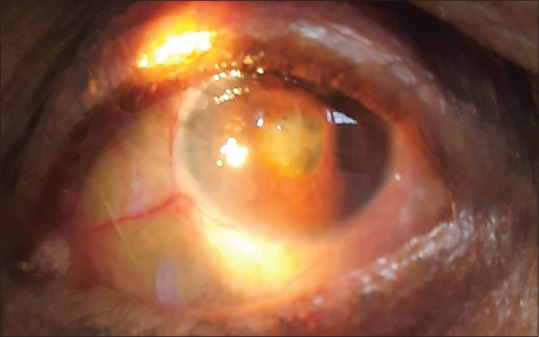
Patient 4 after first intravitreal injection – condensation of fibrinous plaque in the pupillary area
Figure 2.
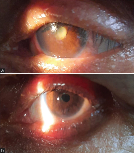
Patient 10 (a) at presentation and (b) after treatment
Figure 3.
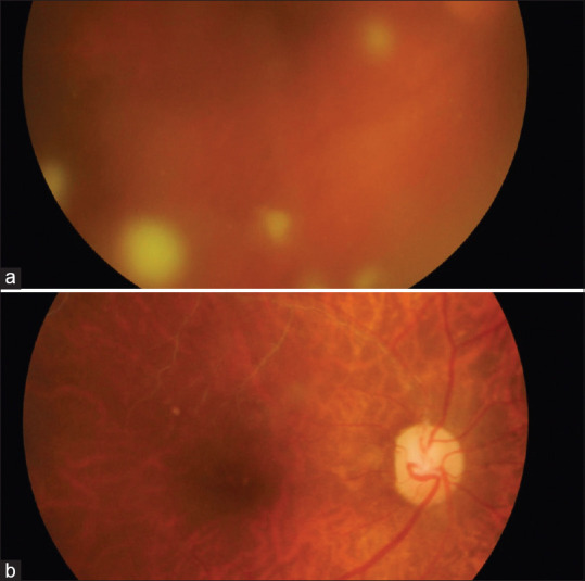
(a) Fundus photograph of patient 2 showing the cotton-wool-like vitreous deposits. (b) Fundus photograph of patient 2 after treatment. Note the old branch retinal vein occlusion (BRVO)
The first two cases received injection (inj.) vancomycin, ceftazidime, and amphotericin B as first the injection. After vitreous culture sensitivity report of patient 2 [Table 1] which showed excellent sensitivity of the fungal pathogen to voriconazole, the following patients received voriconazole and vancomycin as the first and subsequent injections. AC wash and capsular sac flushing were done with voriconazole plus vancomycin. Patients were examined on the 2nd, 5th, and 10th post-injection days and subsequent interventions were carried out when necessary. Follow-up was scheduled at 2, 4, 6, and 8 weeks. In patients 5, 6, 7—who required more than three intravitreal injections—amphotericin B was also added in subsequent injections, whereas in patients 8, 9, 10 amphotericin B was added in the second or third injection itself. Dexamethasone was added to the intravitreal injection after pars plana vitrectomy (PPV). The patients received an average of 3.7 antibiotic injections (range 2–5). Nine patients underwent core PPV. In 6 out of 9 cases, PPV was done as the second intervention. In five eyes, posterior capsulotomy was done during the PPV itself while in two eyes it was done by Nd: YAG laser capsulotomy before giving the subsequent intravitreal injection. In twp patients, IOL explantation with lens capsule removal was done.
Table 1.
Antifungal sensitivity by disc diffusion method for Trichosporon spp. (Patient 2)
| Drug | MIC |
|---|---|
| Fluconazole | |
| Sensitive | 16 microg/ml |
| Voriconazole | |
| Sensitive | 1 microg/ml |
| Itraconazole | |
| Resistant | 64 microg/ml |
| Amphotericin B | |
| Resistant | 4 microg/ml |
MIC was done by Broth dilution method (CLSI M27-A2). “Sensitive” indicates that the organism is inhibited by the usual achievable concentration of antibiotic with standard systemic dosage, while “resistant” implies that the organism is not inhibited by the achievable concentration of the standard dose. MIC: Minimum Inhibitory Concentration
All patients were administered oral fluconazole 150 mg daily for six weeks. Topical regimen included voriconazole eight times per day, dexamethasone (0.1%) plus moxifloxacin (0.5%) hourly, and homatropine 1% twice daily. Vitreous samples were sent for microbiological examination on all the instances of intervention. Gram stain, KOH stain, and culture were done for all samples. The culture was carried out on 5% sheep blood agar, chocolate agar, MacConkey agar, and brain heart infusion (BHI) broth. Further identification and antibiotic sensitivity were done whenever possible.
The onset and duration of symptoms, presenting vision, number of interventions, and final vision are tabulated in Table 2.
Table 2.
Presenting vision, time elapsed since the cataract surgery (days), latent period (days), duration of symptoms (days), number of interventions done, and the final visual outcome of the patients
| Case# | Presenting Vn | Time Since Surgery | Latent Period | No. of Interventions | Final Vn |
|---|---|---|---|---|---|
| 1 | HM | 75 | 45 (30) | 3 | FC-4 m |
| 2 | HM | 70 | 51 (19) | 2 | 6/24 |
| 3 | FC CF | 87 | 62 (25) | 4 | FC CF |
| 4 | HM | 95 | 40 (55) | 4 | FC CF |
| 5 | 6/60 | 98 | 36 (62) | 5 | 6/24 |
| 6 | HM | 108 | 38 (70) | 5 | PL+ |
| 7 | HM | 108 | 38 (70) | 5 | PL± |
| 8 | HM | 110 | 70 (40) | 4 | FC 3 m |
| 9 | 6/60 | 114 | 99 (15) | 2 | 6/18 |
| 10 | HM | 122 | 79 (45) | 3 | 6/24 |
Average latent period is 56 days after surgery (median: 46.5 days). The figures in parenthesis in the third column show the duration of symptoms in days before the patient presented at our clinic. Higher numbers seem associated with poorer prognosis
Results
All 10 eyes achieved infection-free status and maintained globe integrity. Four eyes achieved vision of 6/24 or better. Four eyes had final vision ranging between 6/60 and CF close to face while two eyes had just light perception. In all, seven patients showed improvement over their presenting vision. There was no change in one patient, while two patients had further fall in vision despite the treatment. The patients with the maximum duration of symptoms (70 days) had the most severe signs and unfavorable outcome of treatment while those with minimal duration of symptoms (15 days: patient 9 and patient 2) had better outcomes and faster convalescence.
Eyes that received voriconazole as the initial injection (8 of the 10 patients, patient 3 onwards) had re-appearance of signs (5–7 days) after initial improvement and required multiple injections. The decision to re-treat was based on reappearance of AC cells with hypopyon, fibrinous exudates in the pupillary area on the anterior lens surface [Fig. 4] with symptoms of pain and loss of recovered vision [Fig. 1]. There was recurrence of infection in six of the eight patients who underwent PPV and they were given repeat intravitreal injections with AC lavage. IOL explantation with capsular bag removal was done in two of the cases and helped in achieving an infection-free state.
Figure 4.
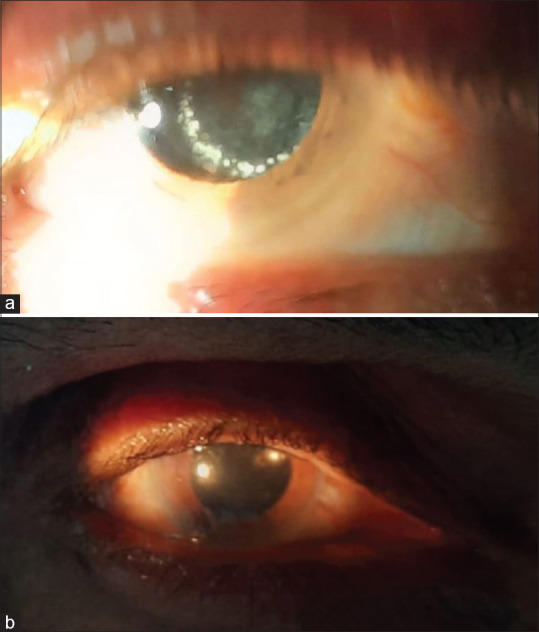
Patient 5. (a) Ring of white deposits on the posterior capsule after first intravitreal injection. (b) Final image after treatment
All the cases were culture positive for yeast on at least one occasion. Identification of Trichosporon spp. was obtained in five patients [Fig. 5] and the remaining five tested positive for “yeast like”, “slow growing” fungal element. Patients 4, 5, 7, 8, and 10 tested culture positive for fungus on more than one occasion, implying the pathogen persisted even after voriconazole injections. Patient 5 tested positive on three occasions [Table 3].
Figure 5.
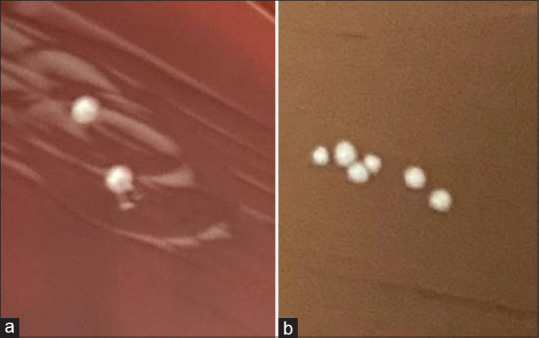
(a) Growth of the Trichosporon colonies on blood agar (patient 2). (b) Chocolate agar (patient 5)
Table 3.
Sensitivity of the Trichosporon spp. in the vitreous culture of the patient no. 5 after two intravitreal injections of voriconazole
| Drug | MIC (microg/ml) |
|---|---|
| Voriconazole | |
| Sensitive | 1 |
| Fluconazole | |
| Sensitive | 4 |
| Amphotericin B | |
| Resistant | 8 |
| Caspofungin | |
| Resistant | 4 |
| Micafungin | |
| Intermediate | 0.5 |
| Flucytosine | |
| Resistant | >64 |
MIC was done by Broth dilution method (CLSI M27-A2). “Sensitive” indicates that the organism is inhibited by usual achievable concentration of antibiotic with standard systemic dosage, while “resistant” implies that the organism is not inhibited by the achievable concentration of the standard dose
Discussion
Trichosporon is ubiquitous in nature: on soil, decomposing wood, stagnant water, rivers, lakes, air, and on rodent and cattle skin. In humans, it is part of the skin flora, especially of the inguinal and perianal region; it may temporarily colonize the respiratory or gastrointestinal (GI) tracts also. It is known to cause respiratory infection, skin infections (white piedra), sepsis, and eye infections.[7] Invasive trichosporonosis (sepsis and eye infections) is documented mostly in patients with hematological malignancies and other immunocompromised conditions, whereas skin infections and allergic pneumonia are documented in predominantly immune-competent hosts.[7] Reported infections of the eye till now were isolated cases in the immunocompromised.[11,12,13] This is the first instance when an incidence of cluster Trichosporon endophthalmitis in healthy individuals undergoing cataract surgery is being reported.
On talking to the surgeon, it was known that a total of 32 patients underwent operation on those three days. Of the 10 cases reported, three were operated on day 1, four on day 2, and three on day 3. The cases showed a mean latent period of over 49 days which is more than the reported 20 days for fungal endophthalmitis by Chakrabarti et al.[1] by almost a month. This highlights the slow replication rate of the causative organism. There was also a wide variation in the number of days between the onset of the symptoms of the patient and the time the first treatment for endophthalmitis was administered. This duration varied from 15 days to 70 days. Longer duration of symptoms was associated with more severe signs (patients 6 and 7) [Table 2] and a more unfavorable final outcome while definitive treatment with shorter duration of symptoms led to better prognosis.
The source of the infection could not be definitively identified due to the long time gap between the surgeries and the first diagnosis of endophthalmitis. The operation theater (OT) cultures turned out to be negative for Trichosporon. The batches of irrigating fluid and viscoelastic had already been consumed completely by the time the possibility of cluster endophthalmitis was conveyed to the surgeon. The ubiquitous presence of the organism makes it difficult to pinpoint the possible source but contamination of disposables with unsterile water was a high possibility. It can only be speculated that the likely culprit was the disposables packed in paper-plastic sealable bags (microblades, the polymethyl methacrylate [PMMA] intraocular lens). One of the sides of the packaging—which is porous paper and enables gas/steam sterilization—can also allow contamination of the contents on getting wet! The paper then invariably dries up but the pathogen may survive in the packaging or on the lens surface.
Systemic infections with Trichosporon spp. are associated with venous, vesical, or peritoneal catheter devices due to the ability of the fungal species to produce an extracellular matrix that allows the aggregation of cells and their adherence to both inorganic and organic surfaces.[5] Some studies suggest that the prosthetic devices act as substrates not only for adhesion but also for growth as colonies, embedded in the “extracellular polymeric substance” or the matrix produced by the fungal cells.[5]
This ability to form biofilms on implants – intraocular lens is the reason many of our patients had a thick adherent fibrous membrane covering the entire lens surfaces, which was extremely difficult to remove completely. The infection in patients 3, 4, 5, 6, 7, 8, and 10 persisted and multiple injections had to be given. It is likely that the biofilm aggregates of fungus on capsular bag and lens surface escaped the antifungals and acted as reservoirs of infection.
Trichosporon asahii biofilms have been found to be resistant to all antifungals, and up to 16,000 times more resistant to voriconazole than non-biofilm cells.[5] Trichosporon spp. exhibit an intrinsic resistance to the echinocandins (caspofungin, micafungin) and a poor susceptibility to the polyenes (amphotericin B), making it a difficult pathogen to manage.[4,7] Some Trichosporon species are also known to show reduced sensitivity to even triazoles![14,15] Yet another factor adding to the virulence of Trichosporon is the ability of the fungus to produce certain enzymes (proteases and phospholipases) that increase fungal pathogenicity by breaking up proteins and disrupting host cell membranes.[16]
Notably voriconazole’s half-life in the eye is in hours, perhaps a reason why symptoms reappeared after the initial improvement; the drug levels fell before the fungus could be eliminated. Its half-life in vitrectomized eyes is even lesser. (It is 6.5 hours in experimental animal models and 2.5 hours in vitrectomized eyes.)[17]
In the final outcome, patients who received amphotericin B (half-life of 8.9 days in vitreous and 1.8 days in vitrectomized eyes in same animal models)[18] did better and required lesser interventions (after receiving amphotericin B) compared to patients who received just voriconazole.
The concentration of amphotericin B given by intravitreal injection is 5 microg/0.1 ml or 50 microg/ml (Minimum Inhibitory Concentration [MIC]: 4 microg/ml) while the concentration of voriconazole given by intravitreal injection is 100 microg/0.1 ml or 1000 microg/ml (MIC: 1 microg/ml) [Table 1]. In accordance with the sensitivity reports, voriconazole should have eliminated the fungal pathogen; yet there were persistent culture-positive samples even after multiple voriconazole injections, while no sample came positive after amphotericin B injection. The fact that amphotericin B was able to eliminate the infection alone in the first two cases (as the subsequent culture came negative, though injection voriconazole was still administered at the time of subsequent intervention) and none of the culture samples came positive after amphotericin B injection in later cases indicate that the in vivo efficacy of amphotericin B against this Trichosporon spp. was much more than the reported in vitro efficacy. The culture positivity even after two intravitreal injections of voriconazole to which the pathogen was sensitive in vitro [Table 3] prompted the addition of another antifungal (amphotericin B) in all subsequent injections.
Literature search yields very few results for exogenous endophthalmitis case series where the pathogens are only yeasts. In most cases of endogenous ophthalmitis, intravitreal injections are used with high intravenous doses of antifungals.[19] Resistance of the pathogen to the administered drug has led to the usage of multiple drugs in combination.[10] On comparing with other such case series of Candida endophthalmitis being treated with voriconazole [Table 4], it is evident that multiple repeat injections of voriconazole (average 4+, range 2–9) were needed to eliminate the infection.
Table 4.
A comparison of reported case series of endophthalmitis caused by yeasts (Candida) treated by intravitreal voriconazole
| # | Case Series | No. of Cases | Pathogen | Antifungal Used | No. of Injections intervention/patient |
|---|---|---|---|---|---|
| 1. | Bienvenu, et al.[20] | 5 | Candida | Voriconazole | 4 (2-9) |
| 2. | Leung, et al.[21] | 16 | Candida (4) | Voriconazole/ | 4.4 ( +/- 2.8) |
| Amphotericin B/Miconazole | |||||
| 3. | Present study | 10 | Trichosporon | Voriconazole/ | 3.7 (2 - 5) |
| Amphotericin B | (PPV in 9/10) |
All patients in the present series received oral fluconazole after the first intravitreal injection but it did not seem to have any effect on the infection as the cultures continued to come positive irrespective. It would be safe to conclude that oral antifungals in the prescribed daily oral doses alone do not have any perceptible effect on the infection in cases of Trichosporon endophthalmitis.
Conclusion
Trichosporon is a multi-drug–resistant ubiquitous yeast that is capable of causing devastating cluster endophthalmitis in immunocompetent patients undergoing cataract surgery. Implants like intraocular lens and catheters in paper-plastic sealed bags can be contaminated by water-borne pathogens as the porous paper of the bag allows permeation of pathogens when wet, thereby increasing the risk of systemic infections including endophthalmitis. There is a definite pharmacokinetic advantage in administering the combination of voriconazole and amphotericin B for Trichosporon (yeast) endophthalmitis. Oral antifungals alone have little role in Trichosporon endophthalmitis.
Financial support and sponsorship
Nil.
Conflicts of interest
There are no conflicts of interest.
Acknowledgements
Dr. Vineeta Khare MD (Microbiology) for providing the microbiology support and sharing the images of the Culture plates.
References
- 1.Chakrabarti A, Shivaprakash MR, Singh R, Tarai B, George VK, Fomda BA, et al. Fungal endophthalmitis:Fourteen years'experience from a center in India. Retina. 2008;28:1400–7. doi: 10.1097/iae.0b013e318185e943. [DOI] [PubMed] [Google Scholar]
- 2.Fardeau C. Endophtalmies chroniques postopératoires [Chronic postoperative endophthalmitis. J Fr Ophtalmol. 2011;34:63–9. doi: 10.1016/j.jfo.2010.08.005. [DOI] [PubMed] [Google Scholar]
- 3.Das T, Joseph J, Jakati S, Sharma S, Velpandian T, Padhy SK, et al. Understanding the science of fungal endophthalmitis-AIOS 2021 SengameduSrinivasBadrinath Endowment Lecture. Indian J Ophthalmol. 2022;70:768–77. doi: 10.4103/ijo.IJO_2329_21. [DOI] [PMC free article] [PubMed] [Google Scholar]
- 4.de Almeida Júnior JN, Hennequin C. Invasive Trichosporon Infection:A systematic review on a Re-emerging Fungal Pathogen. Front Microbiol. 2016;7:1629. doi: 10.3389/fmicb.2016.01629. [DOI] [PMC free article] [PubMed] [Google Scholar]
- 5.Di Bonaventura G, Pompilio A, Picciani C, Iezzi M, D'Antonio D, Piccolomini R. Biofilm formation by the emerging fungal pathogen Trichosporonasahii:Development, architecture, and antifungal resistance. Antimicrob Agents Chemother. 2006;50:3269–76. doi: 10.1128/AAC.00556-06. [DOI] [PMC free article] [PubMed] [Google Scholar]
- 6.Liao Y, Lu X, Yang S, Luo Y, Chen Q, Yang R. Epidemiology and Outcome of Trichosporon Fungemia:A review of 185 reported cases from 1975 to 2014. Open Forum Infect Dis. 2015;2:ofv141. doi: 10.1093/ofid/ofv141. [DOI] [PMC free article] [PubMed] [Google Scholar]
- 7.Colombo AL, Padovan AC, Chaves GM. Current knowledge of Trichosporon spp. And Trichosporonosis. Clin Microbiol Rev. 2011;24:682–700. doi: 10.1128/CMR.00003-11. [DOI] [PMC free article] [PubMed] [Google Scholar]
- 8.Mithal K, Pathengay A, Bawdekar A, Jindal A, Vira D, Relhan N, et al. Filamentous fungal endophthalmitis:Results of combination therapy with intravitreal amphotericin B and voriconazole. Clin Ophthalmol. 2015;9:649–55. doi: 10.2147/OPTH.S80387. [DOI] [PMC free article] [PubMed] [Google Scholar]
- 9.Steinbach WJ, Stevens DA, Denning DW. Combination and sequential antifungal therapy for invasive aspergillosis: Review of published in vitro and in vivo interactions and 6281 clinical cases from 1966 to 2001. Clin Infect Dis. 2003;37((Suppl 3)):S188–224. doi: 10.1086/376524. [DOI] [PubMed] [Google Scholar]
- 10.Ostrosky-Zeichner L. Combination antifungal therapy:A critical review of the evidence. Clin Microbiol Infect. 2008;14((Suppl 4)):65–70. doi: 10.1111/j.1469-0691.2008.01983.x. [DOI] [PubMed] [Google Scholar]
- 11.Slocumb RW, Elner SG, Hall EF. Chronic postoperative fungal endophthalmitis caused by trichosporon asahii. Retin Cases Brief Rep. 2010;4:366–7. doi: 10.1097/ICB.0b013e3181b5ef61. [DOI] [PubMed] [Google Scholar]
- 12.Spirn MJ, Roth DB, Yarian DL, Green SN. Postoperative fungal endophthalmitis caused by Trichosporon beigelii resistant to amphotericin B. Retina. 2003;23:404–5. doi: 10.1097/00006982-200306000-00020. [DOI] [PubMed] [Google Scholar]
- 13.Sheikh HA, Mahgoub S, Badi K. Postoperative endophthalmitis due to Trichosporon cutaneum. Br J Ophthalmol. 1974;58:591–4. doi: 10.1136/bjo.58.6.591. [DOI] [PMC free article] [PubMed] [Google Scholar]
- 14.Iturrieta-González IA, Padovan AC, Bizerra FC, Hahn RC, Colombo AL. Multiple species of Trichosporon produce biofilms highly resistant to triazoles and amphotericin B. PLoS One. 2014;9:e109553. doi: 10.1371/journal.pone.0109553. [DOI] [PMC free article] [PubMed] [Google Scholar]
- 15.Padovan ACB, Rocha WPDS, Toti ACM, Freitas de Jesus DF, Chaves GM, Colombo AL. Exploring the resistance mechanisms in Trichosporon asahii:Triazoles as the last defense for invasive trichosporonosis. Fungal Genet Biol. 2019;133:103267. doi: 10.1016/j.fgb.2019.103267. [DOI] [PubMed] [Google Scholar]
- 16.Ghannoum MA. Potential role of phospholipases in virulence and fungal pathogenesis. Clin Microbiol Rev. 2000;13:122–43. doi: 10.1128/cmr.13.1.122-143.2000. [DOI] [PMC free article] [PubMed] [Google Scholar]
- 17.Shen YC, Wang MY, Wang CY, Tsai TC, Tsai HY, Lee YF, et al. Clearance of intravitreal voriconazole. Invest Ophthalmol Vis Sci. 2007;48:2238–41. doi: 10.1167/iovs.06-1362. [DOI] [PubMed] [Google Scholar]
- 18.Wingard LB, Jr, Zuravleff JJ, Doft BH, Berk L, Rinkoff J. Intraocular distribution of intravitreally administered amphotericin B in normal and vitrectomized eyes. Invest Ophthalmol Vis Sci. 1989;30:2184–9. [PubMed] [Google Scholar]
- 19.Danielescu C, Anton N, Stanca HT, Munteanu M. Endogenous Endophthalmitis:A review of case series published between 2011 and 2020. J Ophthalmol. 2020;2020:8869590. doi: 10.1155/2020/8869590. [DOI] [PMC free article] [PubMed] [Google Scholar]
- 20.Bienvenu AL, Aussedat M, Mathis T, Guillaud M, Leboucher G, Kodjikian L. Intravitreal Injections of Voriconazole for Candida Endophthalmitis:A case series. Ocul Immunol Inflamm. 2020;28:471–8. doi: 10.1080/09273948.2019.1571613. [DOI] [PubMed] [Google Scholar]
- 21.Leung EH, Kuriyan AE, Flynn HW, Jr, Relhan N, Huang LC, Miller D. Persistently vitreous culture-positive exogenous fungal endophthalmitis. Am J Ophthalmol. 2016;172:45–50. doi: 10.1016/j.ajo.2016.09.014. [DOI] [PMC free article] [PubMed] [Google Scholar]