Abstract
Anemia of inflammation (AI) is associated with inflammatory diseases, and inflammation-induced iron metabolism disorder is the major pathogenic factor. Earlier studies have reported a tendency of AI in periodontitis patients, but the explicit relationship and possible pathological mechanisms remain unclear. Here, the analyses of both periodontitis patients and a mouse model of ligature-induced experimental periodontitis showed that periodontitis was associated with lower levels of hemoglobin and hematocrit with evidence of systemic inflammation (increased white blood cell levels) and evidence of iron restriction (low serum iron along with a high serum hepcidin and ferritin levels), in accordance with the current diagnosis criteria for AI. Moreover, periodontal therapy improved the anemia status and iron metabolism disorders. Furthermore, the increased level of hepcidin and significant correlation between hepcidin and key indicators of iron metabolism emphasized the pivotal role of hepcidin in the pathogenesis of periodontitis-related AI. Administration of the signal transducer and activator of transcription 3 (STAT3) inhibitors Stattic suggested that the IL-6–STAT3–hepcidin signaling pathway participated in this regulatory process. Together, these findings demonstrated that periodontitis should be considered an inflammatory disease that contributes to the development of AI; furthermore, IL-6–STAT3–hepcidin signaling pathway plays a key regulatory role in the pathogenesis of periodontitis-related AI. Our study will provide new insights into the systemic effects of periodontitis, while meaningfully expanding the spectrum of inflammatory diseases that contribute to AI.
Keywords: anemia of inflammation, hepcidin, interleukin-6/signal transducer and activator of transcription 3/hepcidin pathway, iron metabolism, periodontal diseases
Introduction
Anemia of inflammation (AI), also referred to as anemia of chronic disease, is the second most prevalent type of anemia affecting hospitalized and chronically ill patients [1]. AI is classically associated with chronic systemic inflammatory disorders, including rheumatoid arthritis, inflammatory bowel disease, and chronic infections [2]. Hepcidin is known as the key mediator of AI [3]. Hepcidin functions by blocking the main iron flows into plasma and is up-regulated by inflammation. Hepcidin binds to ferroportin, the only known cellular iron exporter found on the cell surfaces, which induces its internalization and degradation and inhibits the exporter iron to plasma [4]. Inflammation-induced hepcidin leads to iron metabolism disorder and iron-restricted erythropoiesis, and eventually the development of AI [2]. In addition, shortened erythrocyte half-life and inhibition of erythroid cell differentiation further contribute to AI in a disease-specific pattern [5].
Periodontitis is also an inflammatory disease with the host immune responses against bacterial infections. Globally, the prevalence of severe periodontitis is approximately 10% [6]. Untreated periodontitis is the main cause of tooth loss and considered a significant threat to systemic health [7–9]. In periodontitis, pathogenic microorganisms and their products evoke immune-inflammatory responses in host tissues, resulting in increased white blood cell (WBC) counts as well as serum level of C-reactive protein (CRP) and various inflammatory cytokines, including interleukin (IL), interferons, and members of the tumor necrosis factor superfamily [10]. Recently, a few studies have reported that patients with periodontitis also exhibited the sign of AI [11]. Moreover, Guo et al. reported higher levels of serum hepcidin in periodontitis patients [12]. Our previous study has also shown a tendency of AI in periodontitis patients and also detected AI in experimental periodontitis mice with higher levels hepcidin of mRNA expression and serum concentrations [13]. However, the underlying pathological mechanisms remain unclear.
Therefore, the aims of the present study were to investigate the relationship between periodontitis and AI, and to elucidate the signaling pathways and molecular mechanisms involved in periodontitis-associated AI.
Materials and methods
Study population
The protocol of the human study was approved by the Ethics Committee of Peking University Health Science Center (Approval No. PKUSSIRB-202056090) and registered in the International Clinical Trials Registry Platform under the ID: ChiCTR2000040451. The research had been carried out in accordance with the World Medical Association Declaration of Helsinki, and written informed consent was obtained from all subjects. About 60 patients with periodontitis were recruited from the Department of Periodontology, Peking University School, and Hospital of Stomatology. The diagnosis of periodontitis was based on the diagnostic criteria proposed by 2017 Workshop for the Classification of Periodontal Diseases and Conditions [14]. In addition, 60 healthy subjects were selected with probing depth (PD) ≤3 mm, percentage of sites with bleeding on probing <10%, and no attachment loss (AL). Individuals who were current or previous smokers, and those with any systemic disease, pregnancy, history of iron supplementation or other drugs for the treatment of anemia, periodontal therapy or antimicrobial therapy, or use of any medication (including oral contraceptive drugs) within the previous 6 months were excluded.
After baseline examinations, all patients with periodontitis received non-surgical periodontal therapy by an experienced periodontist, which included oral hygiene instruction, supragingival scaling and quadrant-based subgingival scaling, and root planing under local anesthesia. Clinical re-evaluation and blood tests were conducted at 3 months after non-surgical periodontal therapy. The examinations at baseline and re-evaluation were performed by another calibrated periodontist.
Clinical and laboratory measurements
At baseline, all subjects underwent a full-mouth clinical periodontal examination by a calibrated periodontist, including PD and AL, using a William’s periodontal probe at six sites for each tooth. In addition, the bleeding index (BI) of each tooth was recorded. Periodontal parameters were re-evaluated by the same examiner at 3 months after non-surgical periodontal therapy.
Fasting blood samples were collected from each subject into ethylenediaminetetraacetic acid (EDTA)-containing tubes and analyzed by a technician who was blinded to the case status using a calibrated automated hematology analyzer (Sysmex, Corporation, Kobe, Japan) and a biochemistry automatic analyzer (model 7180; Hitachi High-Technologies Corporation, Tokyo, Japan). Serum was separated and stored immediately at −80°C. Serum concentrations of serum high-sensitivity C-reactive protein (hs-CRP), iron, ferritin and transferrin, in addition to transferrin saturation (TS), and total iron binding capacity (TIBC) were measured with an Automatic Analyzer (model 7170A; Hitachi High-Technologies Corporation). Enzyme-linked immunosorbent assay kits were used to measure serum concentrations of IL-6 (#KE1368; ImmunoWay Biotechnology Company, Plano, TX, U.S.A.) and hepcidin (#DHP250; R&D Systems, Inc., Minneapolis, MN, U.S.A.) in accordance with the manufacturers’ instructions.
Animal model of experimental periodontitis
All of the procedures performed on the animals followed the protocol approved by the Biomedical Ethics Committee of Peking University (approval no. LA2013-32) and conducted in accordance with the Guide for the Care and Use of Laboratory Animals. The animal experiments were conducted in the Animal Laboratory, Peking University School and Hospital of Stomatology, Beijing, China. C57BL/6 male mice 8 weeks of age weighing between 20 and 25 g were randomly allocated to one of four groups of 10 mice each. The mice were placed under general anesthesia via intraperitoneal injection of 1% sodium pentobarbital. The study design was illustrated in Figure 1. Experimental periodontitis was induced by placement of sterilized 5-0 cotton ligatures around the bilateral maxillary and mandibular second molars, followed by mesiobuccal knotting. All of the ligature-placed sites were checked daily until ligature removal or killing. Non-ligatured mice were used as negative controls. After ligation for 10 days, mice in the control group and experimental periodontitis group were killed, meanwhile the ligatures were removed from the other two groups and the mice were allowed to recover for 14 and 28 days prior to killing.
Figure 1. Study design.
Experimental periodontitis was induced by ligature placement around the bilateral maxillary and mandibular second molars. After 10 days of periodontitis progression, the ligatures were removed. Mice recovered for 7 or 14 days after the removal of the ligatures.
Stattic (Selleck Chemicals LLC, Houston, TX, U.S.A.), a small non-peptide molecule that inhibits activation and nuclear translocation of signal transducer and activator of transcription 3 (STAT3), was used for the inhibitor experiment. Mice were allocated to one of four group of 10 mice each: the control group, only Stattic application group, only ligature group, or Stattic + ligature group (n = 10 in each group). Stattic was intraperitoneally injected at 3.75 mg/kg body weight daily for 10 days [15]. Mice were killed by intraperitoneal injection of a sodium pentobarbital overdose.
Histological and immunohistochemical analyses of periodontal tissues
After sacrifice, the jaws of each mouse were fixed in 4% paraformaldehyde for 24 h at 4°C and then decalcified in 10% EDTA (pH 7.2 ± 0.2) at 37°C for 2 weeks (with ×3 solution changes per week). Serial paraffin-embedded sections of 5-μm thickness were obtained from the mesial-distal aspects of the second molars and stained with hematoxylin and eosin for histological evaluation. IL-6 expression in periodontal tissues was detected with a rabbit monoclonal antibody to IL-6 (Beijing Biosynthesis Biotechnology Co. Ltd., Beijing, China) in accordance with the manufacturer’s instructions. Images were captured under a BX51 light microscope equipped with a DP72 high sensitivity digital camera (Olympus Corporation, Tokyo, Japan).
Measurement of blood parameters
The blood samples were analyzed by a technician who was blinded to the experimental grouping using an automated hematology analyzer (MEK-6410C; Nihon Kohden Corporation, Tokyo, Japan). Serum was separated by centrifugation at 10,000 rpm for 5 min. Serum iron levels were measured with an automated biochemistry automatic analyzer (BS-350E; Mindray Medical International Limited, Shenzhen, China). Enzyme-linked immunosorbent assay kits were used to measure serum concentrations of IL-6 (#KE1418; ImmunoWay Biotechnology Company), hepcidin (#KE1286; ImmunoWay Biotechnology Company), ferritin (#KE1279; ImmunoWay Biotechnology Company), and transferrin (#ELM-TF; RayBiotech, Peachtree Corners, GA, U.S.A.) in accordance with the manufacturers' instructions.
RNA extraction and real-time quantitative polymerase chain reaction (RT-qPCR)
Total RNA was isolated from hepatocytes and liver tissues using TRIzol reagent (Invitrogen Corporation, Carlsbad, CA, U.S.A.) in accordance with the manufacturer’s instructions, then reverse-transcribed into complementary DNA using a ReverTra Ace™ reverse transcription kit (Toyobo Co., Ltd., Osaka, Japan) and amplified by RT-qPCR using PowerUp™ SYBR® Green Master Mix (Roche, Basel, Switzerland) and the primers listed in Supplementary Table S1. All reactions were conducted in triplicate. Relative mRNA expression was normalized to that of β-actin (mouse) or glyceraldehyde 3-phosphate dehydrogenase (GAPDH; human) using the 2−ΔΔCt method.
Western blot analysis
Liver tissues were irrigated with sterile phosphate buffer saline (PBS) to remove residual blood and cut into small pieces lysed in radioimmunoprecipitation assay buffer containing 1% phenylmethylsulfonyl fluoride (Sigma-Aldrich Corporation, St. Louis, MO, U.S.A.). Tissue fluid was extracted by ultrasonic degradation at 3000 rpm for 1 min and the supernatant was collected for protein detection and quantification using the Pierce BCA Protein Assay kits (Thermo Fisher Scientific, Waltham, MA, U.S.A.). About 30 μg protein from each sample was separated by electrophoresis and subsequently transferred to polyvinylidene difluoride membranes, which were blocked in Tris-buffered saline with Tween® 20 detergent containing 5% non-fat dried milk for 1 h, then washed and incubated with primary antibodies (dilution, 1:100) against hepcidin (#ab190775; Abcam, Cambridge, MA, U.S.A.), ferroportin (#NBP1-21502; Novus Biologicals, Littleton, CO, U.S.A.), GAPDH (#5174; Cell Signaling Technology, Inc., Danvers, MA, U.S.A.), ferritin (#4393; Cell Signal Technology, Inc.), phosphor-Janus kinase 2 (Jak2) (#3771; Cell Signal Technology, Inc.), Jak2 (#3230; Cell Signal Technology, Inc.), phosphor-STAT3 (#9145; Cell Signal Technology, Inc.), and STAT3 (#30835; Cell Signal Technology, Inc.) overnight at 4°C, followed by appropriate secondary antibodies (#7074; Cell Signaling Technology, Inc.) for 1 h at room temperature. The protein bands were visualized using an enhanced chemiluminescence kit (CoWin Biosciences, Beijing, China). All reactions were conducted in triplicate. The band intensities were analyzed using ImageJ software (https://imagej.nih.gov/ij/) for quantitative calculation. Relative protein expressions were normalized to that of GAPDH for statistical evaluation.
Statistical analysis
The results are presented as mean ± standard deviation (SD, normal distribution). Clinical and blood parameters were compared between the periodontitis group and the control group. Statistical analyses were conducted using the Student’s t-tests and Mann–Whitney tests for variables with normal and abnormal distribution, respectively, while categorical variables were compared with the Chi-square test. Intergroup comparisons of the results of all animal experiments were compared by one‐way analysis of variance followed by Turkey’s test. All statistical analyses were performed with IBM SPSS Statistic, version 20.0. (BM Corporation, Armonk, NY, U.S.A.) software. A two-tailed probability (P)-value below 0.05 was considered statistically significant.
Results
Changes in hematological parameters manifested the tendency of AI and iron metabolic disorders in periodontitis patients
The study cohort included 60 periodontitis patients and 60 healthy controls. All demographic features, periodontal clinical parameters, inflammatory markers, and anemia-related indicators with periodontitis patients and healthy subjects at baseline are shown in Table 1. The mean values of PD, BI, and AL of periodontitis patients were 4.42 ± 0.76 mm, 3.46 ± 0.62 mm, and 3.05 ± 1.10 mm, respectively, which were significantly higher than those of the control group (P<0.001, Table 1).
Table 1. Parameters of the study subjects.
| Variables | Healthy group (n=60) | Periodontitis group (n=60) | P values |
|---|---|---|---|
| Demographic features | |||
| Age | 31.03 ± 6.47 | 33.33 ± 5.45 | 0.081 |
| Gender (M/F) | 26/34 | 28/32 | 0.821 |
| Periodontal clinical parameters | |||
| PD (mm) | 1.56 ± 0.68 | 4.42 ± 0.76 | <0.001*** |
| BI | 1.12 ± 0.59 | 3.46 ± 0.62 | <0.001*** |
| AL (mm) | 0.30 ± 0.13 | 3.05 ± 1.10 | <0.001*** |
| Inflammatory markers | |||
| WBC (109/L) | 5.48 ± 1.24 | 6.40 ± 1.40 | 0.004** |
| hs-CRP (mg/L) | 0.36 ± 0.65 | 1.33 ± 1.75 | 0.002** |
| ESR | 5.89 ± 3.61 | 8.46 ± 4.22 | 0.008** |
| IL-6 (pg/mL) | 14.10 ± 3.99 | 19.89 ± 7.55 | 0.014* |
| Anemia-related indicators | |||
| RBC (1012/L) | 4.62 ± 0.49 | 4.60 ± 0.45 | 0.915 |
| HGB (g/L) | 140.20 ± 15.09 | 129.40 ± 13.08 | 0.003** |
| HCT (L/L) | 0.42 ± 0.04 | 0.40 ± 0.03 | 0.042* |
| MCV (fL) | 88.99 ± 3.82 | 88.34 ± 4.55 | 0.516 |
| MCH (pg/cell) | 30.39 ± 1.89 | 29.98 ± 2.09 | 0.379 |
| MCHC (g/dL) | 341.30 ± 11.85 | 339.00 ± 11.37 | 0.368 |
| Iron metabolism markers | |||
| Hepcidin (ng/ml) | 20.65 ± 16.42 | 38.91 ± 25.48 | <0.001*** |
| Fe (μmol/L) | 16.55 ± 5.43 | 13.16 ± 4.54 | 0.002** |
| Ferritin (ng/ml) | 105.3 ± 81.16 | 153.8 ± 106.50 | 0.044* |
| Transferrin (mg/dL) | 290.50 ± 54.97 | 252.2 ± 54.47 | <0.001*** |
| TS (%) | 27.63 ± 12.04 | 27.63 ± 7.75 | 0.337 |
| TIBC (μmol/L) | 54.37 ± 8.71 | 50.14 ± 10.19 | 0.037* |
Abbreviations: AL, attachment loss; BI, bleeding index; HCT, hematocrit; HGB, hemoglobin; hs-CRP, high-sensitivity C-reactive protein; IL-6, interleukin-6; MCH, mean corpuscular hemoglobin; MCHC, mean corpuscular hemoglobin concentration; MCV, mean corpuscular volume; PD, probing depth; RBC, red blood cell; TIBC, total iron binding capacity; TS, transferrin saturation; WBC, white blood cell. Data are presented as mean ± SD/N. Between-group comparisons were performed using t-test, Chi-square test, or Mann–Whitney U-test; *P<0.05, **P<0.01, ***P<0.001.
There were significant differences between the periodontitis group and control group in serum inflammatory markers, especially IL-6 levels (19.89 ± 7.55 vs. 14.10 ± 3.99 pg/ml, P=0.014), in addition to WBC counts (6.40 ± 1.40 vs. 5.48 ± 1.24 × 10⁹/L, P=0.004), serum hs-CRP (1.33 ± 1.75 vs. 0.36 ± 0.65 mg/L, P=0.002), and the erythrocyte sedimentation rate (ESR; 8.46 ± 4.22 vs. 5.89 ± 3.61, P=0.008).
As for anemia related indicators, mean hemoglobin (HGB) and hematocrit (HCT) values were significantly lower in the periodontitis patients group than the control group (129.40 ± 13.08 vs. 140.20 ± 15.09 g/L and 0.40 ± 0.03 vs. 0.42 ± 0.04 g/L, respectively, P<0.05; Table 1). The red blood cell (RBC) counts were also relatively lower in periodontitis patients, although the difference was not significant. Meanwhile, there was no statistical difference in mean corpuscular volume (MCV), mean corpuscular hemoglobin (MCH), or mean corpuscular hemoglobin concentration (MCHC) between the two groups.
As for iron metabolism markers, the mean serum hepcidin and ferritin were significantly up-regulated in the periodontitis group as compared to the control group (38.91 ± 25.48 vs. 20.65 ± 16.42 ng/ml, 153.8 ± 106.5 vs. 105.3 ± 81.16 ng/ml, respectively, P<0.05; Table 1), while serum iron, transferrin, and TIBC were significantly down-regulated (13.16 ± 4.54 vs. 16.55 ± 5.43 μmol/L, 252.2 ± 54.47 vs. 290.50 ± 54.97 mg/dL, and 50.14 ± 10.19 vs. 54.37 ± 8.71 μmol/L, respectively, P<0.05; Table 1) in periodontitis patients.
Significant correlations between hepcidin and IL-6, Fe, ferritin, and transferrin in periodontitis patients
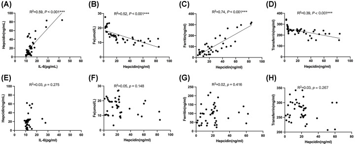
Interestingly, serum hepcidin was significantly correlated with serum IL-6 positively (R2 = 0.59, P<0.001; Figure 2A) but not hs-CRP, WBC, or ESR. In addition, serum hepcidin was positively correlated serum ferritin (R2 = 0.52, P<0.001; Figure 2B) and negatively correlated with serum iron (R2 = 0.74, P<0.001; Figure 2C) and transferrin (R2 = 0.39, P<0.001; Figure 2D).
Figure 2. Correlation analysesCorrelation analyses.
Correlations between IL-6 and hepcidin, hepcidin and Fe, ferritin, transferrin in periodontitis patients (A–D) and in treated periodontitis group (E–H). Scatter plots of IL-6 versus hepcidin (A,E) and hepcidin versus Fe (B,F), ferritin (C,G), and transferrin (D,H). Correlations were calculated by linear regression, ***P<0.001.
Non-surgical periodontal therapy ameliorated AI and dysregulation of iron metabolism
Inflammatory conditions were improved at 3 months after periodontal therapy, as demonstrated by significant reductions in WBC counts and IL-6 expression (5.62 ± 1.50 vs. 6.40 ± 1.40 × 10⁹/L and 14.76 ± 5.43 vs. 19.05 ± 8.55 pg/ml, respectively, P<0.05; Table 2) concomitant with significant decreases in PD and BI.
Table 2. Changes in parameters at 3 months after non- surgical periodontal therapy of periodontitis group.
| Variables | Baseline (n=40) | After therapy (n=40) | P values |
|---|---|---|---|
| Periodontal clinical parameters | |||
| PD (mm) | 4.42 ± 0.76 | 3.06 ± 0.49 | <0.001* |
| BI | 3.44 ± 0.62 | 1.83 ± 0.64 | <0.001* |
| Inflammatory markers | |||
| WBC (109/L) | 6.40 ± 1.40 | 5.62 ± 1.50 | 0.002** |
| hs-CRP (mg/L) | 1.33 ± 1.75 | 0.59 ± 0.48 | 0.025* |
| ESR | 8.46 ± 4.22 | 6.24 ± 3.87 | 0.116 |
| IL-6 (pg/ml) | 19.05 ± 8.55 | 14.76 ± 5.43 | 0.046* |
| Anemia-related indicators | |||
| RBC (1012/L) | 4.60 ± 0.45 | 4.67 ± 0.50 | 0.569 |
| HGB (g/L) | 129.40 ± 13.08 | 139.50 ± 14.85 | 0.002** |
| HCT (L/L) | 0.40 ± 0.03 | 0.42 ± 0.04 | 0.007** |
| MCV (fL) | 88.34 ± 4.55 | 88.61 ± 4.78 | 0.627 |
| MCH (pg/cell) | 29.98 ± 2.09 | 29.90 ± 2.25 | 0.872 |
| MCHC (g/L) | 339.00 ± 11.37 | 337.6 ± 9.01 | 0.594 |
| Iron metabolism markers | |||
| Hepcidin (ng/ml) | 40.25 ± 19.88 | 22.90 ± 24.61 | 0.034* |
| Fe (μmol/L) | 13.16 ± 4.54 | 15.97 ± 7.02 | 0.023* |
| Ferritin (ng/mL) | 153.8 ± 106.5 | 96.50 ± 70.10 | 0.024* |
| Transferrin (mg/dL) | 252.2 ± 54.47 | 283.2 ± 55.82 | 0.021* |
| TS (%) | 27.63 ± 7.75 | 29.17 ± 11.16 | 0.448 |
| TIBC (μmol/L) | 50.14 ± 10.19 | 55.03 ± 11.73 | 0.032* |
Abbreviations: AL, attachment loss; BI, bleeding index; HCT, hematocrit; HGB, hemoglobin; hs-CRP, high-sensitivity C-reactive protein; IL-6, interleukin-6; MCH, mean corpuscular hemoglobin; MCHC, mean corpuscular hemoglobin concentration; MCV, mean corpuscular volume; PD, probing depth; RBC, red blood cell; TIBC, total iron binding capacity; TS, transferrin saturation; WBC, white blood cell. Data are presented as mean ± SD/N. Between-group comparisons were performed using t-test, Chi-square test, or Mann–Whitney U-test; *P<0.05, **P<0.01, ***P<0.001.
Meanwhile, HGB and HCT in periodontitis patients were significantly increased at 3 months after periodontal therapy as compared with baseline values in the periodontitis group (139.50 ± 14.85 vs. 129.40 ± 13.08 g/L and 0.42 ± 0.03 vs. 0.40 ± 0.03 L/L, respectively, P<0.05; Table 2).
Periodontal therapy attenuates the increased serum levels of hepcidin and ferritin and decreased levels of iron and transferrin as compared with baseline values in periodontitis patients (22.90 ± 24.61 vs. 40.25 ± 19.88 ng/ml, 15.97 ± 7.02 vs. 13.16 ± 4.54 μmol/L, 96.50 ± 70.10 vs. 153.8 ± 106.5 ng/ml, and 283.2 ± 55.82 vs. 252.2 ± 54.47 mg/dL, respectively, P<0.05; Table 2).
Moreover, there was no significant correlation between IL-6 and hepcidin, hepcidin and Fe, and ferritin and transferrin at 3 months after periodontal therapy (Figure 2E–H).
Ligature-induced experimental periodontitis caused local inflammation of periodontal tissue and systemic inflammation in mice
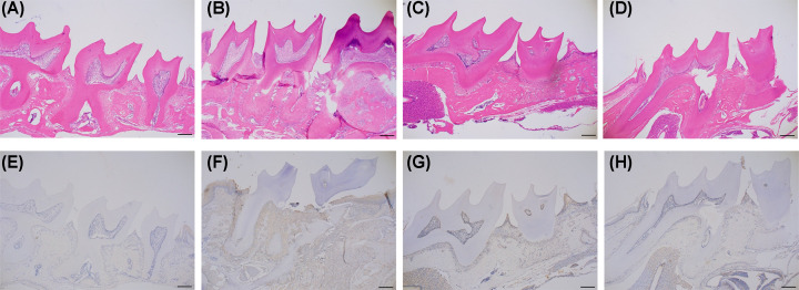
Histological analyses showed that as compared with the periodontal tissues of the control group (Figure 3A), those of the experimental periodontitis group showed dilation of the capillaries, infiltration of inflammatory cells, and breakdown of collagen. The epithelial–connective tissue interfaces and large areas of collagen-depleted connective tissues showed infiltration of numerous inflammatory cells along with intercellular edema (Figure 3B). Histological analyses of periodontal tissues following ligature application revealed infiltration of inflammatory cells with up-regulated levels of IL-6 in the connective tissues of the experimental periodontitis group (Figure 3F) as compared with the control group (Figure 3E).
Figure 3. H&E staining and immunohistochemical localization of IL-6.
H&E staining of periodontal tissues in the control group (A), ligature-induced periodontitis (B), recovery for 14 days (C), recovery for 28 days (D), IL-6 expression in periodontal tissues of control mice (E), and elevated expression of IL-6 in periodontal tissues of mice with ligature-induced periodontitis (F), recovery for 14 days (G), recovery for 28 days (H). Magnification: 40×; scale bar: 200 μm.
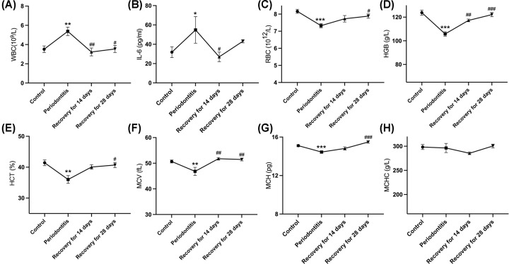
The WBC counts were significantly higher in the experimental periodontitis group than the control group (P<0.05). In the recovery phases, the WBC counts returned to the baseline when removed the ligature for 14 days (Figure 4A). Higher levels of pro-inflammatory cytokine IL-6 were similarly detected in the periodontitis group and similarly decreased to the normal levels in the recovery phases (P<0.05, Figure 4B).
Figure 4. Hematologic analyses.
Mean WBC counts (A), serum IL-6 concentrations (B), RBC (C), HGB (D), HCT (E), MCV (F), MCH (G), and MCHC (H) of mice in the control group, in the ligature-induced periodontitis group, in the recovery for 14 days group and those in the recovery for 28 days (n=10 in each group). Data are presented as mean ± SD. Between-group comparisons were performed using ANOVA test; *P<0.05, **P<0.01, ***P<0.001 as compared with the control group; #P<0.05, ##P<0.01, ###P<0.001 as compared with the periodontitis group.
Mice with ligature-induced experimental periodontitis develop AI and dysregulation of iron metabolism
Ligature-induced experimental periodontitis significant decreased in RBC counts, HGB, HCT, MCV, and MCH (7.32 ± 0.39 vs. 8.16 ± 0.40 × 10¹²/L, 105.8 ± 5.87 vs. 123.89 ± 6.89 g/L, 36.02 ± 3.99 vs. 41.42 ± 3.08%, 46.83 ± 3.85 vs. 50.68 ± 0.1.92 fL, and 14.44 ± 0.40 vs. 15.11 ± 0.37 pg, respectively, P<0.05; Figure 4C–F), although there was no significant change in mean corpuscular hemoglobin concentration (MCHC). The decrease of these parameters returned to the normal levels by day 28 after removal of the ligatures (P<0.05, Figure 4C–F).
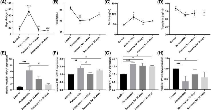
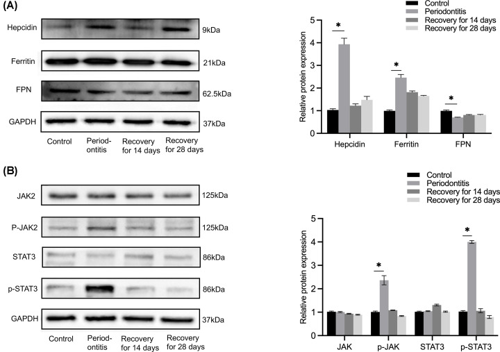
Moreover, higher serum levels of hepcidin were measured in mice with ligature-induced experimental periodontitis (73.21 ± 13.54 vs. 16.65 ± 7.48 ng/ml, P<0.001; Figure 5A). The RT-qPCR results revealed a 6-fold increase in hepcidin mRNA levels in these mice (P<0.01, Figure 5E). Hepcidin protein expression was also significantly increased in the liver specimens of the experimental periodontitis group (Figure 6A). These values were normalized at day 28 after removal of the ligatures. Serum iron concentration decreased after ligature and returned normal after 28 days (P<0.01, Figure 5B). Serum ferritin levels increased (84.62 ± 13.77 vs. 46.72 ± 4.53 ng/ml, P<0.05, Figure 5C) along with mRNA and protein expression levels (Figures 5F,G and 6A) during the 28-day observation period. Transferrin concentrations were also relatively lower in periodontitis group, although the differences were not significant (Figure 5D). Ferroportin mRNA and protein levels were dramatically decreased in the liver tissues of the periodontitis group and returned to the novel levels in the recovery phases (Figures 5H and 6A). In summary, 10 days after ligature, mice developed anemia with lower serum iron levels and higher hepcidin and ferritin levels, thereby fulfilling the criteria of AI. Moreover, all measured parameters returned to baseline levels by day 28 after removal of the ligatures.
Figure 5. Iron metabolism analyses.
Serum hepcidin (A), Fe (B), ferritin (C), transferrin (D) concentrations; mRNA expression levels of iron metabolism genes hepcidin (E), FTH (F), FTL (G), and FPN (H) in livers of the control group, in the ligature-induced periodontitis group, the recovery for 14 days group and those of the recovery for 28 days group (n = 10 in each group). Data are presented as mean ± SD. Between-group comparisons were performed using ANOVA test; *P<0.05, **P<0.01, ***P<0.001 as compared with the control group; #P<0.05 as compared with the ligature group.
Figure 6. Western blot analyses.
(A) Left: Western blot analysis of liver iron metabolism markers in four groups; right: quantification of band intensities. (B) Left: Western blot analysis of JAK-STAT signaling pathway in four groups; right: quantification of band intensities. Data are presented as mean ± SD. *P<0.05 as compared with the control group (n=3 independent experiments).
The JAK-STAT signaling pathway was activated in ligature-induced experimental periodontitis mice
To determine whether the JAK-STAT signaling pathway participates in periodontitis-induced AI, proteins expression levels of related biomarkers were measured. The results of Western blot analysis showed that ligature triggered phosphorylation of JAK2 and STAT3 (Figure 6B), which was down-regulated in the recovery phase.
The STAT3 inhibitor Stattic protected against experimental periodontitis-induced AI
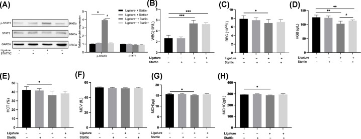
The results of Western blot analysis showed that Stattic had inhibited phosphorylation of STAT3 (Figure 7A) and up-regulated RBC counts and HCT levels equal to those of the control group (P<0.05, Figure 7B,C). HGB levels remained decreased after Stattic application but were greater than in the ligature group (P<0.05, Figure 7D).
Figure 7. Inhibition effect of Stattic and hematologic analyses under the application of Stattic.
(A) Left: Western blot analysis of liver biomarkers of JAK-STAT signaling pathway; right: quantification of band intensities, mean WBC counts (B), RBC (C), HGB(D), HCT (E), MCV (F), MCH (G,) and MCHC (H) of mice in the control group, in the STATTIC group, in the ligature group and those in the combined of ligature and STATTIC application group (n=10 in each group). Data are presented as mean ± SD. Between-group comparisons were performed using ANOVA test; *P<0.05, **P<0.01, ***P<0.001 as compared with the control group; #P<0.05 as compared with the ligature group.
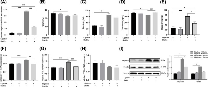
Serum hepcidin concentrations and hepcidin mRNA and protein levels were lower in the Stattic + ligature group as compared with the ligature group, but higher than in the control group (P<0.05, Figure 8A,E,I). Serum ferritin concentrations and FTH mRNA levels were down-regulated after Stattic administration (P<0.05, Figure 8C,F). Ferritin protein levels were relatively lower in the Stattic + ligature group as compared to the ligature group (Figure 8I). The decreased levels of serum iron and transferrin were returned to the normal levels after Stattic administration (Figure 8B,D). Moreover, the disorders of erythrocyte parameters and iron metabolism were improved after the inhibition of the STAT3 signaling pathway by administration of Stattic.
Figure 8. Iron metabolism analyses under the application of Stattic.
Serum hepcidin (A), Fe (B), ferritin (C), transferrin (D) concentrations; mRNA expression levels of iron metabolism genes hepcidin (E), FTH (F), FTL (G,) and FPN (H) in livers and Western blot analysis of liver iron metabolism markers (I left) and quantification of band intensities (I right) in the control group, in the Stattic group, in the ligature group and those in the combined of ligature and Stattic application group (n=10 in each group). Data are presented as mean ± SD. Between-group comparisons were performed using ANOVA test; *P<0.05, **P<0.01, ***P<0.001 as compared with the control group; #P<0.05, ##P<0.01, ###P<0.001 as compared with the ligature group.
Discussion
In summary, we explored the relationship between periodontitis and AI and underlying pathological mechanisms. Both periodontitis patients and the mouse model of ligature-induced experimental periodontitis showed relatively lower levels of HGB and HCT with evidence of systemic inflammation (increased WBC and CRP levels) and evidence of iron restriction (low serum iron with high serum hepcidin and ferritin levels), thereby fulfilling the current diagnostic criteria for AI [2,5]. Moreover, periodontal therapy or the removal of ligatures could improve anemia and iron metabolic disorders. We also found up-regulated level of hepcidin and significant correlation between hepcidin and key indicators of iron metabolism. In addition, the administration of STAT3 inhibitors Stattic could protect against experimental periodontitis induced AI. Togethering, these preliminary results demonstrated that periodontitis should be considered an inflammatory disease that contributes to the development of AI; furthermore, IL-6–STAT3–hepcidin signaling pathway plays a key regulatory role in the pathogenesis of periodontitis-related AI.
Tendencies toward periodontitis-related AI have been reported in some clinical researches and meta-analyses [11,12,16]. A recent meta-analysis enrolled 1423 periodontitis patients demonstrated that lower HGB levels, RBC counts, and MCV were associated with AI [17]. To the best of our knowledge, the present study was the first to report possible association between periodontitis and AI thoroughly, as well as periodontitis between hepcidin, inflammatory markers, and iron metabolism markers. Notably, at 3 months after periodontal therapy, the disorders of AI and iron metabolism improved, as well as disappearance of significant correlations between IL-6 and hepcidin, hepcidin and Fe, ferritin, and transferrin. These results of this study highlight the importance of periodontal therapy to prevent periodontitis-related AI. The resolution of periodontal infection could down-regulate the system inflammation and also improve the anemia and iron metabolic disorders of periodontitis patients over a 3-month period. Likewise, Pradeep et al. reported that nonsurgical periodontal therapy had improved RBC counts and clinical parameters over a 6-month period [18]. Periodontal medicine has recently been created and aims to treat the systemic diseases which are associated with periodontal disease through applying periodontal therapies [19]. It is recognized that such treatments may have a beneficial effect not only on the periodontal disease but also on some major systemic diseases, such as diabetes mellitus and cardiovascular disease [20,21]. Our results indicated that periodontal intervention can prevent periodontitis-related AI, thereby emphasizing the importance of regular periodontal examinations, as the most cost-effective strategy for the treatment and prevention of periodontal diseases and AI.
Hepcidin, a 25-amino acid peptide iron-regulatory hormone secreted by the liver, is known as the key mediator of AI [3]. The results of the present showed that serum hepcidin was dramatically increased in periodontitis patients (35.85 ± 32.23 vs. 17.15 ± 16.58 ng/ml), while serum levels of hepcidin were increased in mice with ligature-induced experimental periodontitis (73.21 ± 13.54 vs. 16.65 ± 7.48 ng/ml), as demonstrated by the qPCR and Western blot results. Hepcidin functions by blocking iron flows into blood. Hepcidin binds to ferroportin, the only known cellular iron exporter found on the cell surfaces, which induces its internalization and degradation and inhibits macrophage iron release and intestinal iron absorption [4]. Moreover, lower ferroportin mRNA and protein levels were detected in the periodontitis group. These preliminary evidences demonstrate that hepcidin plays a pivotal role in the development of periodontitis-related AI.
Hepcidin is also known as iron regulatory hormone and plays the key regulator of iron metabolism [22]. Inflammation-induced iron metabolism disorder is the major pathogenic factors of AI. As for the previous researchers on iron metabolism of periodontitis patients, Guo et al. reported that serum hepcidin levels were increased in chronic periodontitis groups [12]. A few studies have reported the lower serum iron [23] and transferrin levels [24] in periodontitis patients. Chakraborty et al. found the higher concentrations of serum ferritin [25]. However, these studies analyze these correlations individually in periodontitis patients. To the best of our knowledge, the present study was the first to report the iron metabolism disorder of periodontitis patients with higher levels of serum hepcidin and ferritin, lower levels of serum iron and transferrin, the significant positive correlations of ferritin and hepcidin, and negative correlation of hepcidin with serum iron and transferrin, which correspond to the iron metabolism disorder characteristic of AI patients [5]. In addition, the increase in hepcidin production and the disorder in iron distribution are regarded as parts of the host defense mechanism against infection [26]. Our previous studies found that ferritin was up-regulated by the inflammation in periodontitis and may contribute to amplify the innate immune responses of periodontitis [27]. These all indicated that iron metabolism disorder participated in host immune-inflammatory responses evoked by periodontitis. However, as a limitation of this study, we didn’t detect whether periodontal inflammation leads to the alternations of iron homeostasis in spleen. Theurl et al. have reported that AI resulting from chronic arthritis in rat caused increased expression of ferritin and decreased expression of ferroportin in spleen, which led to the impaired release of iron from macrophages and iron retention linked to AI [28]. In the future, we would need to analyze the alternations of iron homeostasis in spleen, together with the alternations of liver and system to complement pathological mechanisms of periodontitis-related AI.
Hepcidin synthesis is predominantly regulated by inflammation, especially by IL-6 [29]. Another convincing finding of the present study is that serum hepcidin levels in periodontitis patient correlated with serum IL-6 levels significantly positively (R2 = 0.47), but not other inflammatory markers, such as hs-CRP, WBC count, and ESR. For periodontitis mice, higher IL-6 expression levels were observed both in local periodontal tissue and serum. These results indicated that IL-6–hepcidin regulatory pathway is involved in periodontitis-related AI. Inflammatory responses via IL-6 are classically mediated via binding of IL6 to the IL6 receptor (p130) to activate the JAK kinases, which activate latent transcription factors, especially members of the STAT family [30]. IL-6 directly regulates hepcidin through induction and subsequent promoter binding of the STAT3 promoter, which is necessary and sufficient for the activation of IL-6 via the hepcidin promoter [31]. Phosphorylation of JAK2 and STAT3 was also observed in the periodontitis mice. To further confirm the pathogenic role of the IL-6-STAT3-hepcidin signaling pathway, the mice were treated with the STAT3 inhibitor Stattic at 3.75 mg/kg body weight for 10 days [32]. The results showed that Stattic protected against AI triggered by ligature-induced experimental periodontitis. These data provide strong evidence that the pro-inflammatory cytokine IL-6 is a primary inducer of increased hepcidin and that the IL-6–STAT3–hepcidin axis is necessary for the development of periodontitis-associated AI. Nevertheless, apart from IL-6, other cytokines and inflammatory pathways including interleukin-4, 10, 13 [33,34], tumor necrosis factors [35], interferon-γ [36], and Toll-like receptors 2 [37], have been reported could affect iron misdistribution in inflammatory diseases contributing to the development of AI, which is a reason why STAT3 inhibition had only a partial compensatory effect. Meanwhile, apart from IL-6, STAT3 also regulated IL-10 involved in iron retention upon infection [38] and erythropoietin involved in erythropoiesis [39]. Future research still needs to clarity whether the protective effect of Stattic involves the inhibition of other cytokines and hormones. Furthermore, other cytokines which could impair erythropoiesis including IL-1β and TNF-α were up-regulated in periodontitis patients [40]. Therefore, further studies are needed to clarify whether other cytokines, hormones, or signaling pathway engage in the periodontitis-related AI.
In conclusion, the results of the present study confirmed a strong association between periodontitis and AI and highlighted the preventive effect of periodontal therapy against periodontitis-related AI. Moreover, IL-6-STAT3-hepcidin signaling pathway plays a key regulatory role in the pathogenesis of periodontitis-related AI. These findings greatly contribute to a better understanding of periodontitis-related AI and supports the hypotheses that periodontitis, like other chronic conditions, can promote AI. The results of this study provide new insights into the systemic effects of periodontitis and meaningfully expand understanding of the contribution of inflammatory diseases to anemia.
Clinical perspectives
Recent studies reported patients with periodontitis exhibit signs of AI. However, the pathological mechanisms remain unclear. This study aimed to investigate the relationship between periodontitis and AI, and to further explore the possible pathogenic mechanisms underlying periodontitis-associated AI.
This study demonstrated that periodontitis should be considered an inflammatory disease that contributes to the development of AI. Moreover, IL-6–STAT3–hepcidin signaling pathway plays a key regulatory role in the pathogenesis of periodontitis-related AI.
This study confirmed a strong association between periodontitis and AI and highlighted the preventative effects of periodontal therapy on periodontitis-related AI. Elucidation of the mechanisms by which IL-6–STAT3–hepcidin signaling pathway interfere with iron metabolism and anemia of inflammation will provide new insights into the systemic effects of periodontitis, while meaningfully expanding the spectrum of inflammatory diseases that contribute to AI.
Supplementary Material
Abbreviations
- AI
anemia of inflammation
- AL
attachment loss
- BI
bleeding index
- CRP
C-reactive protein
- FTH
ferritin heavy polypeptide
- FTL
ferritin light polypeptide
- FPN
ferroportin
- hs-CRP
high-sensitivity C-reactive protein
- IL
interlukin
- Jak2
Janus kinase 2
- MCH
mean corpuscular hemoglobin
- MCHC
mean corpuscular hemoglobin concentration
- MCV
mean corpuscular volume
- PBS
phosphate buffer saline
- PD
probing depth
- STAT3
signal transducer and activator of transcription 3
- TIBC
total iron binding capacity
- TS
transferrin saturation
- WBC
white blood cell
Contributor Information
Yalin Zhan, Email: zhanyalin2014@126.com.
Jianxia Hou, Email: jxhou@bjmu.edu.cn.
Data Availability
Data are available from the corresponding author upon reasonable request.
Competing Interests
The authors declare that there are no competing interests associated with the manuscript.
Funding
This work was supported by the research funds from the National Natural Science Foundation of China [grant number 82071117]; the Natural Science Foundation of Beijing [grant number 7212136]; and the Youth Program of National Natural Science Foundation of China [grant number 81800976].
CRediT Author Contribution
Ye Han: Data curation, Software, Formal analysis, Supervision, Investigation, Methodology, Writing—original draft. Zhiqiang Luo: Resources, Data curation, Software, Validation, Investigation, Visualization. Zhao Guo Yue: Data curation. Li Li Miao: Data curation, Software. Min Xv: Data curation, Software. Shu Chang: Resources, Software, Methodology. Yalin Zhan: Data curation, Supervision, Funding acquisition, Writing—review & editing. Jianxia Hou: Conceptualization, Resources, Data curation, Formal analysis, Supervision, Funding acquisition, Validation, Investigation, Visualization, Methodology, Project administration, Writing—review & editing.
Ethics Approval
The clinical study was approved by the Ethics Committee of Peking University Health Science Center (Approval No. PKUSSIRB-202056090) and registered in the International Clinical Trials Registry Platform under the ID: ChiCTR2000040451. All of the procedures performed on the animals followed the protocol approved by the Experimental Animal Welfare Ethics Branch of Peking University Biomedical Ethics Committee (LA2013-32).
References
- 1.Weiss G. and Goodnough L.T. (2005) Anemia of chronic disease. N. Engl. J. Med. 352, 1011–1023 10.1056/NEJMra041809 [DOI] [PubMed] [Google Scholar]
- 2.Ganz T. (2019) Anemia of inflammation. N. Engl. J. Med. 381, 1148–1157 10.1056/NEJMra1804281 [DOI] [PubMed] [Google Scholar]
- 3.Ganz T. (2011) Hepcidin and iron regulation, 10 years later. Blood 117, 4425–4433 10.1182/blood-2011-01-258467 [DOI] [PMC free article] [PubMed] [Google Scholar]
- 4.Nemeth E., Tuttle M.S., Powelson J., Vaughn M.B., Donovan A., Ward D.M.et al. (2004) Hepcidin regulates cellular iron efflux by binding to ferroportin and inducing its internalization. Science 306, 2090–2093 10.1126/science.1104742 [DOI] [PubMed] [Google Scholar]
- 5.Weiss G., Ganz T. and Goodnough L.T. (2019) Anemia of inflammation. Blood 133, 40–50 10.1182/blood-2018-06-856500 [DOI] [PMC free article] [PubMed] [Google Scholar]
- 6.Frencken J.E., Sharma P., Stenhouse L., Green D., Laverty D. and Dietrich T. (2017) Global epidemiology of dental caries and severe periodontitis: a comprehensive review. J. Clin. Periodontol. 44, S94–S105 10.1111/jcpe.12677 [DOI] [PubMed] [Google Scholar]
- 7.Genco R.J. and Borgnakke W.S. (2013) Risk factors for periodontal disease. Periodontol 2000 62, 59–94 10.1111/j.1600-0757.2012.00457.x [DOI] [PubMed] [Google Scholar]
- 8.Tonetti M.S., Van Dyke T.E. and Working group 1 of the joint EFPAAPw (2013) Periodontitis and atherosclerotic cardiovascular disease: consensus report of the Joint EFP/AAP Workshop on Periodontitis and Systemic Diseases. J. Clin. Periodontol. 40, S24–S29 10.1111/jcpe.12089 [DOI] [PubMed] [Google Scholar]
- 9.de Smit M.J., Westra J., Brouwer E., Janssen K.M., Vissink A. and van Winkelhoff A.J. (2015) Periodontitis and rheumatoid arthritis: what do we know? J. Periodontol. 86, 1013–1019 10.1902/jop.2015.150088 [DOI] [PubMed] [Google Scholar]
- 10.Amano A. (2010) Host-parasite interactions in periodontitis: microbial pathogenicity and innate immunity. Periodontol 2000 54, 9–14 10.1111/j.1600-0757.2010.00376.x [DOI] [PubMed] [Google Scholar]
- 11.Nibali L., Darbar U., Rakmanee T. and Donos N. (2019) Anemia of inflammation associated with periodontitis: analysis of two clinical studies. J. Periodontol. 90, 1252–1259 10.1002/JPER.19-0124 [DOI] [PubMed] [Google Scholar]
- 12.Guo L.N., Yang Y.Z. and Feng Y.Z. (2018) Serum and salivary ferritin and Hepcidin levels in patients with chronic periodontitis and type 2 diabetes mellitus. BMC Oral Health 18, 63 10.1186/s12903-018-0524-4 [DOI] [PMC free article] [PubMed] [Google Scholar]
- 13.Han Y., Huang W., Meng H., Zhan Y. and Hou J. (2021) Pro-inflammatory cytokine interleukin-6-induced hepcidin, a key mediator of periodontitis-related anemia of inflammation. J. Periodontal Res. 56, 690–701 10.1111/jre.12865 [DOI] [PubMed] [Google Scholar]
- 14.Tonetti M.S., Greenwell H. and Kornman K.S. (2018) Staging and grading of periodontitis: framework and proposal of a new classification and case definition. J. Periodontol. 89, S159–S172 10.1002/JPER.18-0006 [DOI] [PubMed] [Google Scholar]
- 15.Scuto A., Kujawski M., Kowolik C., Krymskaya L., Wang L., Weiss L.M.et al. (2011) STAT3 inhibition is a therapeutic strategy for ABC-like diffuse large B-cell lymphoma. Cancer Res. 71, 3182–3188 10.1158/0008-5472.CAN-10-2380 [DOI] [PMC free article] [PubMed] [Google Scholar]
- 16.Franca L.F.C., da Silva F.R.P., di Lenardo D., Alves E.H.P., Nascimento H.M.S., da Silva I.A.T.et al. (2019) Comparative analysis of blood parameters of the erythrocyte lineage between patients with chronic periodontitis and healthy patients: Results obtained from a meta-analysis. Arch. Oral. Biol. 97, 144–149 10.1016/j.archoralbio.2018.10.024 [DOI] [PubMed] [Google Scholar]
- 17.Wu D., Lin Z., Zhang S., Cao F., Liang D. and Zhou X. (2019) Decreased hemoglobin concentration and iron metabolism disorder in periodontitis: systematic review and meta-analysis. Front. Physiol. 10, 1620 10.3389/fphys.2019.01620 [DOI] [PMC free article] [PubMed] [Google Scholar]
- 18.Pradeep A.R. and Anuj S. (2011) Anemia of chronic disease and chronic periodontitis: does periodontal therapy have an effect on anemic status? J. Periodontol. 82, 388–394 10.1902/jop.2010.100336 [DOI] [PubMed] [Google Scholar]
- 19.Loos B.G. (2016) Periodontal medicine: work in progress!. J. Clin. Periodontol. 43, 470–471 10.1111/jcpe.12550 [DOI] [PubMed] [Google Scholar]
- 20.Li C., Lv Z., Shi Z., Zhu Y., Wu Y., Li L.et al. (2017) Periodontal therapy for the management of cardiovascular disease in patients with chronic periodontitis. Cochrane Database Syst. Rev. 11, CD009197 10.1002/14651858.CD009197.pub3 [DOI] [PMC free article] [PubMed] [Google Scholar]
- 21.Baeza M., Morales A., Cisterna C., Cavalla F., Jara G., Isamitt Y.et al. (2020) Effect of periodontal treatment in patients with periodontitis and diabetes: systematic review and meta-analysis. J. Appl. Oral. Sci. 28, e20190248 10.1590/1678-7757-2019-0248 [DOI] [PMC free article] [PubMed] [Google Scholar]
- 22.Ganz T. (2013) Systemic iron homeostasis. Physiol. Rev. 93, 1721–1741 10.1152/physrev.00008.2013 [DOI] [PubMed] [Google Scholar]
- 23.Carvalho R.C.C., Leite S.A.M., Rodrigues V.P., Pereira A.F.V., Ferreira T.C.A., Nascimento F.R.F.et al. (2016) Chronic periodontitis and serum levels of hepcidin and hemoglobin. Oral Dis. 22, 75–76 10.1111/odi.12373 [DOI] [PubMed] [Google Scholar]
- 24.Shirmohamadi A., Chitsazi M.T., Faramarzi M., Salari A., Naser Alavi F. and Pashazadeh N. (2016) Effect of non-surgical periodontal treatment on transferrin serum levels in patients with chronic periodontitis. J. Dental Res. Dental Clin. Dental Prospects. 10, 169–175 10.15171/joddd.2016.027 [DOI] [PMC free article] [PubMed] [Google Scholar]
- 25.Chakraborty S., Tewari S., Sharma R.K. and Narula S.C. (2014) Effect of non-surgical periodontal therapy on serum ferritin levels: an interventional study. J. Periodontol. 85, 688–696 10.1902/jop.2013.130107 [DOI] [PubMed] [Google Scholar]
- 26.Nemeth E., Valore E.V., Territo M., Schiller G., Lichtenstein A. and Ganz T. (2003) Hepcidin, a putative mediator of anemia of inflammation, is a type II acute-phase protein. Blood 101, 2461–2463 10.1182/blood-2002-10-3235 [DOI] [PubMed] [Google Scholar]
- 27.Huang W., Zhan Y., Zheng Y., Han Y., Hu W. and Hou J. (2019) Up-regulated ferritin in periodontitis promotes inflammatory cytokine expression in human periodontal ligament cells through transferrin receptor via ERK/P38 MAPK pathways. Clin. Sci. (Lond.) 133, 135–148 10.1042/CS20180679 [DOI] [PubMed] [Google Scholar]
- 28.Theurl I., Aigner E., Theurl M., Nairz M., Seifert M., Schroll A.et al. (2009) Regulation of iron homeostasis in anemia of chronic disease and iron deficiency anemia: diagnostic and therapeutic implications. Blood 113, 5277–5286 10.1182/blood-2008-12-195651 [DOI] [PubMed] [Google Scholar]
- 29.Nemeth E., Rivera S., Gabayan V., Keller C., Taudorf S., Pedersen B.K.et al. (2004) IL-6 mediates hypoferremia of inflammation by inducing the synthesis of the iron regulatory hormone hepcidin. J. Clin. Invest. 113, 1271–1276 10.1172/JCI200420945 [DOI] [PMC free article] [PubMed] [Google Scholar]
- 30.Heinrich P.C., Behrmann I., Haan S., Hermanns H.M., Müller-Newen G. and Schaper F. (2003) Principles of interleukin (IL)-6-type cytokine signaling and its regulation. Biochem. J. 374, 1–20 10.1042/bj20030407 [DOI] [PMC free article] [PubMed] [Google Scholar]
- 31.Wrighting D.M. and Andrews N.C. (2006) Interleukin-6 induces hepcidin expression through STAT3. Blood 108, 3204–3209 10.1182/blood-2006-06-027631 [DOI] [PMC free article] [PubMed] [Google Scholar]
- 32.Schust J., Sperl B., Hollis A., Mayer T.U. and Berg T. (2006) Stattic: a small-molecule inhibitor of STAT3 activation and dimerization. Chem. Biol. 13, 1235–1242 10.1016/j.chembiol.2006.09.018 [DOI] [PubMed] [Google Scholar]
- 33.Ludwiczek S., Aigner E., Theurl I. and Weiss G. (2003) Cytokine-mediated regulation of iron transport in human monocytic cells. Blood 101, 4148–4154 10.1182/blood-2002-08-2459 [DOI] [PubMed] [Google Scholar]
- 34.Weiss G., Bogdan C. and Hentze M.W. (1997) Pathways for the regulation of macrophage iron metabolism by the anti-inflammatory cytokines IL-4 and IL-13. J. Immunol. 158, 420–425 10.4049/jimmunol.158.1.420 [DOI] [PubMed] [Google Scholar]
- 35.Sharp P. (2008) SNPs linking TNF with anemia. Blood 112, 3923–3924 10.1182/blood-2008-09-177238 [DOI] [PubMed] [Google Scholar]
- 36.Libregts S.F., Gutierrez L., de Bruin A.M., Wensveen F.M., Papadopoulos P., van Ijcken W.et al. (2011) Chronic IFN-gamma production in mice induces anemia by reducing erythrocyte life span and inhibiting erythropoiesis through an IRF-1/PU.1 axis. Blood 118, 2578–2588 10.1182/blood-2010-10-315218 [DOI] [PubMed] [Google Scholar]
- 37.Guida C., Altamura S., Klein F.A., Galy B., Boutros M., Ulmer A.J.et al. (2015) A novel inflammatory pathway mediating rapid hepcidin-independent hypoferremia. Blood 125, 2265–2275 10.1182/blood-2014-08-595256 [DOI] [PMC free article] [PubMed] [Google Scholar]
- 38.Hutchins A.P., Diez D. and Miranda-Saavedra D. (2013) The IL-10/STAT3-mediated anti-inflammatory response: recent developments and future challenges. Brief Funct. Genomics 12, 489–498 10.1093/bfgp/elt028 [DOI] [PMC free article] [PubMed] [Google Scholar]
- 39.Kuo S.C., Li Y., Cheng K.C., Niu C.S., Cheng J.T. and Niu H.S. (2017) Increase in renal erythropoietin receptors in diabetic rats is mainly mediated by hyperglycemia associated with the STAT3/GATA-1 signaling pathway. Biomed. Pharmacother. 96, 1094–1102 10.1016/j.biopha.2017.11.115 [DOI] [PubMed] [Google Scholar]
- 40.Nemeth E. and Ganz T. (2014) Anemia of inflammation. Hematol. Oncol. Clin. North Am. 28, 671–681, vi 10.1016/j.hoc.2014.04.005 [DOI] [PMC free article] [PubMed] [Google Scholar]
Associated Data
This section collects any data citations, data availability statements, or supplementary materials included in this article.
Supplementary Materials
Data Availability Statement
Data are available from the corresponding author upon reasonable request.


