Abstract
We reported this case because calcification of the mitral valve is a common complication of rheumatic fever, but calcification of the left atrium is rare.
Keywords: left atrial calcification, rheumatic fever
We reported this case because calcification of the mitral valve is a common complication of rheumatic fever, but calcification of the left atrium is rare.

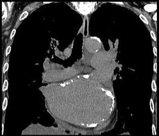
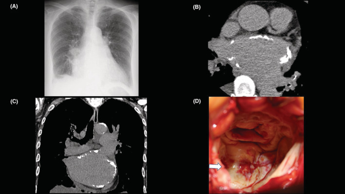
A 73‐year‐old woman with a previous history of cerebral infarction and rheumatic fever was referred for heart failure and atrial fibrillation. A chest X‐ray showed an enlarged cardiac shadow and pleural effusion (A). The plain chest computed tomography (CT) showed massive calcification (B, C) of left atrium (LA). Echocardiography revealed severe calcification of mitral valve and concomitant mitral stenosis (0.43 cm2). Surgical mitral valve replacement was performed. The left atrium was wholly calcified, as in CT (D, white arrow). The patient was discharged from the hospital postoperatively without any complications. The presence of massive LA calcification is extremely rare, and it has been proposed to represent the result of long‐standing rheumatic heart disease. Surgical removal of the LA calcification was associated with difficulty in entering the LA, potential for embolization, and hemostatic failure, with a surgical mortality rate of up to 25% (Figure 1). 1
FIGURE 1.

(A) Chest X‐ray at the admission. (B) The plain chest computed tomography (CT) showed left atrial massive calcification (axial view). (C) The plain chest computed tomography (CT) showed left atrial massive calcification (coronal view). (D) The left atrium was wholly calcified, as in CT.
AUTHOR CONTRIBUTIONS
Masaki Monden: Data curation.
FUNDING INFORMATION
None.
CONFLICT OF INTEREST STATEMENT
We have no potential conflicts of interest related to this manuscript.
CONSENT
Written informed consent was obtained from the patient to publish this report in accordance with the journal's patient consent policy.
ACKNOWLEDGMENTS
The authors thank Dr Makoto Taoka, Dr. Katsuaki Yokoyama, Dr. Michiaki Matsumoto, Dr. Takehiko Washio, Dr. Yasuyuki Suzuki, Dr. Dr. Tsukasa Yagi, Dr Tetsuro Yumikura, Dr Yuro Matsunaga and Dr. Youji Watanabe for contributing to the treatment.
Monden M, Fukamachi D, Matsumoto N, Okumura Y. Massive left atrial calcification. Clin Case Rep. 2023;11:e6919. doi: 10.1002/ccr3.6919
DATA AVAILABILITY STATEMENT
Data available on request due to privacy/ethical restrictions.
REFERENCE
- 1. Leung EC, Hirani N. Pulmonary hypertension caused by a coconut left atrium. Can J Cardiol. 2020;36(10):1691.e1691‐1691.e1692. [DOI] [PubMed] [Google Scholar]
Associated Data
This section collects any data citations, data availability statements, or supplementary materials included in this article.
Data Availability Statement
Data available on request due to privacy/ethical restrictions.


