

Extracorporeal traction-assisted endoscopic submucosal dissection (ESD) for superficial pharyngeal carcinoma has been useful 1 2 3 . However, owing to the anatomical features of the larynx and thyroid cartilage, working spaces within the pharynx are narrow. Furthermore, instruments such as intubation tubes, laryngoscopes, grasping forceps 1 , and thin endoscopes 2 for traction of the lesion interfere with the endoscope, making endoscopic maneuverability difficult. Methods to overcome such difficulties have been reported in recent years 4 5 .
Although clip-and-thread traction is useful 3 , the direction of traction cannot be adjusted. Herein, we report the use of a novel clip–traction band device for intraluminal traction to achieve pharyngeal ESD, overcoming the disadvantages of the conventional traction method 1 2 3 .
A 69-year-old man presented with a flat-elevated lesion extending from the left pyriform sinus to the aryepiglottic fold ( Fig. 1 a ). He had restricted mouth opening due to previous reconstructive surgery and radiation therapy for buccal mucosal carcinoma. The patient underwent ESD under general anesthesia, and laryngeal expansion was performed using a curved laryngoscope to obtain a good view of the entire lesion. After a circumferential incision, a clip–traction band device (Elastic Traction Device; Micro-Tech, Nanjing, China) was deployed. Good traction allowed safe dissection with clear submucosal visualization; however, the traction force gradually decreased as dissection progressed. Therefore, re-traction was attempted using the second ring and en bloc resection was achieved with good traction force maintenance ( Fig. 1 b–d ; Video 1 ).
Fig. 1.
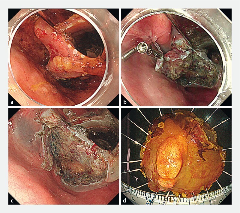
Endoscopic view of clip–traction band device-assisted pharyngeal endoscopic submucosal dissection (ESD). a Lesion after marking. Lack of staining with iodine is observed from the left pyriform sinus to the aryepiglottic fold. b Re-traction of the lesion using a clip–traction band device. c Mucosal defect after ESD. d Resected specimen. The tumor (26 × 22 mm) was diagnosed as squamous cell carcinoma in situ with no lymphovascular invasion and negative margins.
Video 1 Successful pharyngeal endoscopic submucosal dissection using a novel clip–traction band device.
The traction band has two silicone rings which enable the redirection of tension or re-tension. The silicone rings are small and have poor extensibility, making this device suitable for use in the narrow working spaces of the pharynx. Moreover, the maneuverability of the endoscope during submucosal dissection was not restricted because no grasping forceps were required ( Fig. 2 ). Hence, the clip–traction band device may be a useful tool for pharyngeal ESD.
Fig. 2.
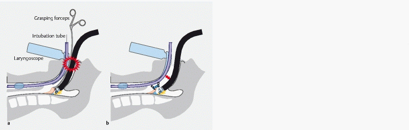
Scheme of the differences in working space within the pharynx between grasping forceps traction-assisted ESD and clip–traction band-assisted ESD. a Grasping forceps traction-assisted ESD. The grasping forceps interfere with the endoscope. b Clip–traction band-assisted ESD. Adequate working space for the endoscope is ensured.
Endoscopy_UCTN_Code_TTT_1AO_2AC
Footnotes
Competing interests The authors declare that they have no conflict of interest.
Endoscopy E-Videos : https://eref.thieme.de/e-videos .
Endoscopy E-Videos is an open access online section, reporting on interesting cases and new techniques in gastroenterological endoscopy. All papers include a high quality video and all contributions are freely accessible online. Processing charges apply (currently EUR 375), discounts and wavers acc. to HINARI are available. This section has its own submission website at https://mc.manuscriptcentral.com/e-videos
References
- 1.Iizuka T, Kikuchi D, Hoteya S et al. A new technique for pharyngeal endoscopic submucosal dissection: peroral countertraction (with video) Gastrointest Endosc. 2012;76:1034–1038. doi: 10.1016/j.gie.2012.07.013. [DOI] [PubMed] [Google Scholar]
- 2.Yoshio T, Tsuchida T, Ishiyama A et al. Efficacy of double-scope endoscopic submucosal dissection and long-term outcomes of endoscopic resection for superficial pharyngeal cancer. Dig Endosc. 2017;29:152–159. doi: 10.1111/den.12712. [DOI] [PubMed] [Google Scholar]
- 3.Minami H, Tabuchi M, Matsushima K et al. Endoscopic submucosal dissection of the pharyngeal region using anchored hemoclip with surgical thread: a novel method. Endosc Int Open. 2016;4:E828–E831. doi: 10.1055/s-0042-108802. [DOI] [PMC free article] [PubMed] [Google Scholar]
- 4.Matsuno K, Miyamoto H, Tanaka M. Novel traction method for pharyngeal endoscopic submucosal dissection using ring-shaped thread and grasping forceps. Dig Endosc. 2020;32:e120–e121. doi: 10.1111/den.13718. [DOI] [PubMed] [Google Scholar]
- 5.Waki K, Kanesaka T, Ishihara R et al. A soft hood improves maneuverability in narrow spaces during pharyngeal endoscopic submucosal dissection. Endoscopy. 2021;53:E384–E385. doi: 10.1055/a-1304-3234. [DOI] [PubMed] [Google Scholar]