Abstract
Case summary
Minimally invasive surgery is an increasingly popular alternative to open surgery in veterinary medicine. Compared with traditional surgical approaches, laparoscopic pancreatectomy provides a less invasive approach and has several potential benefits, including improved visualization, reduced infection rate and decreased postoperative pain. Laparoscopic partial pancreatectomy has been described in humans, dogs and pigs but not cats. Pancreatectomy with or without chemotherapy is a treatment option for exocrine pancreatic carcinoma, a rare but malignant cancer in cats. We report the case of a 16-year-old male neutered domestic longhair cat diagnosed with exocrine pancreatic carcinoma that was treated with laparoscopic partial pancreatectomy, carboplatin and toceranib phosphate. A three-port technique using a 5 mm 0º telescope and bipolar vessel sealing device was performed to remove the entire left limb of the pancreas. No intra- or postoperative complications occurred, and the patient was discharged the following day. Forty days postoperatively, the patient received its first of five doses of carboplatin, which were given every 4–5 weeks over a period of 4 months. A maintenance protocol of toceranib phosphate was started after completion of carboplatin treatment. At the time of this article being submitted, the patient had survived for more than 221 days.
Relevance and novel information
This is the first report of a laparoscopic partial pancreatectomy performed on a feline patient for pancreatic carcinoma.
Keywords: Pancreatic carcinoma, laparoscopy, minimally invasive surgery, partial pancreatectomy
Case description
A 6 kg 16-year-old male neutered domestic longhair cat was presented to the Cornell Small Animal Community Practice for a 3-month history of decreased appetite and lethargy following dental extractions. Its previous medical history included an episode of acute-onset anorexia and lethargy 1 year prior that resolved without treatment. An abdominal ultrasound was performed at that time, which revealed a mild, diffuse, pancreatopathy with several small, hypoechoic nodules in the right limb of the pancreas, and a large (1.8 × 1.3 cm) hypoechoic nodule in the left limb of the pancreas. A fine-needle aspiration (FNA) of the pancreas was recommended but declined. Physical examination at the time of re-presentation revealed a new grade II/VI left systolic murmur but was otherwise unremarkable. Abdominal ultrasound confirmed the presence of pancreatic nodules with mild abdominal lymphadenopathy. An ultrasound-guided FNA of the previously visualized left pancreatic nodule was performed. The right nodules were too small for aspiration. The cytology from the left nodule was interpreted as probable pancreatic exocrine carcinoma.
The patient was referred to Cornell University Hospital for Animals for surgical resection of the pancreatic masses. Bloodwork revealed no relevant abnormalities. Thoracic radiographs were negative for metastasis, and an echocardiogram showed hypertrophic cardiomyopathy without evidence of atrial enlargement. No cardiac medication was prescribed.
CT showed a small (2.5 cm × 3.2 cm × 2.0 cm [length × width × height]), moderately heterogeneous mass in the cranial aspect of the left pancreatic limb, as well as a small (0.8 × 0.8 × 1.0 cm [length × width × height]), poorly marginated, heterogeneous nodule in the caudal extent of the left pancreatic limb (Figure 1). A small, right caudal pulmonary nodule and multifocal nodules within the liver and spleen were also observed on CT but were indeterminate for abdominal or thoracic metastases. The observed pancreatic masses were not invasive into adjacent structures; therefore, surgical excision was recommended. The owner elected to pursue laparoscopic resection of the pancreatic masses owing to the potential for decreased postoperative pain, recovery time and infection rate.
Figure 1.
Postcontrast CT images of the pancreatic masses: (a) left cranial pancreatic limb mass (arrows) in transverse plane; (b) the same left cranial pancreatic limb mass (arrows) in dorsal plane, in relation to the transverse colon; and (c) left caudal pancreatic limb mass (arrows) in transverse plane
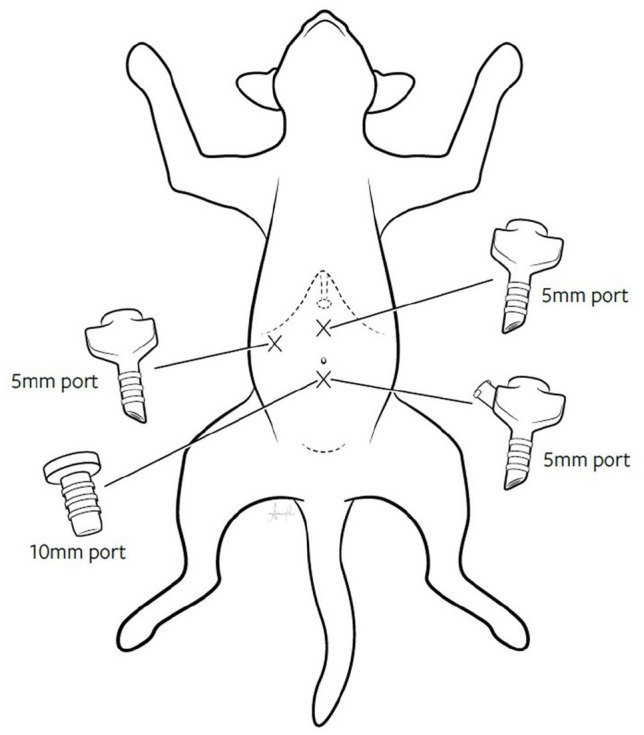
Prior to surgery, the patient was administered cefazolin (22 mg/kg IV), pantoprazole (1 mg/kg IV) and maropitant (1 mg/kg IV). The patient was placed under general anesthesia and positioned in dorsal recumbency. A three-port laparoscopic technique was chosen. The first 5 mm port (Geniport Pyramidal tip trocar and cannula system; Genicon) was placed via modified Hasson technique just caudal to the umbilicus, and the abdomen was insufflated to 6 mmHg with CO2. A 0º laparoscope (5 mm Hopkins telescope; Karl Storz) was inserted to complete a brief intra-abdominal exploration, which showed no peritoneal/serosal metastasis but a diffusely mottled liver parenchyma with multifocal, raised dark red and green nodules. The second 5 mm port was placed 4 cm to the right of midline just caudal to the last rib, and the third 5 mm port was placed 4 cm cranial to the umbilicus on midline under visualization (Figure 2).
Figure 2.

Illustration of the three-port laparoscopic technique. The first 5 mm port was placed just caudal to the umbilicus, the second 5 mm port was placed 4 cm to the right of midline just caudal to the last rib and the third 5 mm port was placed 4 cm cranial to the umbilicus on midline
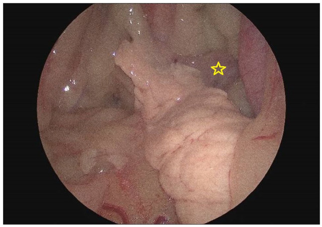
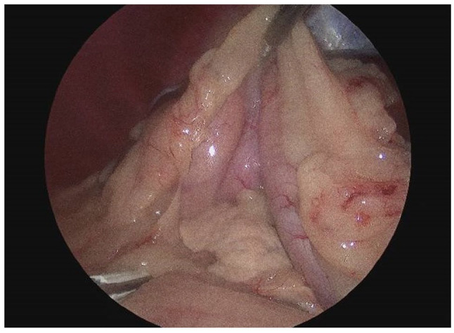
The superficial leaf of the omentum was manipulated with a blunt laparoscopic probe and 5 mm laparoscopic Babcock forceps, and a 5 mm dolphin-tip vessel sealing device (VSD) (Ligasure Dolphin Tip Laparoscopic Sealer/Divider; Covidien) was used to enter the omental bursa to visualize the left limb of the pancreas. The pancreas appeared normal in color, but two obvious nodules were seen within the left limb: one in the mid-limb and one approximately 1 cm from the body of the pancreas (Figure 3). Grasping the mesentery with an atraumatic laparoscopic forceps and starting at the distal tip of the left limb of the pancreas, the VSD was used to deliberately dissect the pancreatic tissue from the mesentery (Figure 4). Specific care was paid to the splenic artery as it coursed near the left limb. Once the left limb was fully dissected, the VSD was used to seal and transect the tissue at the body of the pancreas. The 5 mm umbilical port was exchanged for a 10 mm port and a sterile retrieval bag (Endobag; Coviden) was used to remove the left limb of the pancreas through the 10 mm port.
Figure 3.

Laparoscopic image. The pancreas appeared normal in color, but a dark pink nodule (star) was seen within the left limb approximately 1 cm from the body of the pancreas
Figure 4.

Laparoscopic image. A laparoscopic blunt probe is seen retracting the stomach cranially and ventrally, and the vessel sealing device seen at the bottom of the screen was used to dissect the pancreatic tissue from the mesentery
Laparoscopic cup biopsy forceps were used to take biopsies from multiple nodules within the left medial and lateral lobe of the liver, and hemorrhage was controlled with gentle pressure using a laparoscopic blunt probe. No active hemorrhage was noted at the conclusion of surgery, and all incisions were closed routinely with 3-0 polydioxanone (fascia) and 3-0 Monocryl (intradermal). The total surgery time was 76 mins, and the total anesthesia time was 122 mins.
The patient recovered uneventfully from anesthesia and was placed on a maintenance intravenous fluid (Plasmalyte; 45 ml/kg/day), fentanyl constant rate infusion (2 µg/kg/h), gabapentin (10 mg/kg PO q8h), cefazolin (22 mg/kg IV q8h), maropitant (1 mg/kg IV q24h) and pantoprazole (1 mg/kg IV q12h). The patient remained stable overnight, ate well the next morning and was discharged to the care of its owner 16 h postoperatively with a prescription for buprenorphine (0.02 mg/kg oral transmucosal q8–12h) and gabapentin (10 mg/kg PO q8–12h) for 5 days. The owner administered both medications q12h the for the first 2 days postoperatively and then only once the third day postoperatively. These medications were decreased and then discontinued based on the owner’s perception of the patient’s comfort level, activity and appetite.
Histopathology of the pancreatic mass was consistent with a well-differentiated, multifocal exocrine pancreatic acinar carcinoma with complete margins at the body of the pancreas. Liver changes were benign, revealing a completely excised biliary cyst, completely excised nodular hyperplasia, moderate centrilobular lipofuscinosis and dissecting fibrosis. Owing to the multifocal nature of the carcinoma throughout the left limb and the nodules seen on imaging in the right limb of the pancreas, the decision was made to treat with adjuvant chemotherapy.
At follow-up examination 18 days later, the patient was bright with normal vital parameters and was reported to have increased appetite and energy at home. The patient’s chemotherapy treatment was delayed due to severe constipation, which resolved after the introduction of polyethylene glycol 3350 and multiple enemas over several days.
Forty days postoperatively, the patient received its first of five doses of carboplatin (180 mg/m2 IV) scheduled to be received at 4-week intervals. Aside from a 1-week delay in administration of the second and fourth doses owing to two respective episodes of grade 1 neutropenia (Veterinary Co-operative Oncology Group – Common Terminology Criteria for Adverse Events),1 the patient’s chemotherapy regimen was given without significant interruption. Periodic surveillance imaging and laboratory testing were unremarkable.
Four months from the first carboplatin treatment, the patient had CT of the thorax, abdomen and pelvis that revealed 5–8 small pulmonary nodules most consistent with metastatic carcinoma. The right pancreatic limb was subjectively more nodular compared with previous studies. Five months postoperatively, thoracic radiographs were repeated and showed no metastasis visible in the area of previously identified pulmonary nodules on CT. The patient was started on a maintenance protocol of toceranib phosphate at 2.6 mg/kg PO every Monday, Wednesday and Friday for 2 weeks. At the time of submission, the patient had survived for more than 221 days, was eating and drinking normally, and the owner reported normal activity and interactions.
Discussion
Exocrine pancreatic cancer is responsible for <0.5% of all cancers in dogs and cats, yet of those tumors, most are malignant.2,3 Historically, feline pancreatic carcinoma has carried a grave prognosis owing to metastatic spread and rapid disease progression.4–7 In a 2004 study of eight cats with pancreatic adenocarcinoma, all were euthanized or had died within 7 days of diagnosis, with only supportive care provided.5 The overall metastatic rate has been reported to be between 32% and 50% in retrospective studies of feline patients, and common sites of metastasis include the local lymph nodes, liver, lung, small intestine and peritoneum.4,5,8
While the prognosis of feline pancreatic carcinoma has been guarded historically, early diagnosis and treatment with surgical resection and/or chemotherapy has led to improved survival times.4,6,7 In a study of 34 cats with exocrine pancreatic carcinoma, the median survival time for patients that had received chemotherapy or had their masses surgically removed was 165 days.4 In a recent study of nine cats with exocrine pancreatic carcinoma without identifiable metastatic disease that underwent surgery with or without adjunctive chemotherapy, median overall survival was 316 days.6
Minimally invasive surgery (MIS) is an increasingly popular alternative to open surgery in veterinary medicine. Reported benefits in small animals include decreased postoperative pain, surgical trauma, recovery time and infection rate.3,9–11 In addition, magnification and illumination of the surgical site are superior to traditional surgery.10 Despite this, cats undergo laparoscopic procedures less frequently than dogs owing to instrumentation and working space challenges in smaller patients.10,12
Compared with open laparotomy, laparoscopic partial pancreatectomy for the excision of pancreatic adenocarcinoma in humans appears to be associated with shorter operative and recovery time, reduced blood loss, fewer complications and a similar to decreased overall morbidity and mortality.13,14 An experimental model in dogs revealed quicker recovery of gastrointestinal transit and lower intraoperative cortisol levels in those undergoing laparoscopic partial pancreatectomy compared with open surgery.15 Additionally, a case report documented the first laparoscopic partial pancreatectomy in a dog with a pancreatic beta cell tumor that survived for over 2 years.16 MIS techniques are known to lead to improved patient comfort, but an additional benefit for oncologic patients may include the ability to start adjunctive treatment sooner owing to faster recovery time.17 Unfortunately, that was not the case in this patient, as comorbidities delayed adjuvant therapy. In patients with an oncologic diagnosis, any comorbidity that might delay treatment should be aggressively managed to avoid this postponement.
There is currently no established chemotherapy protocol for feline pancreatic carcinoma patients; therefore, the protocol used for this patient was chosen by the oncology service based on reported outcomes of cats with carcinomas.4,6,7,18–20 One study reported that the gemcitabine–carboplatin combination was moderately tolerated in cats with carcinomas, though side effects such as neutropenia and gastrointestinal toxicity led to treatment delays and minimal overall benefit.18 Toceranib phosphate has been shown to be well tolerated in cats with many types of neoplasia, including pancreatic carcinoma, with toxicity limited to mild gastrointestinal or myelosuppressive effects or, rarely, hepatotoxicity.7,19,20 Two case reports of cats with pancreatic carcinoma treated with toceranib phosphate showed survival rates of 792 days (without surgery) and over 1436 days (with surgery).7,20
To our knowledge, this is the first laparoscopic partial pancreatectomy that has been performed on a feline patient. The advantages of laparoscopic partial pancreatectomy may also be recognized in feline patients.13–15 This surgery occurred without complication, suggesting that a laparoscopic approach may be a viable and perhaps superior alternative compared with open pancreatic procedures.
Conclusions
Laparoscopic partial pancreatectomy is feasible in feline patients with exocrine pancreatic carcinoma. To determine the benefit of laparoscopic pancreatectomy over open laparotomy, further research on a larger feline population is necessary.
Footnotes
The authors declared no potential conflicts of interest with respect to the research, authorship, and/or publication of this article.
Funding: The authors received no financial support for the research, authorship, and/or publication of this article.
Ethical approval: The work described in this manuscript involved the use of non-experimental (owned or unowned) animals. Established internationally recognised high standards (‘best practice’) of veterinary clinical care for the individual patient were always followed and/or this work involved the use of cadavers. Ethical approval from a committee was therefore not specifically required for publication in JFMS Open Reports. Although not required, where ethical approval was still obtained, it is stated in the manuscript.
Informed consent: Informed consent (verbal or written) was obtained from the owner or legal custodian of all animal(s) described in this work (experimental or non-experimental animals, including cadavers) for all procedure(s) undertaken (prospective or retrospective studies). No animals or people are identifiable within this publication, and therefore additional informed consent for publication was not required.
ORCID iD: Jenna Menard  https://orcid.org/0000-0002-4440-2912
Nicole J Buote  https://orcid.org/0000-0003-4623-3582
References
- 1. Veterinary Co-operative Oncology Group (VCOG). Veterinary Co-operative Oncology Group–Common Terminology Criteria for Adverse Events (VCOG-CTCAE) following chemotherapy or biological antineoplastic therapy in dogs and cats v1.0. Vet Comp Oncol 2004; 2: 195–213. [DOI] [PubMed] [Google Scholar]
- 2. Selmic LE. Exocrine pancreatic cancer. In: Vail DM, Thamm DH, Liptak JM. (eds). Withrow and McEwan’s small animal clinical oncology. 6th ed. St Louis, MO: Elsevier, 2020, pp 451–452. [Google Scholar]
- 3. Buishand FO, van Nimwegen SA, Kirpensteijn J. Laparoscopic surgery of the pancreas. In: Fransson BA, Mayhew PD. (eds). Small animal laparoscopy and thoracoscopy. Ames, IA: ACVS Foundation and Wiley-Blackwell, 2015, pp 167–177. [Google Scholar]
- 4. Linderman MJ, Brodsky EM, de Lorimier L-P, et al. Feline exocrine pancreatic carcinoma: a retrospective study of 34 cases. Vet Comp Oncol 2013; 11: 208–218. [DOI] [PubMed] [Google Scholar]
- 5. Seaman RL. Exocrine pancreatic neoplasia in the cat: a case series. J Am Anim Hosp Assoc 2004; 40: 238–245. [DOI] [PubMed] [Google Scholar]
- 6. Nicoletti R, Chun R, Curran KM, et al. Postsurgical outcome in cats with exocrine pancreatic carcinoma: nine cases (2007–2016). J Am Anim Hosp Assoc 2018; 54: 291–295. [DOI] [PubMed] [Google Scholar]
- 7. Todd JE, Nguyen SM. Long-term survival in a cat with pancreatic adenocarcinoma treated with surgical resection and toceranib phosphate. JFMS Open Rep 2020; 6. DOI: 10.1177/2055116920924911. [DOI] [PMC free article] [PubMed] [Google Scholar]
- 8. Knell S, Venzin C. Partial pancreatectomy and splenectomy using a bipolar vessel sealing device in a cat with an anaplastic pancreatic carcinoma. Schweiz Arch Für Tierheilkd 2012; 154: 298–301. [DOI] [PubMed] [Google Scholar]
- 9. Mayhew PD, Freeman L, Kwan T, et al. Comparison of surgical site infection rates in clean and clean-contaminated wounds in dogs and cats after minimally invasive versus open surgery: 179 cases (2007–2008). J Am Vet Med Assoc 2012; 240: 193–198. [DOI] [PubMed] [Google Scholar]
- 10. Case JB, Boscan PL, Monnet EL, et al. Comparison of surgical variables and pain in cats undergoing ovariohysterectomy, laparoscopic-assisted ovariohysterectomy, and laparoscopic ovariectomy. J Am Anim Hosp Assoc 2015; 51. DOI: 10.5326/JAAHA-MS-5886. [DOI] [PubMed] [Google Scholar]
- 11. Gauthier O, Holopherne-Doran D, Gendarme T, et al. Assessment of postoperative pain in cats after ovariectomy by laparoscopy, median celiotomy, or flank laparotomy. Vet Surg 2015; 44 Suppl 1: 23–30. [DOI] [PubMed] [Google Scholar]
- 12. Buote NJ, Hayes G, Bisignano J, et al. Retrospective comparison of open vs minimally invasive cystotomy in 28 cats using a composite outcome score. J Feline Med Surg 2022; 24: 1032–1038. [DOI] [PMC free article] [PubMed] [Google Scholar]
- 13. Chen K, Pan Y, Huang C-J, et al. Laparoscopic versus open pancreatic resection for ductal adenocarcinoma: separate propensity score matching analyses of distal pancreatectomy and pancreaticoduodenectomy. BMC Cancer 2021; 21. DOI: 10.1186/s12885-021-08117-8. [DOI] [PMC free article] [PubMed] [Google Scholar]
- 14. Yang D-J, Xiong J-J, Lu H-M, et al. The oncological safety in minimally invasive versus open distal pancreatectomy for pancreatic ductal adenocarcinoma: a systematic review and meta-analysis. Sci Rep 2019; 9. DOI: 10.1038/s41598-018-37617-0. [DOI] [PMC free article] [PubMed] [Google Scholar]
- 15. Naitoh T, Garcia-Ruiz A, Vladisavljevic A, et al. Gastrointestinal transit and stress response after laparoscopic vs conventional distal pancreatectomy in the canine model. Surg Endosc 2002; 16: 1627–1630. [DOI] [PubMed] [Google Scholar]
- 16. Mcclaran JK, Pavia P, Fischetti AJ, et al. Laparoscopic resection of a pancreatic β cell tumor in a dog. J Am Anim Hosp Assoc 2017; 53: 338–345. [DOI] [PubMed] [Google Scholar]
- 17. Balsa IM, Culp WTN. Use of minimally invasive surgery in the diagnosis and treatment of cancer in dogs and cats. Vet Sci 2019; 6. DOI: 10.3390/vetsci6010033. [DOI] [PMC free article] [PubMed] [Google Scholar]
- 18. Martinez-Ruzafa I, Dominguez PA, Dervisis NG, et al. Tolerability of gemcitabine and carboplatin doublet therapy in cats with carcinomas. J Vet Intern Med 2009; 23: 570–577. [DOI] [PubMed] [Google Scholar]
- 19. Harper A, Blackwood L. Toxicity and response in cats with neoplasia treated with toceranib phosphate. J Feline Med Surg 2017; 19: 619–623. [DOI] [PMC free article] [PubMed] [Google Scholar]
- 20. Dedeaux AM, Langohr IM, Boudreaux BB. Long-term clinical control of feline pancreatic carcinoma with toceranib phosphate. Can Vet J 2018; 59: 751–754. [PMC free article] [PubMed] [Google Scholar]


