Dear Editor,
Tracheostomy causes iatrogenic tracheal stenosis (ITS) in 65% patients and is harder in keloid susceptible patients.[1] This report describes unanticipated ‘cannot ventilate cannot intubate’(CVCI) scenario in such a patient.
A 25-year-old, 60 kg ASA I male patient was scheduled for removal of Montgomery tube (MT), a T tube used as a tracheal stent for a functional trachea. His past history revealed prolonged endotracheal intubation and emergency tracheostomy post extubation for respiratory distress. Six months back, a 14 no MT was inserted through tracheal stoma.
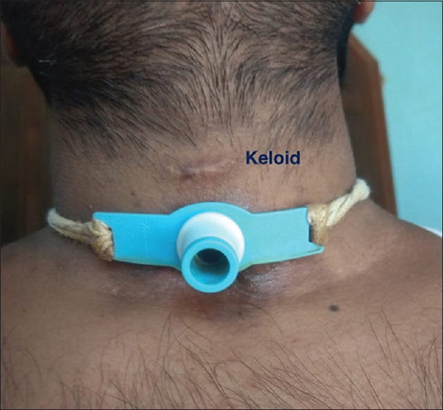
For anesthesia, an orotracheal tube (OTT) 6.0 mm was attached to the anterior limb of MT and fentanyl 2 mcg/kg and sevoflurane 2-6% with air oxygen mixture with a flow rate of 8 L/min was started. On confirming chest expansion with bag mask ventilation, atracurium 0.5 mg/kg was injected. Attempt of MT removal required considerable force and attempted insertion of a flexometallic cuffed orotracheal tube (FCOTT) 7.0 mm through the tracheal stoma was not possible due to extreme resistance of tissues. Subsequent attempts by decreasing sizes of various endotracheal tubes till size 3.0 mm and a rigid ventilating bronchoscope size 0, failed. Needle cricothyrotomy, due to low tracheostomy and surgical widening of stoma, due to extremely hard tissues, was unsuccessful, causing CVCI and fall in SPO2. In a desperate attempt, OTT was placed over the posterior tracheal wall of tracheal stoma, APL valve closed and manual ventilation with high flows of 10 L/min of 100% O2 started. SPO2 slowly increased though no capnograph and chest expansion was visible. Surgeons dissected tracheal tissue, almost like cutting a wire, and managed to first insert OTT 3.0 mm and after further incision of stenotic tissue a COTT 6.5 mm was inserted. Oral bronchoscope revealed hard stenosis of 3 cm from stoma till 4 cm above the carina with sclerosis of the anterior wall of trachea. A keloid was noticed on upper side of neck at the previous suture site for tracheostomy tube [Figure 1]. Rest of the surgery was uneventful. The patient was discharged after a week.
Figure 1.

Keloid in the patient on neck at previous suture line
Anesthesia with tracheostomy and MT is challenging.[2,3] To the best of our knowledge anesthesia in tracheostomy patients with MT and keloid susceptibility has not been described before. Keloid, is an excessive connective tissue, hard in texture that develops in a healing scar.[1] Retrospective data of tracheostomized 2276 patients showed severe tracheal stenosis in patients with keloid with almost 80% cicatricial tissue.
Thus, in patients with tracheostomy scheduled for anesthesia, history for keloid susceptibility in patient and family members should be sought for. In such patients for securing airway a ventilating bougie should first be passed from anterior limb of MT to the lower vertical limb into the trachea, MT then removed over the bougie and OTT railroaded over the bougie into the trachea. In case the stomal fibrosis prevents passage of OTT into the trachea, oxygenation and ventilation can be carried out with the ventilating bougie till definitive measures are taken.
Declaration of patient consent
The authors certify that they have obtained all appropriate patient consent forms. In the form the patient(s) has/have given his/her/their consent for his/her/their images and other clinical information to be reported in the journal. The patients understand that their names and initials will not be published and due efforts will be made to conceal their identity, but anonymity cannot be guaranteed.
Financial support and sponsorship
Nil.
Conflicts of interest
There are no conflicts of interest.
References
- 1.Chang E, Wu L, Masters J, Lu J, Zhou S, Zhao W, et al. Iatrogenic subglottic tracheal stenosis after tracheostomy and endotracheal intubation: A cohort observational study of more severity in keloid phenotype. Acta Anaesthesiol Scand. 2019;63:905–12. doi: 10.1111/aas.13371. [DOI] [PMC free article] [PubMed] [Google Scholar]
- 2.Hu H, Zhang J, Wu F, Chen E. Application of the Montgomery T-tube in subglottic tracheal benign stenosis. J Thorac Dis. 2018;10:3070–7. doi: 10.21037/jtd.2018.05.140. [DOI] [PMC free article] [PubMed] [Google Scholar]
- 3.Kerai S, Gupta R, Wadhawan S, Bhadoria P. Anesthetic management of a patient with Montgomery t-tube in-situ for direct laryngoscopy. J Anaesthesiol Clin Pharmacol. 2013;29:105–7. doi: 10.4103/0970-9185.105815. [DOI] [PMC free article] [PubMed] [Google Scholar]


